Image search results - "artery"
|

Central Retinal Artery Occlusion - 2 months old by history1489 views77-year-old man was doing fine. His left eye was a little worse than the right eye, and then in the 2 months ago he had sudden severe vision loss in the right eye. Vision OD is 3/200; OS is 20/60. IOP: OD 30, OS 15.
OD: Vertical C/D ratio is 0.4. The macula is white from ischemia. The arteries are narrow.
Photos confirm clinical findings.
OCT SCAN: OCT scan of the right eye shows retinal thickening, consistent with macular edema associated with a central retinal artery occlusion. The left eye has severe inferior retinal thinning, consistent with an old hemicentral retinal artery occlusion in the left eye. The fovea in the left eye is a little bit thin but not too bad.
IMPRESSION: 1. CENTRAL RETINAL ARTERY OCCLUSION RIGHT EYE
2 OLD HEMICENTRAL RETINAL ARTERY OCCLUSION LEFT EYE
3 DIABETES WITHOUT RETINOPATHY
4 ELEVATED INTRAOCULAR PRESSURE RIGHT EYE STATUS POST INJECTION RIGHT EYE A WEEK AGO
DISCUSSION: I explained to the patient that the right eye unfortunately has had a central retinal artery and unfortunately we do not have anything to treat that. His vision will probably improve some over time. He needs to be watched though. It is possible he is developing rubeotic glaucoma, although it is probable the intraocular pressure is high from the injection last week. I asked him to restart the Alphagan, to stop the other two drops, and to return here in three weeks to make sure his pressure is more acceptable.
At this point, having lost almost all the retinal circulation in the right eye and half of the retinal circulation in the left eye, he only has half a retina in the left eye he is working off of. I told him it is very important he keep seeing you and anything that can be done to keep his blood pressure low and his cholesterol reasonable would be helpful to what remains of his retinal circulation.
|
|

Central Retinal Artery Occlusion - 2 months old by history802 views77-year-old man was doing fine. His left eye was a little worse than the right eye, and then in the 2 months ago he had sudden severe vision loss in the right eye. Vision OD is 3/200; OS is 20/60. IOP: OD 30, OS 15.
OD: Vertical C/D ratio is 0.4. The macula is white from ischemia. The arteries are narrow.
Photos confirm clinical findings.
OCT SCAN: OCT scan of the right eye shows retinal thickening, consistent with macular edema associated with a central retinal artery occlusion. The left eye has severe inferior retinal thinning, consistent with an old hemicentral retinal artery occlusion in the left eye. The fovea in the left eye is a little bit thin but not too bad.
IMPRESSION: 1. CENTRAL RETINAL ARTERY OCCLUSION RIGHT EYE
2 OLD HEMICENTRAL RETINAL ARTERY OCCLUSION LEFT EYE
3 DIABETES WITHOUT RETINOPATHY
4 ELEVATED INTRAOCULAR PRESSURE RIGHT EYE STATUS POST INJECTION RIGHT EYE A WEEK AGO
DISCUSSION: I explained to the patient that the right eye unfortunately has had a central retinal artery and unfortunately we do not have anything to treat that. His vision will probably improve some over time. He needs to be watched though. It is possible he is developing rubeotic glaucoma, although it is probable the intraocular pressure is high from the injection last week. I asked him to restart the Alphagan, to stop the other two drops, and to return here in three weeks to make sure his pressure is more acceptable.
At this point, having lost almost all the retinal circulation in the right eye and half of the retinal circulation in the left eye, he only has half a retina in the left eye he is working off of. I told him it is very important he keep seeing you and anything that can be done to keep his blood pressure low and his cholesterol reasonable would be helpful to what remains of his retinal circulation.
|
|

Central Retinal Artery Occlusion - 2 months old by history1627 views77-year-old man was doing fine. His left eye was a little worse than the right eye, and then in the 2 months ago he had sudden severe vision loss in the right eye. Vision OD is 3/200; OS is 20/60. IOP: OD 30, OS 15.
OD: Vertical C/D ratio is 0.4. The macula is white from ischemia. The arteries are narrow.
Photos confirm clinical findings.
OCT SCAN: OCT scan of the right eye shows retinal thickening, consistent with macular edema associated with a central retinal artery occlusion. The left eye has severe inferior retinal thinning, consistent with an old hemicentral retinal artery occlusion in the left eye. The fovea in the left eye is a little bit thin but not too bad.
IMPRESSION: 1. CENTRAL RETINAL ARTERY OCCLUSION RIGHT EYE
2 OLD HEMICENTRAL RETINAL ARTERY OCCLUSION LEFT EYE
3 DIABETES WITHOUT RETINOPATHY
4 ELEVATED INTRAOCULAR PRESSURE RIGHT EYE STATUS POST INJECTION RIGHT EYE A WEEK AGO
DISCUSSION: I explained to the patient that the right eye unfortunately has had a central retinal artery and unfortunately we do not have anything to treat that. His vision will probably improve some over time. He needs to be watched though. It is possible he is developing rubeotic glaucoma, although it is probable the intraocular pressure is high from the injection last week. I asked him to restart the Alphagan, to stop the other two drops, and to return here in three weeks to make sure his pressure is more acceptable.
At this point, having lost almost all the retinal circulation in the right eye and half of the retinal circulation in the left eye, he only has half a retina in the left eye he is working off of. I told him it is very important he keep seeing you and anything that can be done to keep his blood pressure low and his cholesterol reasonable would be helpful to what remains of his retinal circulation.
|
|
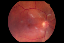
Bilateral Retinal Arteriol Occlusions - Possible Susac Syndrome 1208 views80-year-old woman one month ago had vision loss and a vascular occlusion in the right eye. Vision loss occurred in the left eye about 9 years ago with cotton wool spots. Patients has a 30 year history of tinnitis.
|
|

Bilateral Retinal Arteriol Occlusions - Possible Susac Syndrome 743 views80-year-old woman one month ago had vision loss and a vascular occlusion in the right eye. Vision loss occurred in the left eye about 9 years ago with cotton wool spots. Patients has a 30 year history of tinnitis.
|
|
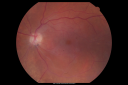
Bilateral Retinal Arteriol Occlusions - Possible Susac Syndrome 763 views80-year-old woman one month ago had vision loss and a vascular occlusion in the right eye. Vision loss occurred in the left eye about 9 years ago with cotton wool spots. Patients has a 30 year history of tinnitis.
|
|
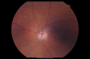
Bilateral Retinal Arteriol Occlusions - Possible Susac Syndrome 676 views80-year-old woman one month ago had vision loss and a vascular occlusion in the right eye. Vision loss occurred in the left eye about 9 years ago with cotton wool spots. Patients has a 30 year history of tinnitis.
|
|

Bilateral Retinal Arteriol Occlusions - Possible Susac Syndrome 776 views80-year-old woman one month ago had vision loss and a vascular occlusion in the right eye. Vision loss occurred in the left eye about 9 years ago with cotton wool spots. Patients has a 30 year history of tinnitis.
|
|
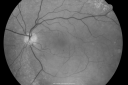
Bilateral Retinal Arteriol Occlusions - Possible Susac Syndrome 624 views80-year-old woman one month ago had vision loss and a vascular occlusion in the right eye. Vision loss occurred in the left eye about 9 years ago with cotton wool spots. Patients has a 30 year history of tinnitis.
|
|

Bilateral Retinal Arteriol Occlusions - Possible Susac Syndrome 689 views80-year-old woman one month ago had vision loss and a vascular occlusion in the right eye. Vision loss occurred in the left eye about 9 years ago with cotton wool spots. Patients has a 30 year history of tinnitis.
|
|

Bilateral Retinal Arteriol Occlusions - Possible Susac Syndrome 575 views80-year-old woman one month ago had vision loss and a vascular occlusion in the right eye. Vision loss occurred in the left eye about 9 years ago with cotton wool spots. Patients has a 30 year history of tinnitis.
|
|

Bilateral Retinal Arteriol Occlusions - Possible Susac Syndrome 595 views80-year-old woman one month ago had vision loss and a vascular occlusion in the right eye. Vision loss occurred in the left eye about 9 years ago with cotton wool spots. Patients has a 30 year history of tinnitis.
|
|
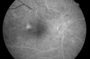
Bilateral Retinal Arteriol Occlusions - Possible Susac Syndrome 564 views80-year-old woman one month ago had vision loss and a vascular occlusion in the right eye. Vision loss occurred in the left eye about 9 years ago with cotton wool spots. Patients has a 30 year history of tinnitis.
|
|

Bilateral Retinal Arteriol Occlusions - Possible Susac Syndrome 611 views80-year-old woman one month ago had vision loss and a vascular occlusion in the right eye. Vision loss occurred in the left eye about 9 years ago with cotton wool spots. Patients has a 30 year history of tinnitis.
|
|
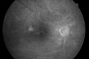
Bilateral Retinal Arteriol Occlusions - Possible Susac Syndrome 587 views80-year-old woman one month ago had vision loss and a vascular occlusion in the right eye. Vision loss occurred in the left eye about 9 years ago with cotton wool spots. Patients has a 30 year history of tinnitis.
|
|
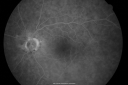
Bilateral Retinal Arteriol Occlusions - Possible Susac Syndrome 554 views80-year-old woman one month ago had vision loss and a vascular occlusion in the right eye. Vision loss occurred in the left eye about 9 years ago with cotton wool spots. Patients has a 30 year history of tinnitis.
|
|

Bilateral Retinal Arteriol Occlusions - Possible Susac Syndrome 598 views80-year-old woman one month ago had vision loss and a vascular occlusion in the right eye. Vision loss occurred in the left eye about 9 years ago with cotton wool spots. Patients has a 30 year history of tinnitis.
|
|

Bilateral Retinal Arteriol Occlusions - Possible Susac Syndrome 519 views80-year-old woman one month ago had vision loss and a vascular occlusion in the right eye. Vision loss occurred in the left eye about 9 years ago with cotton wool spots. Patients has a 30 year history of tinnitis.
|
|

Bilateral Retinal Arteriol Occlusions - Possible Susac Syndrome 579 views80-year-old woman one month ago had vision loss and a vascular occlusion in the right eye. Vision loss occurred in the left eye about 9 years ago with cotton wool spots. Patients has a 30 year history of tinnitis.
|
|

Bilateral Retinal Arteriol Occlusions - Possible Susac Syndrome 580 views80-year-old woman one month ago had vision loss and a vascular occlusion in the right eye. Vision loss occurred in the left eye about 9 years ago with cotton wool spots. Patients has a 30 year history of tinnitis.
|
|

Bilateral Retinal Arteriol Occlusions - Possible Susac Syndrome 712 views80-year-old woman one month ago had vision loss and a vascular occlusion in the right eye. Vision loss occurred in the left eye about 9 years ago with cotton wool spots. Patients has a 30 year history of tinnitis.
|
|

475 views
|
|

Inferotemporal and Inferonasal BRAO1531 viewsBranch Retinal Artery Occlusions - Multiple
|
|

Cilioretinal artery occlusion630 viewsCilioretinal artery occlusion with early venous stasis retinopathy
|
|

Branch Retinal Artery Occlusion with Hollenhorst Plaques549 viewsBranch Retinal Artery Occlusion with Hollenhorst Plaques
|
|

Cilio-Retinal Artery Occlusion with "Box Carring" and Cherry Red Spot1538 viewsCilioretinal Artery Occlusion with Cherry Red Spot
|
|

CRAO422 viewsCRAO in OD of a 37 yr old Male, Transit Phase
|
|

Iris Neovascularization secondary to Central Retinal Artery Occlusion1299 views
|
|

FA: CRAO751 viewsCRAO with cilio artery perfusion
|
|

Branch Retinal Artery Occlusion with Hollenhorst Plaque562 views
|
|

CRAO WITH SPARING OF CILIORETINAL ARTERY510 viewsThis patient was found to have retinal edema from a Central Retinal Artery Occlusion with sparing of the cilioretinal artery. There were no discernible Hollenhorst plaques
|
|

Branch Retinal Artery Occlusion with Hollenhorst Plaque465 viewsThis patient was found to have retinal edema along the superotemporal arcade secondary to Branch Retinal Artery Occlusion. A Hollenhorst plaque is visible at the first bifurcation of central artery.
|
|
|
|


