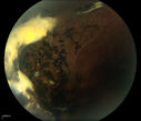
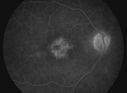
Acute Multifocal Placoid Pigment Epitheliopathy (AMPPE) related scarringvista 1217 veces (1 votos)
Nonproliferative Diabetic Retinopathy with Circinate Hard Exudatesvista 1393 veces (1 votos)
Choriodal Rupture with Hemorrhagevista 1000 veces (1 votos)
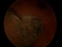
Retinal Tear Surrounded By Laser Bordervista 913 veces (1 votos)
Central Retinal Vein Occlusion with Disc Collateralsvista 1077 veces (1 votos)
Epiretinal Membrane with Flecks of Hemorrhagevista 1148 veces (1 votos)
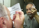
Intraocular Self injectionvista 913 vecesThe treatment of wet macular degeneration was revolutionized in late 2013 when Geneneron introduced its innovative and inexpensive take-home kit. Now, millions of patients around the world treat themselves on a PRN basis, using instructions written in 10 different languages.
April Fool! (1 votos)
Traumatic Chorioretinal Scarvista 684 veces (1 votos)
Choroidal Rupture Secondary to Ocular Traumavista 857 veces (1 votos)
Molteno Drainage Tubevista 933 veces (1 votos)
ERM with Pseudoholevista 782 veces (1 votos)
Central Retinal Venous Occlusion with Papilledema, Intraretinal Hemorrhage and Macular Edemavista 844 veces (1 votos)
Pseudophakic Cystoid Macular Edema with Disc Hyperfluorescencevista 781 veces (1 votos)
Pseudophakic Cystoid Macular Edema FA late framevista 1016 veces (1 votos)
Epiretinal Membrane with Pseudoholevista 1120 veces (1 votos)
Retinoschisisvista 860 veces (1 votos)
Acute Multifocal Placoid Pigment Epitheliopathyvista 821 veces (1 votos)
Melanomavista 707 veces (1 votos)
Ocular Trauma with Retinal Detachmentvista 814 vecesPatient suffered blunt force trauma to the eye resulting total retinal detachment. The retina is visible in the pupillary space. (1 votos)
Iris Neovascularization secondary to Central Retinal Artery Occlusionvista 1299 veces (1 votos)
Eales Disease - Fluorescein Angiogramvista 1376 veces (1 votos)
Central Serous Choroidopathy with Smoke Stack and Mushroom Cloud - Fluorescein Angiogramvista 1196 veces (1 votos)
Choroidal Rupture Secondary to Ocular Traumavista 1069 veces (1 votos)
Cilio-Retinal Artery Occlusion with "Box Carring" and Cherry Red Spotvista 1538 vecesCilioretinal Artery Occlusion with Cherry Red Spot (1 votos)
Retinal Tearvista 1013 veces (1 votos)
Retinal Detachment Secondary to Irregular Retinal Tearvista 899 vecesRetinal Detachment with Irregular Retinal Tear (1 votos)