am meisten angesehen - Dystrophies
|
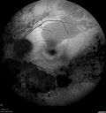
Retinitis Pigmentosa - Autosomal Recessive - 82 year old man with Good Vision508 x angesehenVA 20/25, 20/32 - Bone spicules in periphery - FAF is very symmetric
|
|
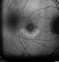
Cone Dystrophy - Autosomal Recessive508 x angesehen74 year old man with 20/25 vision OD and subtle bull's eye on FAF. Left eye is 20/200 with atrophy of the outer retina centrally
|
|
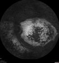
Enhanced S Cone Syndrome - Goldmann Favre - NR2E3 Mutation508 x angesehen82 year old man with poor vision for many years. VA HM OD, 5/200 OS. Diagnosed at age 12 with retinitis pigmentosa. Nystagmus.
|
|
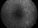
Pigmented Maculae - Mother and Daughter - Normal Visual Acuity507 x angesehen12-year-old had a dilated macular exam and the macula look abnormal. Interestingly, her mother, who is with her at this visit, was told about a year ago that her maculae are abnormal too. Her mother’s vision is fine. In the family tree, there are no known retinal dystrophies. They are both healthy.
VISUAL ACUITY: Her vision is 20/20 in both eyes.
|
|
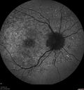
Stargardt's Macular Dystrophy ABCA4 positive507 x angesehen Fundus Autofluorescence shows central atrophy with Hyper FAF pisciform triradiate lesions. Images show some progression over 3 years of macular dystrophy.
|
|
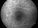
Pigmented Maculae - Mother and Daughter - Normal Visual Acuity506 x angesehen12-year-old had a dilated macular exam and the macula look abnormal. Interestingly, her mother, who is with her at this visit, was told about a year ago that her maculae are abnormal too. Her mother’s vision is fine. In the family tree, there are no known retinal dystrophies. They are both healthy.
VISUAL ACUITY: Her vision is 20/20 in both eyes.
|
|
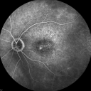
Fluorescein Angiogram - Enhanced S Cone Syndrome - Goldmann Favre - 506 x angesehen55-year-old woman while in college her vision was poor even with glasses and she sought evaluation for that. She was told after she had an electroretinogram at USF 15 years ago, that she had something with her blue cones. She does have poor night vision, but her reading vision is pretty good.
VISUAL ACUITY: OD 20/40, OS 20/40
|
|
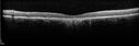
Stargardt's Macular Dystrophy ABCA4 positive506 x angesehenImages show some progression over 3 years of macular dystrophy.
|
|
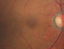
Cone Dystrophy - Autosomal Recessive506 x angesehen74 year old man with 20/25 vision OD and subtle bull's eye on FAF. Left eye is 20/200 with atrophy of the outer retina centrally
|
|

Fundus Albipunctatus505 x angesehen12 year old female with normal vision. She has 4 siblings all of whom have either white spots or spots on IR. Genetic testing by parents was deferred.
|
|
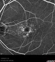
Non-specific (uncharaterized / unknown) Macular Dystrophy 504 x angesehen59-year-old man has macular dystrophy in both eyes. He had some vision changes in medical school in the 1980s and saw Dr. Gass for that. He had some pigment epithelial irregularities in both eyes. He had strabismus as a child and had muscle surgery. He is color blind, along with many people in his family, and as far as he knows, it is congenital.
20/25, 20/30
|
|
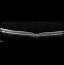
Stargardt's Macular Dystrophy ABCA4 positive504 x angesehenImages show some progression over 3 years of macular dystrophy.
|
|
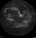
Enhanced S Cone Syndrome - Goldmann Favre - NR2E3 Mutation504 x angesehen82 year old man with poor vision for many years. VA HM OD, 5/200 OS. Diagnosed at age 12 with retinitis pigmentosa. Nystagmus.
|
|
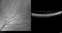
Fundus Albipunctatus504 x angesehen12 year old female with normal vision. She has 4 siblings all of whom have either white spots or spots on IR. Genetic testing by parents was deferred.
|
|

Stargardt's Macular Dystrophy ABCA4 positive503 x angesehenImages show some progression over 3 years of macular dystrophy.
|
|
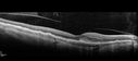
Late Onset Retinal Degeneration (L-ORD)503 x angesehen55 year old with acute vision loss from a CNVM in the right eye. He responded to Lucentis therapy. His mother and her family has been confirmed genetically to have L-ORD and were part of the early reports.
|
|
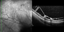
X-linked Juvenille Retinoschisis - Peripheral Retinal Vascular Anomalies - Vitreous Hemorrhage - 8 Year Old Boy502 x angesehen8-year-old child OD 20/40, OS 20/50
OD: Vertical C/D ratio is 0.2. There are foveal cysts. There is also a retinal elevation inferiorly and there are patchy peripheral retinal hemorrhages.
OS: Vertical C/D ratio is 0.2. There are foveal cysts. There is peripheral retinal issues with some hemorrhage in some areas of peripheral retinoschisis.
|
|
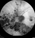
Retinitis Pigmentosa - Autosomal Recessive - 82 year old man with Good Vision502 x angesehenVA 20/25, 20/32 - Bone spicules in periphery - FAF is very symmetric
|
|

Chorodial Sclerosis501 x angesehen74 year old female diagnosed with Chorodial Sclerosis OU and ARMD OU. Va 20/400 OD 2'200 OS
|
|
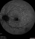
Stargardt's Macular Dystrophy ABCA4 positive501 x angesehen Fundus Autofluorescence shows central atrophy with Hyper FAF pisciform triradiate lesions. Images show some progression over 3 years of macular dystrophy.
|
|
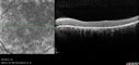
Stargardt's Macular Dystrophy ABCA4 positive501 x angesehenImages show some progression over 3 years of macular dystrophy.
|
|

Late Onset Cone Dystrophy - Very Mild501 x angesehenOnly IR and OCT show very mild bull's eye. 67 year old female with difficulty seeing in bright light
|
|
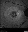
Non-specific (uncharaterized / unknown) Macular Dystrophy 500 x angesehen59-year-old man has macular dystrophy in both eyes. He had some vision changes in medical school in the 1980s and saw Dr. Gass for that. He had some pigment epithelial irregularities in both eyes. He had strabismus as a child and had muscle surgery. He is color blind, along with many people in his family, and as far as he knows, it is congenital.
20/25, 20/30
|
|
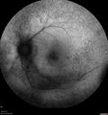
Diffuse outer retinal atrophy in 51 year old woman with 22 years plaquenil (hydroxychlorquine) use500 x angesehenRetinitis pigmentosa or similar dystrophy vs plaquenil toxicity
|
|

Cone Dystrophy - Autosomal Recessive500 x angesehen74 year old man with 20/25 vision OD and subtle bull's eye on FAF. Left eye is 20/200 with atrophy of the outer retina centrally
|
|
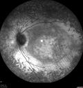
Enhanced S Cone Syndrome - Goldmann Favre - NR2E3 Mutation500 x angesehen82 year old man with poor vision for many years. VA HM OD, 5/200 OS. Diagnosed at age 12 with retinitis pigmentosa. Nystagmus.
|
|
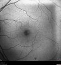
Late Onset Cone Dystrophy - Very Mild500 x angesehenOnly IR and OCT show very mild bull's eye. 67 year old female with difficulty seeing in bright light
|
|

Retinitis Pigmentosa - Autosomal Recessive - 82 year old man with Good Vision499 x angesehenVA 20/25, 20/32 - Bone spicules in periphery - FAF is very symmetric
|
|
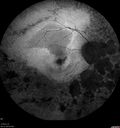
Retinitis Pigmentosa - Autosomal Recessive - 82 year old man with Good Vision499 x angesehenVA 20/25, 20/32 - Bone spicules in periphery - FAF is very symmetric
|
|
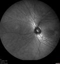
Fundus Albipunctatus499 x angesehen12 year old female with normal vision. She has 4 siblings all of whom have either white spots or spots on IR. Genetic testing by parents was deferred.
|
|

Non-specific (uncharaterized / unknown) Macular Dystrophy VIDEO TAKES TIME TO LOAD498 x angesehen59-year-old man has macular dystrophy in both eyes. He had some vision changes in medical school in the 1980s and saw Dr. Gass for that. He had some pigment epithelial irregularities in both eyes. He had strabismus as a child and had muscle surgery. He is color blind, along with many people in his family, and as far as he knows, it is congenital.
20/25, 20/30
|
|
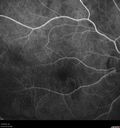
Late Onset Retinal Degeneration (L-ORD)498 x angesehen55 year old with acute vision loss from a CNVM in the right eye. He responded to Lucentis therapy. His mother and her family has been confirmed genetically to have L-ORD and were part of the early reports.
|
|
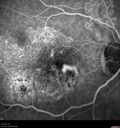
Late Onset Retinal Degeneration (L-ORD)498 x angesehen55 year old with acute vision loss from a CNVM in the right eye. He responded to Lucentis therapy. His mother and her family has been confirmed genetically to have L-ORD and were part of the early reports.
|
|
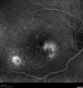
Late Onset Retinal Degeneration (L-ORD)498 x angesehen55 year old with acute vision loss from a CNVM in the right eye. He responded to Lucentis therapy. His mother and her family has been confirmed genetically to have L-ORD and were part of the early reports.
|
|
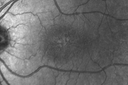
X-linked Juvenille Retinoschisis - Peripheral Retinal Vascular Anomalies - Vitreous Hemorrhage - 8 Year Old Boy497 x angesehen8-year-old child OD 20/40, OS 20/50
OD: Vertical C/D ratio is 0.2. There are foveal cysts. There is also a retinal elevation inferiorly and there are patchy peripheral retinal hemorrhages.
OS: Vertical C/D ratio is 0.2. There are foveal cysts. There is peripheral retinal issues with some hemorrhage in some areas of peripheral retinoschisis.
|
|
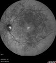
Stargardt's Macular Dystrophy ABCA4 positive497 x angesehenImages show some progression over 3 years of macular dystrophy.
|
|

Diffuse outer retinal atrophy in 51 year old woman with 22 years plaquenil (hydroxychlorquine) use497 x angesehenRetinitis pigmentosa or similar dystrophy vs plaquenil toxicity
|
|
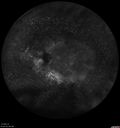
Enhanced S Cone Syndrome - Goldmann Favre - NR2E3 Mutation497 x angesehen82 year old man with poor vision for many years. VA HM OD, 5/200 OS. Diagnosed at age 12 with retinitis pigmentosa. Nystagmus.
|
|
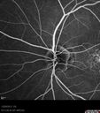
Non-specific (uncharaterized / unknown) Macular Dystrophy 496 x angesehen59-year-old man has macular dystrophy in both eyes. He had some vision changes in medical school in the 1980s and saw Dr. Gass for that. He had some pigment epithelial irregularities in both eyes. He had strabismus as a child and had muscle surgery. He is color blind, along with many people in his family, and as far as he knows, it is congenital.
20/25, 20/30
|
|

Stargardt's Macular Dystrophy ABCA4 positive496 x angesehenImages show some progression over 3 years of macular dystrophy.
|
|
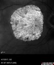
Central Areolar Choroidal Sclerosis496 x angesehen66 year old woman with 20/160 vision - loss of central vision 11 years ago. Strong family history of vision loss.
|
|
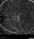
Non-specific (uncharaterized / unknown) Macular Dystrophy - ICG495 x angesehen59-year-old man has macular dystrophy in both eyes. He had some vision changes in medical school in the 1980s and saw Dr. Gass for that. He had some pigment epithelial irregularities in both eyes. He had strabismus as a child and had muscle surgery. He is color blind, along with many people in his family, and as far as he knows, it is congenital.
20/25, 20/30
|
|
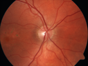
Pattern Dystrophy - Probably Early Butterfly495 x angesehen33 year old with normal vision and no visual complaints - normal color vision
|
|
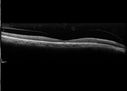
Cone Dystrophy - Autosomal Recessive495 x angesehen74 year old man with 20/25 vision OD and subtle bull's eye on FAF. Left eye is 20/200 with atrophy of the outer retina centrally
|
|
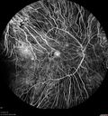
Choroideremia - Adult495 x angesehen69 year old man with no family history of choroideremia and gradual vision loss in both eyes. VVA 20/125 OD; 20/63 OS
|
|
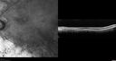
Retinitis Pigmentosa - Autosomal Recessive - 82 year old man with Good Vision494 x angesehenVA 20/25, 20/32 - Bone spicules in periphery - FAF is very symmetric
|
|
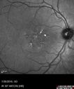
Pattern Dystrophy - Probably Early Butterfly494 x angesehen33 year old with normal vision and no visual complaints - normal color vision
|
|

Late Onset Cone Dystrophy - Very Mild494 x angesehenOnly IR and OCT show very mild bull's eye. 67 year old female with difficulty seeing in bright light
|
|

Late Onset Cone Dystrophy - Very Mild494 x angesehenOnly IR and OCT show very mild bull's eye. 67 year old female with difficulty seeing in bright light
|
|
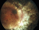
Enhanced S Cone Syndrome - Goldmann Favre - NR2E3 Mutation494 x angesehen82 year old man with poor vision for many years. VA HM OD, 5/200 OS. Diagnosed at age 12 with retinitis pigmentosa. Nystagmus.
|
|
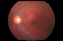
Diffuse outer retinal atrophy in 51 year old woman with 22 years plaquenil (hydroxychlorquine) use493 x angesehenRetinitis pigmentosa or similar dystrophy vs plaquenil toxicity
|
|
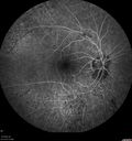
Retinitis Pigmentosa with Cystoid Macular Edema responsive to topical carbonic anhydrase inhibitors493 x angesehen67 year old female She thought it was time to have her glasses changed.She has no family history of reitnal disease and has never been diagnosed with a problem. She started having night vision trouble the last year. Two sisters and a brother with normal vision.
VA OD: Dcc20/40-2 NccJ3
VA OS: Dcc20/20 NccJ1
IOP: TP: OD:9
|
|
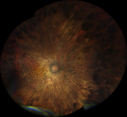
Choroideremia - Adult493 x angesehen69 year old man with no family history of choroideremia and gradual vision loss in both eyes. VVA 20/125 OD; 20/63 OS
|
|

Stargardt's Macular Dystrophy492 x angesehen62 year old man with good vision
|
|

Retinitis Pigmentosa Newly Diagnosed 55 Year Old490 x angesehen55-year-old noticed his vision declining recently, over the last few weeks, particularly in the right eye. He said previous to that the vision was not normal but was pretty good. OD is 20/100, OS is 20/50
OCT SCAN: There is macular thickening in both eyes with intraretinal cysts.
|
|
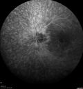
Diffuse outer retinal atrophy in 51 year old woman with 22 years plaquenil (hydroxychlorquine) use490 x angesehenRetinitis pigmentosa or similar dystrophy vs plaquenil toxicity
|
|
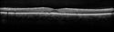
Pattern Dystrophy - Probably Early Butterfly490 x angesehen33 year old with normal vision and no visual complaints - normal color vision
|
|
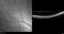
Fundus Albipunctatus490 x angesehen12 year old female with normal vision. She has 4 siblings all of whom have either white spots or spots on IR. Genetic testing by parents was deferred.
|
|

Retinitis Pigmentosa - Autosomal Recessive - 82 year old man with Good Vision489 x angesehenVA 20/25, 20/32 - Bone spicules in periphery - FAF is very symmetric
|
|

Diffuse outer retinal atrophy in 51 year old woman with 22 years plaquenil (hydroxychlorquine) use489 x angesehenRetinitis pigmentosa or similar dystrophy vs plaquenil toxicity
|
|
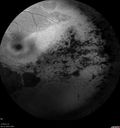
Retinitis Pigmentosa - Autosomal Recessive - 82 year old man with Good Vision488 x angesehenVA 20/25, 20/32 - Bone spicules in periphery - FAF is very symmetric
|
|
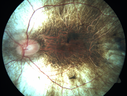
Choroideremia - Complete CHM gene deletion488 x angesehenVision loss since age 20 - now age 36 VA 20/160 (about) OU
|
|
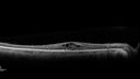
Retinitis Pigmentosa with Cystoid Macular Edema responsive to topical carbonic anhydrase inhibitors488 x angesehen67 year old female She thought it was time to have her glasses changed.She has no family history of reitnal disease and has never been diagnosed with a problem. She started having night vision trouble the last year. Two sisters and a brother with normal vision.
VA OD: Dcc20/40-2 NccJ3
VA OS: Dcc20/20 NccJ1
IOP: TP: OD:9
|
|
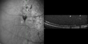
X-linked Juvenille Retinoschisis - Peripheral Retinal Vascular Anomalies - Vitreous Hemorrhage - 8 Year Old Boy487 x angesehen8-year-old child OD 20/40, OS 20/50
OD: Vertical C/D ratio is 0.2. There are foveal cysts. There is also a retinal elevation inferiorly and there are patchy peripheral retinal hemorrhages.
OS: Vertical C/D ratio is 0.2. There are foveal cysts. There is peripheral retinal issues with some hemorrhage in some areas of peripheral retinoschisis.
|
|
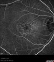
Non-specific (uncharaterized / unknown) Macular Dystrophy 487 x angesehen59-year-old man has macular dystrophy in both eyes. He had some vision changes in medical school in the 1980s and saw Dr. Gass for that. He had some pigment epithelial irregularities in both eyes. He had strabismus as a child and had muscle surgery. He is color blind, along with many people in his family, and as far as he knows, it is congenital.
20/25, 20/30
|
|

Diffuse outer retinal atrophy in 51 year old woman with 22 years plaquenil (hydroxychlorquine) use487 x angesehenRetinitis pigmentosa or similar dystrophy vs plaquenil toxicity
|
|
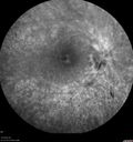
Retinitis Pigmentosa with Cystoid Macular Edema responsive to topical carbonic anhydrase inhibitors487 x angesehen67 year old female She thought it was time to have her glasses changed.She has no family history of reitnal disease and has never been diagnosed with a problem. She started having night vision trouble the last year. Two sisters and a brother with normal vision.
VA OD: Dcc20/40-2 NccJ3
VA OS: Dcc20/20 NccJ1
IOP: TP: OD:9
|
|
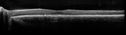
Pattern Dystrophy - Probably Early Butterfly486 x angesehen33 year old with normal vision and no visual complaints - normal color vision
|
|
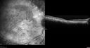
Enhanced S Cone Syndrome - Goldmann Favre - NR2E3 Mutation486 x angesehen82 year old man with poor vision for many years. VA HM OD, 5/200 OS. Diagnosed at age 12 with retinitis pigmentosa. Nystagmus.
|
|
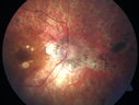
Retinitis Pigmentosa - RP1 Mutation486 x angesehen78 year old man: 20/32 OD and 20/50 OS - RP1 Gene is Bad -- > RP with Novel Amino Acid Change in GLY723Stop sequence of the RP1 gene - consistent with AD RP
|
|
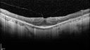
Retinitis Pigmentosa - RP1 Mutation486 x angesehen78 year old man: 20/32 OD and 20/50 OS - RP1 Gene is Bad -- > RP with Novel Amino Acid Change in GLY723Stop sequence of the RP1 gene - consistent with AD RP
|
|
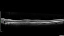
Retinitis Pigmentosa with Cystoid Macular Edema responsive to topical carbonic anhydrase inhibitors486 x angesehen67 year old female She thought it was time to have her glasses changed.She has no family history of reitnal disease and has never been diagnosed with a problem. She started having night vision trouble the last year. Two sisters and a brother with normal vision.
VA OD: Dcc20/40-2 NccJ3
VA OS: Dcc20/20 NccJ1
IOP: TP: OD:9
|
|
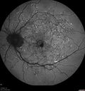
Stargardt's Macular Dystrophy ABCA4 positive485 x angesehen Fundus Autofluorescence shows central atrophy with Hyper FAF pisciform triradiate lesions. Images show some progression over 3 years of macular dystrophy.
|
|

Non-specific (uncharaterized / unknown) Macular Dystrophy 484 x angesehen59-year-old man has macular dystrophy in both eyes. He had some vision changes in medical school in the 1980s and saw Dr. Gass for that. He had some pigment epithelial irregularities in both eyes. He had strabismus as a child and had muscle surgery. He is color blind, along with many people in his family, and as far as he knows, it is congenital.
20/25, 20/30
|
|

Diffuse outer retinal atrophy in 51 year old woman with 22 years plaquenil (hydroxychlorquine) use483 x angesehenRetinitis pigmentosa or similar dystrophy vs plaquenil toxicity
|
|
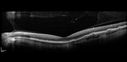
Late Onset Retinal Degeneration (L-ORD)483 x angesehen55 year old with acute vision loss from a CNVM in the right eye. He responded to Lucentis therapy. His mother and her family has been confirmed genetically to have L-ORD and were part of the early reports.
|
|
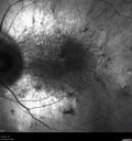
Choroideremia - Complete CHM gene deletion483 x angesehenVision loss since age 20 - now age 36 VA 20/160 (about) OU
|
|

Central Areolar Choroidal Sclerosis482 x angesehen66 year old woman with 20/160 vision - loss of central vision 11 years ago. Strong family history of vision loss.
|
|
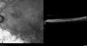
Retinitis Pigmentosa - Autosomal Recessive - 82 year old man with Good Vision481 x angesehenVA 20/25, 20/32 - Bone spicules in periphery - FAF is very symmetric
|
|
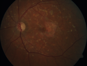
Stargardt's Macular Dystrophy481 x angesehen62 year old man with good vision
|
|
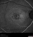
Non-specific (uncharaterized / unknown) Macular Dystrophy 480 x angesehen59-year-old man has macular dystrophy in both eyes. He had some vision changes in medical school in the 1980s and saw Dr. Gass for that. He had some pigment epithelial irregularities in both eyes. He had strabismus as a child and had muscle surgery. He is color blind, along with many people in his family, and as far as he knows, it is congenital.
20/25, 20/30
|
|

Diffuse outer retinal atrophy in 51 year old woman with 22 years plaquenil (hydroxychlorquine) use480 x angesehenRetinitis pigmentosa or similar dystrophy vs plaquenil toxicity
|
|
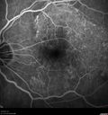
Late Onset Retinal Degeneration (L-ORD)480 x angesehen55 year old with acute vision loss from a CNVM in the right eye. He responded to Lucentis therapy. His mother and her family has been confirmed genetically to have L-ORD and were part of the early reports.
|
|
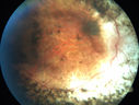
Enhanced S Cone Syndrome - Goldmann Favre - NR2E3 Mutation480 x angesehen82 year old man with poor vision for many years. VA HM OD, 5/200 OS. Diagnosed at age 12 with retinitis pigmentosa. Nystagmus.
|
|
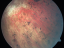
Retinitis Pigmentosa - RP1 Mutation479 x angesehen78 year old man: 20/32 OD and 20/50 OS - RP1 Gene is Bad -- > RP with Novel Amino Acid Change in GLY723Stop sequence of the RP1 gene - consistent with AD RP
|
|
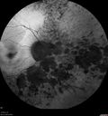
Retinitis Pigmentosa - Autosomal Recessive - 82 year old man with Good Vision478 x angesehenVA 20/25, 20/32 - Bone spicules in periphery - FAF is very symmetric
|
|
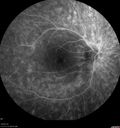
Diffuse outer retinal atrophy in 51 year old woman with 22 years plaquenil (hydroxychlorquine) use478 x angesehenRetinitis pigmentosa or similar dystrophy vs plaquenil toxicity
|
|
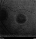
Cone Dystrophy - Autosomal Recessive478 x angesehen74 year old man with 20/25 vision OD and subtle bull's eye on FAF. Left eye is 20/200 with atrophy of the outer retina centrally
|
|
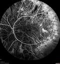
Choroideremia - Adult478 x angesehen69 year old man with no family history of choroideremia and gradual vision loss in both eyes. VVA 20/125 OD; 20/63 OS
|
|
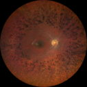
Retinitis Pigmentosa - Simplex - 16 year old female good vision478 x angesehenDifficulty with night vision
No family history
VA 20/25 OD, 20/16 OS
Negative for RPE65
|
|
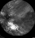
Retinitis Pigmentosa - Autosomal Recessive - 82 year old man with Good Vision476 x angesehenVA 20/25, 20/32 - Bone spicules in periphery - FAF is very symmetric
|
|
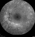
Retinitis Pigmentosa with Cystoid Macular Edema responsive to topical carbonic anhydrase inhibitors476 x angesehen67 year old female She thought it was time to have her glasses changed.She has no family history of reitnal disease and has never been diagnosed with a problem. She started having night vision trouble the last year. Two sisters and a brother with normal vision.
VA OD: Dcc20/40-2 NccJ3
VA OS: Dcc20/20 NccJ1
IOP: TP: OD:9
|
|
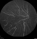
Choroideremia - Adult476 x angesehen69 year old man with no family history of choroideremia and gradual vision loss in both eyes. VVA 20/125 OD; 20/63 OS
|
|
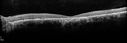
Stargardt's Macular Dystrophy ABCA4 positive475 x angesehenImages show some progression over 3 years of macular dystrophy.
|
|
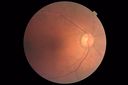
Late Onset Cone Dystrophy - Very Mild475 x angesehenOnly IR and OCT show very mild bull's eye. 67 year old female with difficulty seeing in bright light
|
|
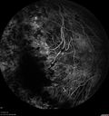
Choroideremia - Adult475 x angesehen69 year old man with no family history of choroideremia and gradual vision loss in both eyes. VVA 20/125 OD; 20/63 OS
|
|
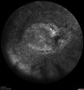
Enhanced S Cone Syndrome - Goldmann Favre - NR2E3 Mutation475 x angesehen82 year old man with poor vision for many years. VA HM OD, 5/200 OS. Diagnosed at age 12 with retinitis pigmentosa. Nystagmus.
|
|

Stargardt's Macular Dystrophy474 x angesehen62 year old man with good vision
|
|
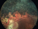
Enhanced S Cone Syndrome - Goldmann Favre - NR2E3 Mutation474 x angesehen82 year old man with poor vision for many years. VA HM OD, 5/200 OS. Diagnosed at age 12 with retinitis pigmentosa. Nystagmus.
|
|
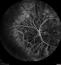
Choroideremia - Adult473 x angesehen69 year old man with no family history of choroideremia and gradual vision loss in both eyes. VVA 20/125 OD; 20/63 OS
|
|
| 1242 Dateien auf 13 Seite(n) |
 |
|
|
|
|
8 | |
|
|
|
|


