Most viewed - Dystrophies
|

Fundus Flavimaculatus - Stargardt Disease - 20/50 OD 20/200 OS 61 Year old Fundus Autofluorescence od2274 views61-year-old decreasing vision for about the last five years. OD 20/50, OS 20/200.
Pisciform Lesions and Macular Atrophy
|
|

Fundus Flavimaculatus - Stargardt Disease - 20/50 OD 20/200 OS 61 Year old - SD - OCT OS2138 views61-year-old decreasing vision for about the last five years. OD 20/50, OS 20/200.
Pisciform Lesions and Macular Atrophy
|
|

Malattia leventinese2078 viewsBasal laminar drusen in a perifoveal radial distribution are noted in this rare condition. Inheritance is AD with variable expressivity but full penetrance.
|
|

Stargardt's Juvenille Macular Dystrophy - Fundus Flavimaculatus - Famlial1971 views55-year-old woman was seen in the office on 11/18/08. She has a half sister with Stargardt’s disease and seven other siblings who are fine. 20/120 OD, 20/160 OS.
|
|

Best Disease - Left Eye Treated Previously, Elsewhere, with PDT and Avastin1860 views11-year-old has Best’s disease that runs in his family on his mothers side. At another retina specialists office, he had photodynamic laser in April of 2009 and Avastin subsequently in June of 2009. Unfortunately with that his vision is substantially declining. OD 20/30, OS 20/100
|
|

Retinitis Pigmentosa1815 viewsPatient comes in with cloudy vision. VA is 20/200 in both eyes. Patient has had retinal pigmentosa since 1986. Patient to consider cataract surgery to help her visual symptoms...
|
|

Fundus Flavimaculatus - Stargardt Disease - 20/50 OD 20/200 OS 61 Year old Color Photo1804 views61-year-old decreasing vision for about the last five years. OD 20/50, OS 20/200.
Pisciform Lesions and Macular Atrophy
|
|

Fundus Flavimaculatus - Stargardt Disease - 20/50 OD 20/200 OS 61 Year old Fundus Autofluorescence os1722 views61-year-old decreasing vision for about the last five years. OD 20/50, OS 20/200.
Pisciform Lesions and Macular Atrophy
|
|

Pigmented Peripheral Retinal Degeneration1697 views42-year old male comes in for routine eye exam and to follow up on peripheral retinal degeneration in both eyes. VA is 20/20, right eye and 20/25, left eye. Patient is asymptomatic with no visual complaints.
|
|

Cone-Rod Dystrophy 1582 views34-year-old woman who is having some difficulty seeing at night, especially navigating through dark movie theaters and dark restaurants. OD is 20/30, OS is 20/30
|
|
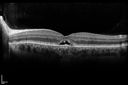
Best Disease - SD OCT Line Scan1560 views
|
|
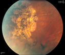
Pigmented Peripheral Retinal Degeneration1519 views42-year old male comes in for routine eye exam and to follow up on peripheral retinal degeneration in both eyes. VA is 20/20, right eye and 20/25, left eye. Patient is asymptomatic with no visual complaints.
|
|
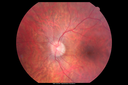
Retinitis Pigmentosa Sine Pigmento 1452 views
|
|

Fundus Flavimaculatus - Stargardt Disease - 20/50 OD 20/200 OS 61 Year old Color Photo1446 views61-year-old decreasing vision for about the last five years. OD 20/50, OS 20/200.
Pisciform Lesions and Macular Atrophy
|
|
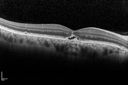
Best Disease - SD OCT Line Scan1441 views
|
|
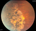
Pigmented Peripheral Retinal Degeneration1430 views42-year old male comes in for routine eye exam and to follow up on peripheral retinal degeneration in both eyes. VA is 20/20, right eye and 20/25, left eye. Patient is asymptomatic with no visual complaints.
|
|
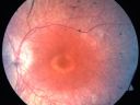
Cone-Rod Dystrophy 1428 views34-year-old woman who is having some difficulty seeing at night, especially navigating through dark movie theaters and dark restaurants. OD is 20/30, OS is 20/30
|
|
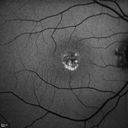
Best Disease Fundus Autofluorescence1400 views
|
|
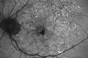
Fundus Flavimaculatus - Stargardt Disease - 20/50 OD 20/200 OS 61 Year old Fundus Autofluorescence os1399 views61-year-old decreasing vision for about the last five years. OD 20/50, OS 20/200.
Pisciform Lesions and Macular Atrophy
|
|

Retinitis Pigmentosa - Leber's Congenital Amaurosis - 7 Year Old Child -Fundus Photo Right Eye - Periphery1393 views7-year-old child who trips over things that the other children do not have trouble seeing. She is the only person in the family tree that the mother knows has any vision problems. Vision is 20/20 in each eye
|
|
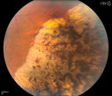
Pigmented Peripheral Retinal Degeneration1381 views42-year old male comes in for routine eye exam and to follow up on peripheral retinal degeneration in both eyes. VA is 20/20, right eye and 20/25, left eye. Patient is asymptomatic with no visual complaints.
|
|
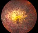
Central Areolar Choroidal Dystrophy1373 viewsPatient wanted second opinion for atrophic macular degeneration. VA is 20/400, right eye and 20/100. left eye. Patient has very poor vision and also hearing impaired. Fundiscopic exam reveals very atrophy in the macula. FAF shows a central hole to the choroid with no neovascularization present.
|
|

Best Disease - Left Eye Treated Previously, Elsewhere, with PDT and Avastin1364 views11-year-old has Best’s disease that runs in his family on his mothers side. At another retina specialists office, he had photodynamic laser in April of 2009 and Avastin subsequently in June of 2009. Unfortunately with that his vision is substantially declining. OD 20/30, OS 20/100
|
|

Probable Measles Retinopathy1362 views52-year old female wanted second opinion on retinal eval. Patient had PK and PDT in the left eye. VA was 20/30, right eye was count fingers. Pinhole was NI, both eyes. Patient's mother had measles. Notice pigment changes in both the color and FAF photo.
|
|

Best Disease - Left Eye Treated Previously, Elsewhere, with PDT and Avastin1358 views11-year-old has Best’s disease that runs in his family on his mothers side. At another retina specialists office, he had photodynamic laser in April of 2009 and Avastin subsequently in June of 2009. Unfortunately with that his vision is substantially declining. OD 20/30, OS 20/100
|
|
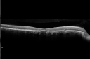
Fundus Flavimaculatus - Stargardt Disease - 20/50 OD 20/200 OS 61 Year old - SD OCT OD1356 views61-year-old decreasing vision for about the last five years. OD 20/50, OS 20/200.
Pisciform Lesions and Macular Atrophy
|
|

Fundus Photograph - Enhanced S Cone Syndrome - Goldmann Favre - 1315 views55-year-old woman while in college her vision was poor even with glasses and she sought evaluation for that. She was told after she had an electroretinogram at USF 15 years ago, that she had something with her blue cones. She does have poor night vision, but her reading vision is pretty good.
VISUAL ACUITY: OD 20/40, OS 20/40
|
|
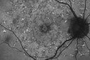
Fundus Flavimaculatus - Stargardt Disease - 20/50 OD 20/200 OS 61 Year old Fundus Autofluorescence od1313 views61-year-old decreasing vision for about the last five years. OD 20/50, OS 20/200.
Pisciform Lesions and Macular Atrophy
|
|

Best Disease - Left Eye Treated Previously, Elsewhere, with PDT and Avastin1308 views11-year-old has Best’s disease that runs in his family on his mothers side. At another retina specialists office, he had photodynamic laser in April of 2009 and Avastin subsequently in June of 2009. Unfortunately with that his vision is substantially declining. OD 20/30, OS 20/100
|
|

Stargardt's Juvenille Macular Dystrophy - Fundus Flavimaculatus - Famlial1302 views55-year-old woman was seen in the office on 11/18/08. She has a half sister with Stargardt’s disease and seven other siblings who are fine. 20/120 OD, 20/160 OS.
|
|

Best Disease, Vitelliform Macular Dystrophy - 6 year old child1298 viewsVISUAL ACUITY: Vision OD is 20/30, OS is 20/100.
VISUAL ACUITY: Vision OD is 20/30, OS is 20/100.
|
|
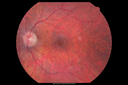
Retinitis Pigmentosa Sine Pigmento 1294 views
|
|
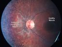
Retinitis Pigmentosa - Leber's Congenital Amaurosis - 7 Year Old Child - Fundus Photo Left Eye1274 views7-year-old child who trips over things that the other children do not have trouble seeing. She is the only person in the family tree that the mother knows has any vision problems. Vision is 20/20 in each eye
|
|

Fundus Flavimaculatus - Stargardt Disease - 20/50 OD 20/200 OS 61 Year old Infrared Image OD1269 views61-year-old decreasing vision for about the last five years. OD 20/50, OS 20/200.
Pisciform Lesions and Macular Atrophy
|
|

Best Disease - Left Eye Treated Previously, Elsewhere, with PDT and Avastin1248 views11-year-old has Best’s disease that runs in his family on his mothers side. At another retina specialists office, he had photodynamic laser in April of 2009 and Avastin subsequently in June of 2009. Unfortunately with that his vision is substantially declining. OD 20/30, OS 20/100
|
|

Chorodial Sclerosis1247 views74 year old female diagnosed with Chorodial Sclerosis OU and ARMD OU. Va 20/400 OD 2'200 OS
|
|
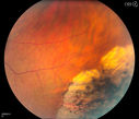
Pigmented Peripheral Retinal Degeneration1247 views42-year old male comes in for routine eye exam and to follow up on peripheral retinal degeneration in both eyes. VA is 20/20, right eye and 20/25, left eye. Patient is asymptomatic with no visual complaints.
|
|

Choroideremia1241 views62-year old female who has extensive retinal damage in both eyes. VA 20/50 right eye, HM, left eye. Spot in vision, which is PSC, both eyes.
|
|

Pigmented Peripheral Retinal Degeneration1239 views42-year old male comes in for routine eye exam and to follow up on peripheral retinal degeneration in both eyes. VA is 20/20, right eye and 20/25, left eye. Patient is asymptomatic with no visual complaints.
|
|

Fundus Albipunctatus1239 views12 year old female with normal vision. She has 4 siblings all of whom have either white spots or spots on IR. Genetic testing by parents was deferred.
|
|

Fundus Flavimaculatus - Stargardt Disease - 20/50 OD 20/200 OS 61 Year old Color Photo1232 views61-year-old decreasing vision for about the last five years. OD 20/50, OS 20/200.
Pisciform Lesions and Macular Atrophy
|
|

Central Areolar Choroidal Sclerosis1217 viewsElderly man with poor central vision starting in his 50's
|
|

Stargardt's Juvenille Macular Dystrophy - Fundus Flavimaculatus - Famlial1205 views55-year-old woman was seen in the office on 11/18/08. She has a half sister with Stargardt’s disease and seven other siblings who are fine. 20/120 OD, 20/160 OS.
|
|
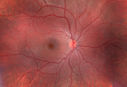
Cone Rod Dystrophy1202 viewsCone Rod Dystrophy in OD of seven year old girl
|
|

Alphamethlyacyl COA Racemase Deficiency1201 views
|
|

Bullseye Maculopathy without Plaquinel1176 viewsFundus photograph reveals Bull's-eye maculopathy without the use of plaquinel. Patients VA is 20/20, Right eye and 20/25, Left eye. There are reported cases of Scleroderma patients with retinal pigment epithelial atrophy. Will return for follow up in 6-months.
|
|

Retinitis Pigmentosa - Leber's Congenital Amaurosis - 7 Year Old Child - Left Eye1175 views7-year-old child who trips over things that the other children do not have trouble seeing. She is the only person in the family tree that the mother knows has any vision problems. Vision is 20/20 in each eye
|
|
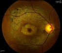
Bullseye Maculopathy without Plaquinel1161 viewsFundus photograph reveals Bull's-eye maculopathy without the use of plaquinel. Patients VA is 20/20, Right eye and 20/25, Left eye. There are reported cases of Scleroderma patients with retinal pigment epithelial atrophy. Will return for follow up in 6-months.
|
|

Best Disease, Vitelliform Macular Dystrophy - 6 year old child1153 viewsVISUAL ACUITY: Vision OD is 20/30, OS is 20/100.
|
|

Retinitis Pigmentosa1150 viewsRetinitis Pigmentosa
|
|
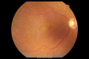
Fundus Photograph - Enhanced S Cone Syndrome - Goldmann Favre - 1131 views55-year-old woman while in college her vision was poor even with glasses and she sought evaluation for that. She was told after she had an electroretinogram at USF 15 years ago, that she had something with her blue cones. She does have poor night vision, but her reading vision is pretty good.
VISUAL ACUITY: OD 20/40, OS 20/40
|
|

Central Areolar Choroidal Sclerosis1126 views66 year old woman with 20/160 vision - loss of central vision 11 years ago. Strong family history of vision loss.
|
|
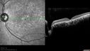
Macular possible Cone Dystrophy - SD-OCT1119 views
|
|
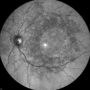
Fundus Flavimaculatus - Stargardt Disease - 20/50 OD 20/200 OS 61 Year old Infrared Image OS1118 views61-year-old decreasing vision for about the last five years. OD 20/50, OS 20/200.
Pisciform Lesions and Macular Atrophy
|
|

Best Disease- Fundus Autofluorescence1118 views
|
|

Fundus Flavimaculatus - Stargardt Disease - 20/50 OD 20/200 OS 61 Year old Color Photo1112 views61-year-old decreasing vision for about the last five years. OD 20/50, OS 20/200.
Pisciform Lesions and Macular Atrophy
|
|
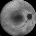
Fundus Autofluorescence - Enhanced S Cone Syndrome - Goldmann Favre - 1105 views55-year-old woman while in college her vision was poor even with glasses and she sought evaluation for that. She was told after she had an electroretinogram at USF 15 years ago, that she had something with her blue cones. She does have poor night vision, but her reading vision is pretty good.
VISUAL ACUITY: OD 20/40, OS 20/40
|
|

Fundus Flavimaculatus - Asymptomatic - Fundus Autofluorescence1103 views45 year old man was told to come in because of ‘white spots on their retina’. - FAF image shows that spots are strongly autofluorescent
|
|

Fundus Albipunctatus1088 views12 year old female with normal vision. She has 4 siblings all of whom have either white spots or spots on IR. Genetic testing by parents was deferred.
|
|

Fundus Flavimaculatus - Stargardt Disease - 20/50 OD 20/200 OS 61 Year old Color Photo1087 views61-year-old decreasing vision for about the last five years. OD 20/50, OS 20/200.
Pisciform Lesions and Macular Atrophy
|
|

Best Disease, Vitelliform Macular Dystrophy - 6 year old child1082 viewsVISUAL ACUITY: Vision OD is 20/30, OS is 20/100.
|
|

Best Disease - Left Eye Treated Previously, Elsewhere, with PDT and Avastin1081 views11-year-old has Best’s disease that runs in his family on his mothers side. At another retina specialists office, he had photodynamic laser in April of 2009 and Avastin subsequently in June of 2009. Unfortunately with that his vision is substantially declining. OD 20/30, OS 20/100
|
|

Macular possible Cone Dystrophy - Fundus Autofluorescence1076 views
|
|

Albinism and Foveal Hypoplasia1068 views65-year-old man was told about twenty years ago by a retina specialist that he spent so much time in the sun that he burned out his maculae. He does have two brothers who have dark brown hair and he has always had white hair. He had eye exercises when he was eight or nine because of a lazy eye and his brothers, when he was little, used to call him an albino. He has been diagnosed by dermatologists as having possible albinism but he has been told by other people his skin is just fair.
VA 20/70 OU
|
|

Fundus Flavimaculatus - Asymptomatic - Spectral Domain OCT Line Scan1066 views45 year old man was told to come in because of ‘white spots on their retina’.
|
|

Fundus Albipunctatus1065 views12 year old female with normal vision. She has 4 siblings all of whom have either white spots or spots on IR. Genetic testing by parents was deferred.
|
|

Gyrate Atrophy - 15 year old male - 20/25 vision both eyes - Positive Family History1059 views15 year old male was diagnosed with “retinitis pigmentosa†at age seven. The patient reports that his night vision is poor. He reports he does not notice a visual field change. He reports that his maternal grandfather has lost vision and a cousin has lost vision as well. He reports he has an uncle with some visual changes.
The most significantly affected relative is a 26-year-old legally blind cousin.
20/25+ OD, 20/25 OS.
|
|

Best Disease, Vitelliform Macular Dystrophy - 6 year old child1048 viewsVISUAL ACUITY: Vision OD is 20/30, OS is 20/100.
|
|

Central Areolar Choroidal Sclerosis1035 viewsElderly man with poor central vision starting in his 50's
|
|

Stargardt's Disease - Macular Dystrophy - 69 Year Old African American Male - 20/200 both eyes1029 views69-year-old man has had vision loss in both eyes starting 20 years ago. He was diagnosed at one point with Stargardt’s macular dystrophy. When I saw him, he had retinal atrophy in each eye.
Vision is 20/200 in each eye
|
|

Retinitis Pigmentosa - Leber's Congenital Amaurosis - 7 Year Old Child1027 views7-year-old child who trips over things that the other children do not have trouble seeing. She is the only person in the family tree that the mother knows has any vision problems. Vision is 20/20 in each eye
|
|
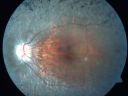
Retinitis Pigmentosa 32 Year Old Woman - 20/25 vision both eyes1022 views32-year-old woman was seen in the office on 6/26/2009. She has had retinitis pigmentosa since 2003. She has difficulty with night vision but overall she is doing pretty well.
Vision OU is 20/25.
|
|

Congenital Achromatopsia 1021 views26-year-old man was diagnosed with congenital achromatopsia by ERG as a child. He has family members who also have this disorder. OD 20/200, OS 20/200.
|
|

Fundus Flavimaculatus - Stargardt Disease - 20/50 OD 20/200 OS 61 Year old Color Photo1020 views61-year-old decreasing vision for about the last five years. OD 20/50, OS 20/200.
Pisciform Lesions and Macular Atrophy
|
|
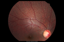
Fundus Albipunctatus1019 views12 year old female with normal vision. She has 4 siblings all of whom have either white spots or spots on IR. Genetic testing by parents was deferred.
|
|

Gyrate Atrophy - 15 year old male - 20/25 vision both eyes - Positive Family History1017 views15 year old male was diagnosed with “retinitis pigmentosa†at age seven. The patient reports that his night vision is poor. He reports he does not notice a visual field change. He reports that his maternal grandfather has lost vision and a cousin has lost vision as well. He reports he has an uncle with some visual changes.
The most significantly affected relative is a 26-year-old legally blind cousin.
20/25+ OD, 20/25 OS.
|
|
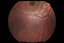
Fundus Albipunctatus1015 views12 year old female with normal vision. She has 4 siblings all of whom have either white spots or spots on IR. Genetic testing by parents was deferred.
|
|

Retinal Pigmintosa1014 views28-year old female with no light perception in the right eye.
|
|

Best Disease, Vitelliform Macular Dystrophy - 6 year old child1009 viewsVISUAL ACUITY: Vision OD is 20/30, OS is 20/100.
|
|
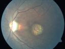
Stargardt's Disease - Macular Dystrophy - 69 Year Old African American Male - 20/200 both eyes1009 views69-year-old man has had vision loss in both eyes starting 20 years ago. He was diagnosed at one point with Stargardt’s macular dystrophy. When I saw him, he had retinal atrophy in each eye.
Vision is 20/200 in each eye
|
|

Retinitis Pigmentosa 32 Year Old Woman - 20/25 vision both eyes1002 views32-year-old woman was seen in the office on 6/26/2009. She has had retinitis pigmentosa since 2003. She has difficulty with night vision but overall she is doing pretty well.
Vision OU is 20/25.
|
|

1001 views
|
|

Concentric Annular Macular Dystrophy 4/200 2/200 vision 61 Year Old Macular Crystals1001 views61-year-old woman has concentric annular macular dystrophy. She has had vision loss for 10 years who is no longer able to function even with the magnifiers and vision aids that she has. Her brother who is ten years younger than her, is recently diagnosed with the same macular dystrophy and her father and grandfather may have had similar problems in the past.
VISUAL ACUITY: OD 4/200, OS 2/200.
|
|
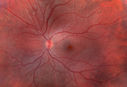
Cone Rod Dystrophy999 viewsCone Rod Dystrophy in OS of seven year old girl
|
|

Retinitis Pigmentosa Sine Pigmento 999 views
|
|

Albinism and Foveal Hypoplasia990 views65-year-old man was told about twenty years ago by a retina specialist that he spent so much time in the sun that he burned out his maculae. He does have two brothers who have dark brown hair and he has always had white hair. He had eye exercises when he was eight or nine because of a lazy eye and his brothers, when he was little, used to call him an albino. He has been diagnosed by dermatologists as having possible albinism but he has been told by other people his skin is just fair.
VA 20/70 OU
|
|

Stargardt's Disease - Macular Dystrophy - 69 Year Old African American Male - 20/200 both eyes990 views69-year-old man has had vision loss in both eyes starting 20 years ago. He was diagnosed at one point with Stargardt’s macular dystrophy. When I saw him, he had retinal atrophy in each eye.
Vision is 20/200 in each eye
|
|
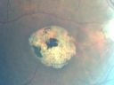
Stargardt's Disease - Macular Dystrophy - 69 Year Old African American Male - 20/200 both eyes986 views69-year-old man has had vision loss in both eyes starting 20 years ago. He was diagnosed at one point with Stargardt’s macular dystrophy. When I saw him, he had retinal atrophy in each eye.
Vision is 20/200 in each eye
|
|

Fundus Flavimaculatus - Stargardt Disease - 20/50 OD 20/200 OS 61 Year old Color Photo977 views61-year-old decreasing vision for about the last five years. OD 20/50, OS 20/200.
Pisciform Lesions and Macular Atrophy
|
|

Best Disease - Color Fundus Photo965 views
|
|
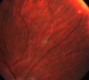
Benign retinal flecks964 viewsAsymptomatic, well nourished 16 yo presents with unilateral retinal flecks
|
|

Retinitis Pigmentosa Sine Pigmento 964 views
|
|

Pigmented Peripheral Retinal Degeneration958 views42-year old male comes in for routine eye exam and to follow up on peripheral retinal degeneration in both eyes. VA is 20/20, right eye and 20/25, left eye. Patient is asymptomatic with no visual complaints.
|
|

Stargardt's Disease - Macular Dystrophy - 69 Year Old African American Male - 20/200 both eyes956 views69-year-old man has had vision loss in both eyes starting 20 years ago. He was diagnosed at one point with Stargardt’s macular dystrophy. When I saw him, he had retinal atrophy in each eye.
Vision is 20/200 in each eye
|
|
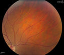
Pigmented Peripheral Retinal Degeneration949 views42-year old male comes in for routine eye exam and to follow up on peripheral retinal degeneration in both eyes. VA is 20/20, right eye and 20/25, left eye. Patient is asymptomatic with no visual complaints.
|
|
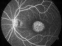
Stargardt's Disease - Macular Dystrophy - 69 Year Old African American Male - 20/200 both eyes944 views69-year-old man has had vision loss in both eyes starting 20 years ago. He was diagnosed at one point with Stargardt’s macular dystrophy. When I saw him, he had retinal atrophy in each eye.
Vision is 20/200 in each eye
|
|
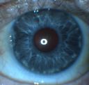
Albinism and Foveal Hypoplasia939 views65-year-old man was told about twenty years ago by a retina specialist that he spent so much time in the sun that he burned out his maculae. He does have two brothers who have dark brown hair and he has always had white hair. He had eye exercises when he was eight or nine because of a lazy eye and his brothers, when he was little, used to call him an albino. He has been diagnosed by dermatologists as having possible albinism but he has been told by other people his skin is just fair.
VA 20/70 OU
|
|
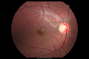
Fundus Albipunctatus939 views12 year old female with normal vision. She has 4 siblings all of whom have either white spots or spots on IR. Genetic testing by parents was deferred.
|
|

Retinitis Pigmentosa Sine Pigmento 935 views
|
|

X-Linked Juvenille Retinoschisis 10 Year Old Child treated with Trusopt933 views10-year-old child has x-linked juvenile retinoschisis VA 20/60, OS is 20/100. Macula and Vision improved with Trusopt drops.
|
|
| 1242 files on 13 page(s) |
1 |
 |
|
|
|
|
|
|
|
|
|


