Most viewed - Macular Diseases - Acquired
|

Stage 4 Macular Hole765 views57 year old female with 20/160 vision and stage 4 macular hole
|
|
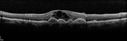
Macular Edema and dry AMD - excellent example of why FA and ICG are necessary in management of wet AMD761 viewsThis 72 year old woman has an OCT that looks like wet AMD. Only her FA and ICG show that she has inflammatory macular edema. She had no clinical signs or symptoms of uveitis. Nevertheless, the macular edema responded to subtenon's kenalog.
|
|

Central Serous Retinopathy760 viewsOCT scan showing serous RD
|
|

Macular Pucker in eye following giant retinal tear RD repair - photo of tear758 views 42-year old man has noticed his vision is changing now that the bubble is gone, but he is starting to notice distortion of central vision. He had a giant retinal tear retinal detachment repair emergently done on November 8, 2007, and subsequent laser on November 15th.
VISUAL ACUITY: Vision OD 20/400.
6 weeks post-op VA 20/40
|
|

Juxtafoveal Telangiectasis - MacTel - Retinal Telangiectasis - Both Eyes - Crystals757 views61-year-old man has idiopathic juxtafoveal retinal telangiectasis type II in both eyes OD is 20/40, OS is 20/25
|
|

Post Retinal Detachment Repair Macular Pucker - VA 20/400 Pre-op 20/25 Post-op757 views72 year old woman who is 6 months post macula-on retinal detachment repair developed a 20/400 macular pucker. 8 months after surgery vision had improved to 20/25
|
|
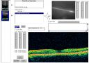
Idiopathic Central Serous Retinopathy - Bilateral - Acute - 4 months later VA 20/40 OU756 viewsNo treatment and fluid under fovea in left eye is competely absorbed at 4 months.
|
|
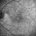
Macular Edema and dry AMD - excellent example of why FA and ICG are necessary in management of wet AMD756 viewsThis 72 year old woman has an OCT that looks like wet AMD. Only her FA and ICG show that she has inflammatory macular edema. She had no clinical signs or symptoms of uveitis. Nevertheless, the macular edema responded to subtenon's kenalog.
|
|

Plaquenil Toxicity - Bulls Eye Maculopathy755 views70-year-old woman with systemic Lupus erythematosus and clotting problems. She was on the Plaquenil for about eight years and then off the Plaquenil for the last eight years because she developed macular toxicity. Although her vision was hazy, it was stable. Recent deceased vision left eye: OD 20/60, OS 20/100. IOP: OD 18, OS 19.
|
|

Macular Telangiectasia - Stage 5 left eye - 20/40 vision after 2 years of Avastin 755 viewsSpectral Domain OCT Line Scan
|
|

Juxtafoveal Telangiectasis - Stage 5 Left Eye - New Blood and Neovascularization MacTel FAF753 views60-year-old man has juxtafoveal retinal telangiectasis recent vision loss OD 20/50, OS 20/70 : Hemorrhage OS and new SRNVM
|
|

Central Serous Retinopathy751 viewsEarly FA
|
|
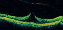
Vitreomacular Traction - Macular Pucker - 20/50 Vision751 views75-year-old woman decreasing vision in the right eye a few months ago. She had her cataract removed and unfortunately her vision is still hazy in the right eye. Her left eye seems a little hazy from the cataract.
VISUAL ACUITY: OD 20/50, OS 20/20. IOP: OD 17, OS 18.
SLIT-LAMP EXAMINATION
|
|
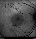
Chronic Central Serous Retinopathy - FAF751 views
|
|

Central Serous Retinopathy750 viewsFundus photo showing serous RD
|
|

Chronic Central Serous Chorioretinopathy Both Eyes750 views60 year old man with 20 year history of chronic CSR. He has had laser and PDT in the past. The left eye has been 20/100 for decades. The right eye is now declining. Was 20/32 and is down to 20/50. There is chronic low grade subretinal fluid away from the center in the right eye. (focal laser in the right eye in 2005, left eye 2001 at BPEI). Refampin tried in 2012, Aldactone in 2013. PDT in 204 and 2017 Images are from 2017
|
|
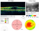
Chronic Pseudophakic Cystoid Macular Edema in a Patient with Diabetes749 views74-year-old woman has chronic cystoid macular edema in the left eye. She has had vision loss intermittently in that eye for 2 years, the right eye, unfortunately, lost vision from a central retinal vein occlusion in 6 years ago.
|
|

Macular Hole - Asymptomatic - 20/400 Vision747 views75-year-old woman in a recent exam it was noticed her vision in the left eye had substantially declined. She is not clear as to when that exactly happened. She had not noticed a vision change in the left eye. VISUAL ACUITY: OD 20/30, OS 20/400.
|
|

Group 2a Juxtafoveal Telangiectasis - MacTel - Idiopathic Juxtafoveal Macular Telangiectasia - Macular Hole745 views80-year-old man has juxtafoveal retinal telangiectasis in both eyes. He also has a true full-thickness macular hole in the left eye. The vision OD is 20/40, OS is 20/160
Group 2a Juxtafoveal Retinal Telangiectasis (telangiectasia), MacTel
|
|

Outer Retinal Holes - Probably Solar Retinopathy (history is negative)745 views
|
|

Macular Hole - Asymptomatic - 20/400 Vision744 views75-year-old woman in a recent exam it was noticed her vision in the left eye had substantially declined. She is not clear as to when that exactly happened. She had not noticed a vision change in the left eye. VISUAL ACUITY: OD 20/30, OS 20/400.
|
|
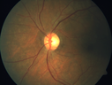
Multifocal Bilateral Central Serous Retinopathy in African American Male744 views67 year old african american male with : Blurred Vision OD. Duration of Problem: 4-6 weeks. VA 20/32 OD 20/20 OS
|
|
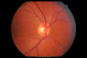
Idiopathic Central Serous Retinopathy - Bilateral - Acute 743 views49-year-old man decreasing vision in the left eye for one week. He is not taking any steroid or steroid-related products. His blood pressure as far as he knows is under control.
VISUAL ACUITY: OD 20/30, OS 20/400
|
|
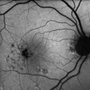
Group 2a Juxtafoveal Telangiectasis - MacTel - Stage 5 left eye - Macular Hole Right eye743 views76-year-old woman has juxtafoveal retinal telangiectasis. She had Stage five neovascularization in the left eye I treated with injections between September of 2007 and October of 2008 with Avastin (3 years ago). OCT scan today shows macular hole right eye. VISUAL ACUITY: OD 20/400, OS 20/40.
|
|

Cystoid Macular Edema - Xalatan - ECCE was done 20 years previously742 views71 Year old woman who noticed about a month ago the central vision in the right eye fogged up. OD 20/200 POSSIBLE XALATAN INDUCED CYSTOID MACULAR EDEMA
|
|
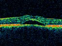
Chronic Central Serous Retinopathy treated with Photodynamic Laser and Visudyne742 views48-year-old man has had waxing and waning vision in the left eye for the last year from central serous retinopathy. OD is 20/16, OS is 20/30.
|
|

ERM OCT Composite741 viewsMultiple OCT scan Composite to show ERM
|
|
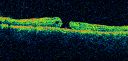
Macular Hole - Traumatic - 11 year old boy (also retinal whitening Berlins Edema)740 views11-year-old boy with a history of blunt trauma to the left eye yesterday. He reports he lost vision immediately but it has gotten somewhat better. He reports he sees “red streamers†and he has very blurry visual acuity., 20/200 OS
1 week post injury and 1 month later (not shown here) mature full thickness macular hole.
|
|
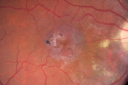
Juxtfoveal Telangiectasis Group 2a - Type II - MacTel - also Juxtapapillary Asymptomatic CNVM Right Eye739 views82-year-old woman who has juxtafoveal retinal telangiectasis in both eyes. She recently had cataract surgery in the right eye and the vision in the right eye is still limited. OD 20/100, OS 20/40.
|
|

Macular Pucker in eye following giant retinal tear RD repair pre-op Macular Photo Pucker738 viewsnow that the bubble is gone, but he is starting to notice distortion of central vision. He had a giant retinal tear retinal detachment repair emergently done on November 8, 2007, and subsequent laser on November 15th.
VISUAL ACUITY: Vision OD 20/400.
6 weeks post-op VA 20/40
|
|
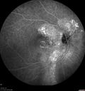
Chronic Central Serous Chorioretinopathy Both Eyes738 views60 year old man with 20 year history of chronic CSR. He has had laser and PDT in the past. The left eye has been 20/100 for decades. The right eye is now declining. Was 20/32 and is down to 20/50. There is chronic low grade subretinal fluid away from the center in the right eye. (focal laser in the right eye in 2005, left eye 2001 at BPEI). Refampin tried in 2012, Aldactone in 2013. PDT in 204 and 2017 Images are from 2017
|
|

737 views48-year-old man has had waxing and waning vision in the left eye for the last year from central serous retinopathy. OD is 20/16, OS is 20/30.
|
|

Central Serous Retinopathy - Acute735 views49-year-old man sees a spot in the central vision with a red halo for 2 weeks OD is 20/50, OS is 20/20
|
|
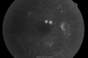
Acute Central Serous Chorioretinopathy with Symptoms in Hazy Vision Both Eyes735 views48-year-old man with hazy vision in the right eye for about three days. He has a little more of a headache than usual. He is under a fair amount of stress. He does not take any steroids that he knows of. He does use a nasal spray, which he is not sure if that is a steroid spray or not. He is going to call us with that medicine. I checked his blood pressure today is was 115/95.
VISUAL ACUITY: OD 20/15, OS 20/25
|
|
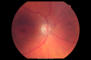
Plaquenil (Hydroxycholorquine) Toxicity - Bull's Eye Maculopathy - 73 year old womand 25 Years therapy - 64 inches tall - 110 lbs735 views73-year-old woman has been on Plaquenil for twenty five years. She noticed loss of paracentral vision a few months ago. VISUAL ACUITY: OD 20/70, OS 20/30. Her color vision was 0 out 10 correct in both eyes and she also missed the controls.
|
|
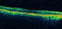
Chronic Central Serous Retinopathy - Gutter in Both Eyes - Treated with PDT - POST PDT Line Scan733 views69-year-old man with blurred vision in the right eye for about three weeks. He had a similar episode about twenty years ago which lasted about six weeks. He is not taking any steroids. He takes Lupron injections for his prostate cancer every three months.
VISUAL ACUITY: Vision OD is 20/80
FU: Treated with PDT after 6 months of observation with success. Treatment was done to area between nerve and fovea at 1/2 fluence.
|
|
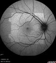
Outer Retinal Holes - Probably Solar Retinopathy (history is negative)733 views
|
|
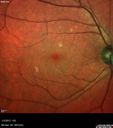
Travatan Induced CME Left eye733 views63 year old woman 4 years post ECCE and 2 years post PPV for floaters. She developed CME in the left eye after 1 year of travatan. The travatan was stopped and the CME and serous retinal detachment resolved.
|
|
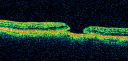
Group 2a Juxtafoveal Telangiectasis - MacTel - Idiopathic Juxtafoveal Macular Telangiectasia - Macular Hole732 views80-year-old man has juxtafoveal retinal telangiectasis in both eyes. He also has a true full-thickness macular hole in the left eye. The vision OD is 20/40, OS is 20/160
Group 2a Juxtafoveal Retinal Telangiectasis (telangiectasia), MacTel
|
|

Stage 2 Macular Hole with 20/70 Vision731 views57-year-old woman blurred central vision in the right eye for about two to three weeks. OD 20/70
|
|
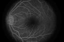
Central Serous Retinopathy730 viewsLater FA
|
|
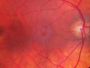
Juxtafoveal Telangiectasis - Stage 5 Left Eye - New Blood and Neovascularization MacTel 730 views60-year-old man has juxtafoveal retinal telangiectasis recent vision loss OD 20/50, OS 20/70 : Hemorrhage OS and new SRNVM
|
|
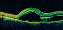
Idiopathic Central Serous Retinopathy - Bilateral - Acute 724 views49-year-old man decreasing vision in the left eye for one week. He is not taking any steroid or steroid-related products. His blood pressure as far as he knows is under control.
VISUAL ACUITY: OD 20/30, OS 20/400
|
|

Macular Hole - Traumatic - 11 year old boy (also retinal whitening Berlins Edema)724 views11-year-old boy with a history of blunt trauma to the left eye yesterday. He reports he lost vision immediately but it has gotten somewhat better. He reports he sees “red streamers†and he has very blurry visual acuity., 20/200 OS
|
|

Cystoid Macular Edema - Xalatan - ECCE was done 20 years previously720 views71 Year old woman who noticed about a month ago the central vision in the right eye fogged up. OD 20/200 POSSIBLE XALATAN INDUCED CYSTOID MACULAR EDEMA
|
|

Central Serous Retinopathy - Acute720 views49-year-old man sees a spot in the central vision with a red halo for 2 weeks OD is 20/50, OS is 20/20
|
|
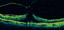
Vitreomacular Traction - Macular Pucker - 20/50 Vision720 views75-year-old woman decreasing vision in the right eye a few months ago. She had her cataract removed and unfortunately her vision is still hazy in the right eye. Her left eye seems a little hazy from the cataract.
VISUAL ACUITY: OD 20/50, OS 20/20. IOP: OD 17, OS 18.
SLIT-LAMP EXAMINATION
|
|
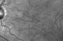
Macular Telangiectasis (Group 2a Juxtafoveal Telangiectasis) IR719 views63-year-old woman has juxtafoveal retinal telangiectasis in both eyes. She notices her vision a little worse with more distortion and change over the last six months.
VISUAL ACUITY: OD 20/40, OS 20/40.
|
|

Cystoid Macular Edema - Xalatan - ECCE was done 20 years previously718 views71 Year old woman who noticed about a month ago the central vision in the right eye fogged up. OD 20/200 POSSIBLE XALATAN INDUCED CYSTOID MACULAR EDEMA
|
|
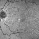
Juxtafoveal Retinal Telangiectasia (Telangiectasis) - MacTel - Striking Gray Ring in Macula718 views59-year-old woman decreasing vision in the right eye for a few years. She has had diabetes for five years. OD 20/100, Pinhole 20/40. OS 20/20
|
|
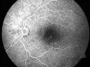
Chronic Central Serous Retinopathy treated with Photodynamic Laser and Visudyne716 views48-year-old man has had waxing and waning vision in the left eye for the last year from central serous retinopathy. OD is 20/16, OS is 20/30.
|
|
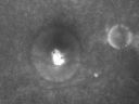
Acute Central Serous Retinopathy (Chorioretinopathy) Right Eye716 views45-year-old man decreased vision in the right eye starting about two days ago. VISUAL ACUITY: Vision OD is 20/60, OS is 20/20.
|
|

3-D OCT Image of Tractional Maculopathy716 views
|
|

Central Serous Retinopathy - Acute715 views49-year-old man sees a spot in the central vision with a red halo for 2 weeks OD is 20/50, OS is 20/20
|
|

Plaquenil (Hydroxycholorquine) Toxicity - Bull's Eye Maculopathy - 73 year old womand 25 Years therapy - 64 inches tall - 110 lbs715 views73-year-old woman has been on Plaquenil for twenty five years. She noticed loss of paracentral vision a few months ago. VISUAL ACUITY: OD 20/70, OS 20/30. Her color vision was 0 out 10 correct in both eyes and she also missed the controls.
|
|

Acute Central Serous Chorioretinopathy with Symptoms in Hazy Vision Both Eyes714 views48-year-old man with hazy vision in the right eye for about three days. He has a little more of a headache than usual. He is under a fair amount of stress. He does not take any steroids that he knows of. He does use a nasal spray, which he is not sure if that is a steroid spray or not. He is going to call us with that medicine. I checked his blood pressure today is was 115/95.
VISUAL ACUITY: OD 20/15, OS 20/25
|
|
714 views
|
|

Idiopathic Central Serous Retinopathy - Bilateral - Acute 714 views49-year-old man decreasing vision in the left eye for one week. He is not taking any steroid or steroid-related products. His blood pressure as far as he knows is under control.
VISUAL ACUITY: OD 20/30, OS 20/400
|
|

Macular Pucker - 1 year after Vitrectomy - 20/25 Vision - Postoperative Images713 views60-year-old man had vitrectomy for a macular pucker in the right eye a year ago. His vision is good.
VISUAL ACUITY: Vision OD is 20/25 Eye is now pseudophakic
|
|
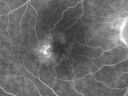
Group 2a Juxtafoveal Telangiectasis - MacTel - Idiopathic Juxtafoveal Macular Telangiectasia - Macular Hole712 views80-year-old man has juxtafoveal retinal telangiectasis in both eyes. He also has a true full-thickness macular hole in the left eye. The vision OD is 20/40, OS is 20/160
Group 2a Juxtafoveal Retinal Telangiectasis (telangiectasia), MacTel
|
|

Macular Telangiectasis (Group 2a Juxtafoveal Telangiectasis) IR712 views63-year-old woman has juxtafoveal retinal telangiectasis in both eyes. She notices her vision a little worse with more distortion and change over the last six months.
VISUAL ACUITY: OD 20/40, OS 20/40.
|
|

Central Serous Retinopathy - Acute711 views49-year-old man sees a spot in the central vision with a red halo for 2 weeks OD is 20/50, OS is 20/20
|
|
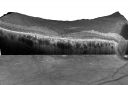
Plaquenil Toxicity - Bull's Eye Maculopathy - 25 Years Therapy - OCT Video - TAKES TIME TO LOAD711 views73-year-old woman has been on Plaquenil for twenty five years. Plaquenil has now been stopped for 6 months. VA continues to decline as does visual function - 20/80 OD , 20/40 OS
|
|

Group 2a Juxtafoveal Retinal Telangiectasis (type 2)710 views58-year-old man decreased vision right eye and not better since cataract surgery one year ago. OD 20/40, OS 20/40.
|
|

HD Grayscale OCT of Macular Hole710 views
|
|
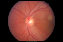
Idiopathic Central Serous Retinopathy - Bilateral - Acute 709 views49-year-old man decreasing vision in the left eye for one week. He is not taking any steroid or steroid-related products. His blood pressure as far as he knows is under control.
VISUAL ACUITY: OD 20/30, OS 20/400
|
|

Plaquenil Toxicity - Bulls Eye Maculopathy708 views70-year-old woman with systemic Lupus erythematosus and clotting problems. She was on the Plaquenil for about eight years and then off the Plaquenil for the last eight years because she developed macular toxicity. Although her vision was hazy, it was stable. Recent deceased vision left eye: OD 20/60, OS 20/100. IOP: OD 18, OS 19.
|
|

Outer Retinal Holes - Probably Solar Retinopathy (history is negative)708 views
|
|
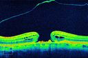
HD OCT of Macular Hole with Operculum706 views
|
|
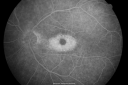
Plaquenil (Hydroxycholorquine) Toxicity - Bull's Eye Maculopathy - 73 year old womand 25 Years therapy - 64 inches tall - 110 lbs705 views73-year-old woman has been on Plaquenil for twenty five years. She noticed loss of paracentral vision a few months ago. VISUAL ACUITY: OD 20/70, OS 20/30. Her color vision was 0 out 10 correct in both eyes and she also missed the controls.
|
|

Acute Central Serous Retinopathy - CSR - Steroid Induced (probably) - Good Vision - ICG Affected Eye - Choroidal Leakage705 views42-year-old man was seen in the office on October 5, 2011. He had noticed starting in August after a course of antibiotic and steroids, that he developed new spots in his vision in the right eye. He may have had an episode like this sometime in the past. He did take steroids a few years ago and his vision did change at that time, but then returned.
VISUAL ACUITY: OD 20/32, OS 20/32. The OCT scan of the right eye confirms subretinal fluid.
|
|
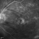
Macular Edema and dry AMD - excellent example of why FA and ICG are necessary in management of wet AMD705 viewsThis 72 year old woman has an OCT that looks like wet AMD. Only her FA and ICG show that she has inflammatory macular edema. She had no clinical signs or symptoms of uveitis. Nevertheless, the macular edema responded to subtenon's kenalog.
|
|

Cystoid Macular Edema - Xalatan - ECCE was done 20 years previously704 views71 Year old woman who noticed about a month ago the central vision in the right eye fogged up. OD 20/200 POSSIBLE XALATAN INDUCED CYSTOID MACULAR EDEMA
|
|

Group 2a Juxtafoveal Telangiectasis - MacTel - Idiopathic Juxtafoveal Macular Telangiectasia - Macular Hole704 views80-year-old man has juxtafoveal retinal telangiectasis in both eyes. He also has a true full-thickness macular hole in the left eye. The vision OD is 20/40, OS is 20/160
Group 2a Juxtafoveal Retinal Telangiectasis (telangiectasia), MacTel
|
|

Acute Central Serous Retinopathy - CSR - Steroid Induced (probably) - Good Vision 699 views42-year-old man was seen in the office on October 5, 2011. He had noticed starting in August after a course of antibiotic and steroids, that he developed new spots in his vision in the right eye. He may have had an episode like this sometime in the past. He did take steroids a few years ago and his vision did change at that time, but then returned.
VISUAL ACUITY: OD 20/32, OS 20/32. The OCT scan of the right eye confirms subretinal fluid.
|
|
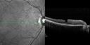
Psuedomacular Hole - Epiretinal Membrane - Vision unchanged 20/40 for 6 months698 views
|
|

Plaquenil Toxicity - Bulls Eye Maculopathy697 views70-year-old woman with systemic Lupus erythematosus and clotting problems. She was on the Plaquenil for about eight years and then off the Plaquenil for the last eight years because she developed macular toxicity. Although her vision was hazy, it was stable. Recent deceased vision left eye: OD 20/60, OS 20/100. IOP: OD 18, OS 19.
|
|

Plaquenil Toxicity - Bulls Eye Maculopathy697 views70-year-old woman with systemic Lupus erythematosus and clotting problems. She was on the Plaquenil for about eight years and then off the Plaquenil for the last eight years because she developed macular toxicity. Although her vision was hazy, it was stable. Recent deceased vision left eye: OD 20/60, OS 20/100. IOP: OD 18, OS 19.
|
|
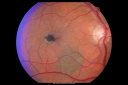
Juxtafoveal Telangiectasis - MacTel - Group 2a - Stage IV - Pigment Plaque 89 Year Old Man (Also Nevus)697 views89-year-old man has stage IV juxtafoveal retinal telangiectasis in both eyes with background diabetic retinopathy. His vision is stable since he was here last which is about 6 months ago. OD 20/50, OS 20/70.
|
|
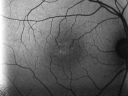
Juxtafoveal Telangiectasis - Stage 5 Left Eye - New Blood and Neovascularization MacTel FAF694 views60-year-old man has juxtafoveal retinal telangiectasis recent vision loss OD 20/50, OS 20/70 : Hemorrhage OS and new SRNVM
|
|

Idiopathic Cystoid Macular Edema - CME - Responded to Subtenons Kenalog - Red Free Photos Shows Foveal Cysts693 views49-year-old man had a flu two weeks ago the right eye has been throbbing and his vision in the right eye has been foggy. OD 20/80, OS 20/40. IOP: 14 OU.
|
|

Chronic Pseudophakic Cystoid Macular Edema - Haptic Migrated into Anterior Chamber Through Peripheral Iridotomy691 views57-year-old man with congenital nystagmus. He had cataract surgery done 30 years ago and has decreased vision left eye for 6 months. OD is 20/50, OS is 20/100
|
|

Idiopathic Cystoid Macular Edema - CME - Responded to Subtenons Kenalog - Early Fluorescein Angiogram Showing Dilated Capillaries690 views49-year-old man had a flu two weeks ago the right eye has been throbbing and his vision in the right eye has been foggy. OD 20/80, OS 20/40. IOP: 14 OU.
|
|

Acute Central Serous Retinopathy - CSR - Steroid Induced (probably) - Good Vision - FA Affected Eye688 views42-year-old man was seen in the office on October 5, 2011. He had noticed starting in August after a course of antibiotic and steroids, that he developed new spots in his vision in the right eye. He may have had an episode like this sometime in the past. He did take steroids a few years ago and his vision did change at that time, but then returned.
VISUAL ACUITY: OD 20/32, OS 20/32. The OCT scan of the right eye confirms subretinal fluid.
|
|
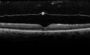
Lamellar Hole Epiretinal Proliferation - LHEP688 views
|
|
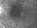
Group 2a Juxtafoveal Telangiectasis - MacTel - Idiopathic Juxtafoveal Macular Telangiectasia - Macular Hole687 views80-year-old man has juxtafoveal retinal telangiectasis in both eyes. He also has a true full-thickness macular hole in the left eye. The vision OD is 20/40, OS is 20/160
Group 2a Juxtafoveal Retinal Telangiectasis (telangiectasia), MacTel
|
|
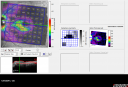
Plaquenil Toxicity - Bull's Eye Maculopathy - 25 Years Therapy - OCT Posterior Pole MAP687 views73-year-old woman has been on Plaquenil for twenty five years. Plaquenil has now been stopped for 6 months. VA continues to decline as does visual function - 20/80 OD , 20/40 OS
|
|
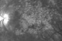
Chronic Pseudophakic Cystoid Macular Edema - Haptic Migrated into Anterior Chamber Through Peripheral Iridotomy687 views57-year-old man with congenital nystagmus. He had cataract surgery done 30 years ago and has decreased vision left eye for 6 months. OD is 20/50, OS is 20/100
|
|

Chronic Central Serous Retinopathy 1 month after Photodynamic Therapy (Visudyne)687 viewsSerous Retinal Detachment is Gone (Absorbed)
|
|

Plaquenil Toxicity - Bulls Eye Maculopathy685 views70-year-old woman with systemic Lupus erythematosus and clotting problems. She was on the Plaquenil for about eight years and then off the Plaquenil for the last eight years because she developed macular toxicity. Although her vision was hazy, it was stable. Recent deceased vision left eye: OD 20/60, OS 20/100. IOP: OD 18, OS 19.
|
|
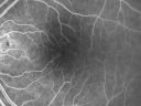
Group 2a Juxtafoveal Telangiectasis - MacTel - Idiopathic Juxtafoveal Macular Telangiectasia - Macular Hole685 views80-year-old man has juxtafoveal retinal telangiectasis in both eyes. He also has a true full-thickness macular hole in the left eye. The vision OD is 20/40, OS is 20/160
Group 2a Juxtafoveal Retinal Telangiectasis (telangiectasia), MacTel
|
|

Macular Edema and dry AMD - excellent example of why FA and ICG are necessary in management of wet AMD684 viewsThis 72 year old woman has an OCT that looks like wet AMD. Only her FA and ICG show that she has inflammatory macular edema. She had no clinical signs or symptoms of uveitis. Nevertheless, the macular edema responded to subtenon's kenalog.
|
|

Plaquenil Toxicity - Bulls Eye Maculopathy683 views70-year-old woman with systemic Lupus erythematosus and clotting problems. She was on the Plaquenil for about eight years and then off the Plaquenil for the last eight years because she developed macular toxicity. Although her vision was hazy, it was stable. Recent deceased vision left eye: OD 20/60, OS 20/100. IOP: OD 18, OS 19.
|
|
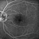
Lumigan (Bimatoprost) Cystoid Macular Edema - Eye Better 1 month after Stopping Lumigan683 views 82-year-old man had cataract surgery in 1993. He has had glaucoma for seven or eight years for a long time. He is taking Lumigan only in the left eye. About a year ago he started in the right eye and then starting just about a few months ago he has noticed intermittent blurring in the vision in the right eye. Vision 20/25 OD, 20/20 OS
|
|

Central Serous Retinopathy - Subretinal Fibrin683 views66 year old female with 20 year history of single episode of CSR. Now with vision loss for a month but improving - VA 20/63 OD; 20/32 OS
|
|

Plaquenil (Hydroxycholorquine) Toxicity - Bull's Eye Maculopathy - 73 year old womand 25 Years therapy - 64 inches tall - 110 lbs682 views73-year-old woman has been on Plaquenil for twenty five years. She noticed loss of paracentral vision a few months ago. VISUAL ACUITY: OD 20/70, OS 20/30. Her color vision was 0 out 10 correct in both eyes and she also missed the controls.
|
|
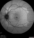
Outer Retinal Holes - Probably Solar Retinopathy (history is negative)682 views
|
|
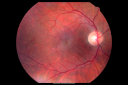
Macular Pucker Pre-OP 20/200 680 views
|
|
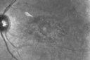
Plaquenil Toxicity - Bull's Eye Maculopathy - 25 Years Therapy - Infra-red Image680 views73-year-old woman has been on Plaquenil for twenty five years. Plaquenil has now been stopped for 6 months. VA continues to decline as does visual function - 20/80 OD , 20/40 OS
|
|
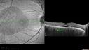
Acute Central Serous Retinopathy - CSR - Steroid Induced (probably) - Good Vision 680 views42-year-old man was seen in the office on October 5, 2011. He had noticed starting in August after a course of antibiotic and steroids, that he developed new spots in his vision in the right eye. He may have had an episode like this sometime in the past. He did take steroids a few years ago and his vision did change at that time, but then returned.
VISUAL ACUITY: OD 20/32, OS 20/32. The OCT scan of the right eye confirms subretinal fluid.
|
|
| 2319 files on 24 page(s) |
 |
3 | |
|
|
|
|
|
|
|
|


