Most viewed - Tumors, Masses, Pigmented Lesions
|

Choroidal Nevus with Overlying Drusen4283 views48-year-old woman Vision OD is 20/16, OS is 20/20.
|
|

CHRPE - Gardner Syndrome - FAMILIAL COLORECTAL POLYPOSIS - 11 year old female4107 views11-year-old child with CHRPE OS has six undescended adult teeth and also she has had a cyst removed from her arm pit and she has three more in her arm pit and one on her face and she is seeing a dermatologist for that in the near future. The mother reports no health problems, however, the patient’s grandmother does have a history of intestinal polyps, as does the patient’s grandmother’s brothers son.
|
|

Optic Nerve Tumor3896 views21 year old male: Presents with a history of Von Hippel-Lindau Syndrome, diagnosed in 2005 affecting his left Optic Nerve, Brain, Spine, Kidney and Pancreas. He has undergone Laser for Retinal Hemangioma measuring 2/3-4/5DD -vs_ 1/3DD when originally diagnosed. However his visual acuity remains good 20/25. Patient has also undergone Neurosurgery in 2005 and Spinal cord in 2006. Recent MRI of spinal cord hemangioma showed stable tumors.
|
|

Grouped pigmentation - Bear Tracks2797 views71 year old caucasian woman with incidental finding of extensive grouped RPE hypertrophy. Routine colonoscopy performed in the preceding year was normal.
|
|

CHRPE - Gardner Syndrome - FAMILIAL COLORECTAL POLYPOSIS - 11 year old female2146 views11-year-old child with CHRPE OS has six undescended adult teeth and also she has had a cyst removed from her arm pit and she has three more in her arm pit and one on her face and she is seeing a dermatologist for that in the near future. The mother reports no health problems, however, the patient’s grandmother does have a history of intestinal polyps, as does the patient’s grandmother’s brothers son.
|
|

Amelanotic Choroidal Nevus 2112 views45-year-old man OD 20/20, OS 20/20. 5x5 mm flat, yellow choroidal lesion just inferonasal to the optic nerve, two disc-diameters from the nerve with scalloped edges. It looks like a confluent drusen, but it is not elevated.
|
|

Red Free Hemangioma left eye2061 views
|
|

Retinoblastoma2046 viewsYoung male who has a retinal tumor inferior with a calcium core in the left eye. This is a regressed retinoblastoma.
|
|

Multiple - Congenital Hypertrophy of the Pigment Epithelium - CHRPE - Gardner Syndrome1972 views63 Year Old woman with a history of colon cancer at age 32. She has also had thyroid cancer. Two of her 6 siblings died of cancer, one of sarcoma and one of lung cancer. Vision 20/30, 20/25. Fundus shows CHRPE in both eyes.
|
|

Retinoblastoma1842 views
|
|

Amelanotic Choroidal Nevus 1741 views45-year-old man OD 20/20, OS 20/20. 5x5 mm flat, yellow choroidal lesion just inferonasal to the optic nerve, two disc-diameters from the nerve with scalloped edges. It looks like a confluent drusen, but it is not elevated.
|
|

Macular Choroidal Nevus1740 views78-year-old woman has choroidal nevus in the macula of the left eye. You asked her to come in here for an evaluation and nevus was noted in the past about 7 years ago. She has not noticed any change in vision in that eye.
VISUAL ACUITY: OU 20/30.
|
|

CHRPE - Gardner Syndrome - FAMILIAL COLORECTAL POLYPOSIS - 11 year old female1734 views11-year-old child with CHRPE OS has six undescended adult teeth and also she has had a cyst removed from her arm pit and she has three more in her arm pit and one on her face and she is seeing a dermatologist for that in the near future. The mother reports no health problems, however, the patient’s grandmother does have a history of intestinal polyps, as does the patient’s grandmother’s brothers son.
|
|

Choroidal Nevus with Overlying Drusen1713 views48-year-old woman Vision OD is 20/16, OS is 20/20.
|
|

CHRPE - Gardner Syndrome - FAMILIAL COLORECTAL POLYPOSIS - 11 year old female1684 views11-year-old child with CHRPE OS has six undescended adult teeth and also she has had a cyst removed from her arm pit and she has three more in her arm pit and one on her face and she is seeing a dermatologist for that in the near future. The mother reports no health problems, however, the patient’s grandmother does have a history of intestinal polyps, as does the patient’s grandmother’s brothers son.
|
|

Melanoma1681 viewsYoung male with large Melanoma
|
|

Melanoma1641 views40 Year old male
|
|

Choroidal Melanoma1598 views
|
|

Amelanotic Nevus Initial Visit1589 views
|
|

Choroidal Metastases - Both Eyes - Exudative Retinal Detachment1511 views58-year-old man smoke a pack of cigarettes a day. He has had some ribcage pain for about the last two weeks. He also had a sinus infection for about two weeks, and he has also had hazy vision on the superior visual field in the left eye for about the last two weeks. vision is 20/25 in both eyes
|
|

Choroidal Melanoma1469 views
|
|

Choroidal Osteoma1460 viewsPatient with decreased vision within the past 2-years. VA 20/100 in the right eye. Fundus photo shows atrophic scar temporal to the macula in the right eye.
|
|
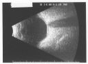
Pigmented Choroidal Mass Right Eye - Melanoma vs. Nevus vs. Unilateral Melanocytic Proliferation - Initial Visit B Scan1454 views71-year-old man is vision has been bothering him for about a month. He does not notice one eye particularly worse than the other. OD is 20/40, OS is 20/25 - Initial Visit
|
|

Retinocytoma - Retinoma - Regressed Retinoblastoma 43 year old male incidental finding 1429 views
|
|

Choroidal Melanoma with Retinal Detachment1400 views57 year old female who underwent proton beam therapy for Choroidal Melanoma. VA was 20/400 at post op visit.
|
|

Chorodial Melanoma1380 views80 year old male came in for retinal evaluation and presented with a melanoma.
|
|
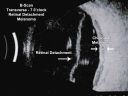
Choroidal Melanoma - Exudative Retinal Detachment 82 Year Old Man B Scan - Transverse1363 views82-year-old man who had 3 falls 2 weeks ago. After the falls he started checking his vision and noticed there was a veil over the left eye, which he had seen for about two weeks in the superior visual field. OD 20/25, OS 20/40
(patient was medically never well enough for brachytherapy and died 6 months later from heart disease)
|
|
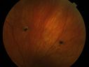
Multiple - Congenital Hypertrophy of the Pigment Epithelium - CHRPE - Gardner Syndrome1321 views63 Year Old woman with a history of colon cancer at age 32. She has also had thyroid cancer. Two of her 6 siblings died of cancer, one of sarcoma and one of lung cancer. Vision 20/30, 20/25. Fundus shows CHRPE in both eyes.
|
|
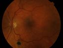
Multiple - Congenital Hypertrophy of the Pigment Epithelium - CHRPE - Gardner Syndrome1313 views63 Year Old woman with a history of colon cancer at age 32. She has also had thyroid cancer. Two of her 6 siblings died of cancer, one of sarcoma and one of lung cancer. Vision 20/30, 20/25. Fundus shows CHRPE in both eyes.
|
|
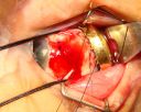
Radioactive Plaque Placement for Choroidal Melanoma Plaque Placed but not Sutured to Eye Yet1300 views
|
|

Choroidal Melanoma with Radiation Retinopathy1286 viewsPatient comes with follow up on Choroidal Melanoma, Right Eye that was treated back in June of 2009 with Radioactive Implant. Vein Occlusion is also present with VA - Hand Motion. Hemorrhages visible with hard exudates from the Radiation Retinopathy.
|
|
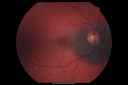
Peripapillary Choroidal Nevus1283 views37-year-old woman was seen in the office on July 27, 2011. She has photographs from 2009 and 2010 showing a peripapillary choroidal nevus in the right eye. VISUAL ACUITY: OD 20/20
|
|
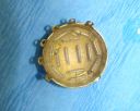
Radioactive Plaque Placement for Choroidal Melanoma Iodine Radioactive Seeds in Plaque1249 views
|
|
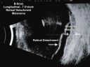
Choroidal Melanoma - Exudative Retinal Detachment 82 Year Old Man B Scan Ultrasound Longitudinal1243 views82-year-old man who had 3 falls 2 weeks ago. After the falls he started checking his vision and noticed there was a veil over the left eye, which he had seen for about two weeks in the superior visual field. OD 20/25, OS 20/40
(patient was medically never well enough for brachytherapy and died 6 months later from heart disease)
|
|

Retinocytoma - Retinoma - Regressed Retinoblastoma 43 year old male incidental finding 1235 views
|
|

Radioactive Plaque Placement for Choroidal Melanoma Eye is Anesthetized Prior to Surgery1223 views
|
|

CHOROIDAL Melanoma1206 views
|
|
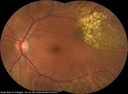
Choroidal Nevus with Drusen1195 viewsLow risk choroidal nevus with many overlying drusen
|
|

Choroidal Melanoma with Sub Retinal Fluid1188 viewsFundus photography shows choroidal melanoma superiorily between 11-12-o'clock in the left eye. VA is 20/150 in the left eye.
|
|

Choroidal Tumor consistent with Uveal Melanoma1176 viewsPatient comes in for second opinion for 2-melanomas in the right eye. Patient's VA is 20/20, both eyes. Fundus exam reveals melanoma superior to the macula which measured at 0.7-mm thick. Second melanoma is nasally which measured at 4.1-mm thick and has low internal reflectivity. Patient will be followed up with MRI and ocular protocol.
|
|

Amelanotic Choroidal Nevus 1161 views45-year-old man OD 20/20, OS 20/20. 5x5 mm flat, yellow choroidal lesion just inferonasal to the optic nerve, two disc-diameters from the nerve with scalloped edges. It looks like a confluent drusen, but it is not elevated.
|
|

Choroidal Melanoma1160 viewsPatient comes in with spot in vision in the right eye. Fundus photography shows medium size melanoma adjacent to the optic nerve and measures 13mm across horizontally and 6mm diameter in thickness. Patient will undergo proton beam therapy.
|
|

Retinocytoma - Retinoma - Regressed Retinoblastoma 43 year old male incidental finding 1155 views
|
|

Radioactive Plaque Placement for Choroidal Melanoma Plaque Sutured to Globe1151 views
|
|
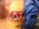
Radioactive Plaque Placement for Choroidal Melanoma Radioactive Plaque Placement for Choroidal Melanoma 1146 views
|
|
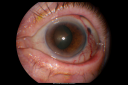
Melanoma - Large Ciliary Body Photo shows tumor visible through the pupil1143 views
|
|

Melanoma of Optic Nerve1138 viewsPatient comes in for evaluation on Optic Nerve Melanoma in the right eye. Patient's VA is 20/40 in the right eye. Fundus photograph reveals melanoma invading optic nerve and ultrasound reveals behind the optic nerve as well.
|
|

Metastatic Choroidal Lesion1135 viewsYoung female with choroidal lesion with subretinal fluid in the left eye. VA is finger count in the left eye.
|
|

Radioactive Plaque Placement for Choroidal Melanoma Eye After Surgery with Conjunctiva Closed1123 views
|
|

Radioactive Plaque Placement for Choroidal Melanoma Plaque Placed but not Sutured to Eye Ye1119 views
|
|

Choroidal Melanoma1109 viewsFemale patient comes in with a visual defect in the right eye. VA is 20/40 in the right eye. Fundus photography shows choroidal melanoma with retinal detachment temporally at 3-o'clock in the right eye.
|
|

Peripapillary Choroidal Nevus1106 views37-year-old woman was seen in the office on July 27, 2011. She has photographs from 2009 and 2010 showing a peripapillary choroidal nevus in the right eye. VISUAL ACUITY: OD 20/20
|
|

Retinocytoma - Retinoma - Regressed Retinoblastoma 43 year old male incidental finding 1099 views
|
|
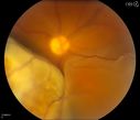
Choroidal Melanoma with Retinal Detachment1098 views57 year old female who underwent proton beam therapy for Choroidal Melanoma. VA was 20/400 at post op visit.
|
|

Peripapillary Choroidal Nevus1090 views37-year-old woman was seen in the office on July 27, 2011. She has photographs from 2009 and 2010 showing a peripapillary choroidal nevus in the right eye. VISUAL ACUITY: OD 20/20
|
|

Choroidal Metastasis Tumor 1086 viewsPatient that a history of breast cancer. She has a metastasis tumor superiority in the right eye. VA was 20/20, right eye. Patient underwent radiation and chemotherapy and mass has not changed in size for the past year.
|
|
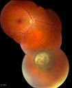
Choroidal Nevus1084 viewsPatient comes in with pigmented spot in the right eye. VA is 20/25. Fundus photography shows elevated choroidal nevus inferiorily in the right eye. Will be reevaluated in 3-months...
|
|

Amelanotic Choroidal Nevus 1078 views45-year-old man OD 20/20, OS 20/20. 5x5 mm flat, yellow choroidal lesion just inferonasal to the optic nerve, two disc-diameters from the nerve with scalloped edges. It looks like a confluent drusen, but it is not elevated.
|
|

Treated Choroidal Melanoma1073 viewsFollow up patient for melanoma in the right eye that was treated with proton beam therapy. VA is 20/50 in the right eye. Fundus photo shows scattered hemorrhages inferiorly with tumor temporal inferior at 8-o'clock. Patient will be followed up in 6-months
|
|

Choroidal Nevus with Overlying Drusen1072 views48-year-old woman Vision OD is 20/16, OS is 20/20.
|
|

Radioactive Plaque Placement for Choroidal Melanoma Plaque Placed but not Sutured to Eye Ye1069 views
|
|

Retinoblastoma - Bilateral - Regressed - Chemotherapy, Thermal Therapy, Cryotherapy1067 views10-year-old child was seen in the office on August 6, 2008. She was treated with a combination of chemotherapy at 10 months old as well as thermal therapy and cryotherapy for bilateral retinoblastoma tumors. She reportedly had nine tumors in the left eye and five tumors in the right eye
|
|

Pigmented Choroidal Mass Right Eye - Melanoma vs. Nevus vs. Unilateral Melanocytic Proliferation - Initial Visit and 4 Month Follow-Up - Color Photo1054 viewsPigmented lesion is visible in the superior half of the macular on the follow-up visit.
|
|

Amelanotic Nevus - 1 year Follow-up Visit - No Change1053 views
|
|

Radioactive Plaque Placement for Choroidal Melanoma Transillumination of Globe to Localize Melanoma Arrow Points to Tumor Shadow1038 views
|
|

Optic Nerve Tumor1037 viewsYoung male patient with a tumor present on the optic nerve in the right eye. VA is hand motion in the right eye
|
|

Amelanotic Choroidal Nevus 1034 views45-year-old man OD 20/20, OS 20/20. 5x5 mm flat, yellow choroidal lesion just inferonasal to the optic nerve, two disc-diameters from the nerve with scalloped edges. It looks like a confluent drusen, but it is not elevated.
|
|
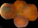
Uveal Choroidal Melanoma1023 viewsPatient comes in for evaluation on a Choroidal Melanoma in the right eye. VA was 20/25 in both eyes. The melanoma is in the temporal aspect of the right eye. It measured at 0.7mm elevated after doing a BSCAN Ultrasound.
|
|

Radioactive Plaque Placement for Choroidal Melanoma Transillumination of Globe to Localize Melanoma 1019 views
|
|

Radioactive Plaque Placement for Choroidal Melanoma Gold Plaque about the size of a Quarter1017 views
|
|
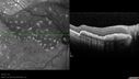
Primary Ocular Lymphoma - 020513 - SD OCT - SD OCT1014 views
|
|

Large Choroidal Melanoma with Retinal Detachment1014 viewsPatient notices a decline in her vision for the past 3-weeks in her right eye. VA was light perception in the right eye. Fundus photo shows a very large melanoma temporal to the optic nerve and retinal detachment inferiorly. Patient will under go proton beam therapy.
|
|

Radioactive Plaque Placement for Choroidal Melanoma Plaque Sutured to Globe1007 views
|
|

Radioactive Plaque Placement for Choroidal Melanoma Lead Patch after Surgery1003 views
|
|

Hemangioma999 viewsFP Composite of Hemangioma in OD
|
|

Radioactive Plaque Placement for Choroidal Melanoma Transillumination of Globe to Localize Melanoma 998 views
|
|

Choroidal Metastases - Both Eyes - Exudative Retinal Detachment995 views58-year-old man smoke a pack of cigarettes a day. He has had some ribcage pain for about the last two weeks. He also had a sinus infection for about two weeks, and he has also had hazy vision on the superior visual field in the left eye for about the last two weeks. vision is 20/25 in both eyes
|
|

Radioactive Plaque Placement for Choroidal Melanoma Transillumination of Globe to Localize Melanoma 992 views
|
|

Melanoma - Large Ciliary Body B-Scan ultrasound shows tumore filling much of the eye979 views
|
|

Melanoma - Large Ciliary Body Photo shows tumor visible throught the pupil967 views
|
|

6mm x 11mm Melanoma967 views76 year old male present with a 6mm x 1mm melanoma. Large melanoma very nasal OS. Asteroid Hyalosis present OS only. VA 20/40 OD 20/40 OS.
|
|
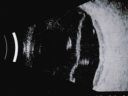
Choroidal Melanoma - Exudative Retinal Detachment 82 Year Old Man B Scan Ultrasound Transverce964 views82-year-old man who had 3 falls 2 weeks ago. After the falls he started checking his vision and noticed there was a veil over the left eye, which he had seen for about two weeks in the superior visual field. OD 20/25, OS 20/40
(patient was medically never well enough for brachytherapy and died 6 months later from heart disease)
|
|

Choroidal Nevus with Drusen963 viewsLow risk choroidal nevus with many overlying drusen
|
|
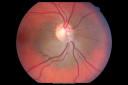
Peripapillary Choroidal Nevus952 views37-year-old woman was seen in the office on July 27, 2011. She has photographs from 2009 and 2010 showing a peripapillary choroidal nevus in the right eye. VISUAL ACUITY: OD 20/20
|
|

Multiple - Congenital Hypertrophy of the Pigment Epithelium - CHRPE - Gardner Syndrome946 views63 Year Old woman with a history of colon cancer at age 32. She has also had thyroid cancer. Two of her 6 siblings died of cancer, one of sarcoma and one of lung cancer. Vision 20/30, 20/25. Fundus shows CHRPE in both eyes.
|
|
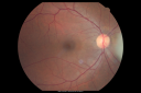
Retinocytoma - Retinoma - Regressed Retinoblastoma 43 year old male incidental finding 938 views
|
|
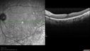
Primary Ocular Lymphoma - 020513 - SD OCT937 views
|
|
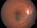
Choroidal Nevus in Macula 935 views
|
|

Treated Melanoma929 viewsTreated melanoma in the right eye. VA is light perception in the right eye.
|
|

Ciliary Body Melanoma925 views71-year old male who was recently diagnosed with a large ciliary body melanoma that is pushing into the anterior chamber of the left eye. Patient is going to proceed with proton therapy.
|
|
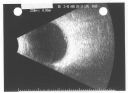
Pigmented Choroidal Mass Right Eye - Melanoma vs. Nevus vs. Unilateral Melanocytic Proliferation - Initial Visit B Scan919 views71-year-old man is vision has been bothering him for about a month. He does not notice one eye particularly worse than the other. OD is 20/40, OS is 20/25 - Initial Visit
|
|

Choroidal Melanoma919 viewsPost op Radioactive Implant with hard exudates surrounding the Melanoma with hemorrhage present.
|
|
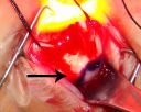
Radioactive Plaque Placement for Choroidal Melanoma Transillumination of Globe to Localize Melanoma Arrow Points to Tumor Shadow914 views
|
|

Retinoblastoma - Bilateral - Regressed - Chemotherapy, Thermal Therapy, Cryotherapy911 views10-year-old child was seen in the office on August 6, 2008. She was treated with a combination of chemotherapy at 10 months old as well as thermal therapy and cryotherapy for bilateral retinoblastoma tumors. She reportedly had nine tumors in the left eye and five tumors in the right eye
|
|
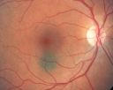
Choroidal Nevus in Macula 909 views
|
|

Retinoma - Retinocytoma - B Scan Ultrasound906 viewsUltrasound shows shadowing from the calcifications in the tumor.
|
|
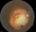
Choroidal Osteoma903 viewsYoung girl with vision loss from osteoma.
|
|
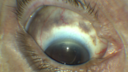
Melanosis Oculi and Choroidal Melanoma900 views88 year old female with 2.5 mm elevated choroidal melanoma and melanosis oculi. She also has geographic atrophy from AMD. The right eye is the better eye with 20/40 vision. After observation with growth, brachytherapy was done.
|
|
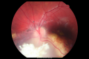
Retinoblastoma - Bilateral - Regressed - Chemotherapy, Thermal Therapy, Cryotherapy893 views10-year-old child was seen in the office on August 6, 2008. She was treated with a combination of chemotherapy at 10 months old as well as thermal therapy and cryotherapy for bilateral retinoblastoma tumors. She reportedly had nine tumors in the left eye and five tumors in the right eye
|
|

Retinoblastoma - Bilateral - Regressed - Chemotherapy, Thermal Therapy, Cryotherapy890 views10-year-old child was seen in the office on August 6, 2008. She was treated with a combination of chemotherapy at 10 months old as well as thermal therapy and cryotherapy for bilateral retinoblastoma tumors. She reportedly had nine tumors in the left eye and five tumors in the right eye
|
|
| 1098 files on 11 page(s) |
1 |
 |
|
|
|
|
|
|
|
|


