am meisten angesehen - Non-infectious Posterior Uveitis
|

Choroidal Neovascular Membrane - Multifocal Scars - PIC vs MFC vs OHS 524 x angesehen
|
|
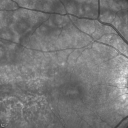
Acute Annular Outer Retinopathy for 3 Months - 25 YO Female InfraRed Image523 x angesehen25-year-old female who, about three months ago, put on her sunglasses and noticed a smudge in the vision and at that point she noticed the right eye was hazy. She had a cold a week or two before the vision loss, but she didn’t have any fever. She has no focal neurological problems in her past history. VA 20/20 OU
|
|

AZOOR - unilateral pseudo-retinitis pigmentosa523 x angesehen74 year old woman with unilateral vision loss 40 years ago.
|
|

Punctate Inner Choroidopathy (PIC) Mild523 x angesehen24 year old female with high myopia. Noticing some distortion on grid. Vision 20/20 OU. Was followed frequently for several months with no progression and no CNVM.
|
|

Multiple Evanescent White Dot Syndrome - Atypical Yellow Foveal Spot Right Eye522 x angesehen12-year-old had decreased vision in the left eye from reported amblyopia since around the year 2000. His right eye was doing fine until two days ago, when he had sudden severe vision loss. He sees a blurred spot in the middle of the vision in the right eye wherever he looks.
OD 20/60 with eccentric movement, OS 20/200. Pinhole 20/70. IOP: OD 14, OS 10.
|
|
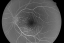
Multiple Evanescent White Dot Syndrome - Acute Idiopathic Blind Spot Enlargement522 x angesehen33-year-old woman one week ago she awoke and saw a red spots flashing out of her right eye. She notices them to the right side, which is the temporal visual field out of the right eye, then they changed to bursts of white lights without colors and then she notices straight ahead a couple spots. She has headaches in the back of her head. OD 20/20, OS 20/20
|
|

Acute Annular Outer Retinopathy for 3 Months - 25 YO Female Spectral Domain Optical Coherence Tomgraphy522 x angesehen25-year-old female who, about three months ago, put on her sunglasses and noticed a smudge in the vision and at that point she noticed the right eye was hazy. She had a cold a week or two before the vision loss, but she didn’t have any fever. She has no focal neurological problems in her past history. VA 20/20 OU
|
|

Multifocal Choroiditis Panuveitis with Subretinal Fibrosis Acute Phase522 x angesehen36 year old latin american female 3 weeks of vision loss and new headaches. MRI was normal. VA initially 20/32 OD; 20/20 OS Final vision was 20/63 OD; 20/20 OS
|
|

Choroidal Neovascular Membrane - Multifocal Scars - PIC vs MFC vs OHS 521 x angesehen
|
|

Birdshot Chorioretinitis - Fresh Presentation521 x angesehenUltawidefield ICG shows best the multifocal lesions. 61 year old woman with floaters for less than 6 months. HLA-A29 Positive
|
|
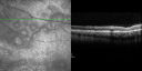
Acute Annular Outer Retinopathy for 3 Months - 25 YO Female Spectral Domain Optical Coherence Tomgraphy519 x angesehen25-year-old female who, about three months ago, put on her sunglasses and noticed a smudge in the vision and at that point she noticed the right eye was hazy. She had a cold a week or two before the vision loss, but she didn’t have any fever. She has no focal neurological problems in her past history. VA 20/20 OU
|
|

Choroidal Neovascular Membrane - Multifocal Scars - PIC vs MFC vs OHS 519 x angesehen
|
|

Recurrent MEWDS Left Eye - Teenage Girl519 x angesehenPatient presented in 2014 (first images) 2 months following vision loss left eye. Vision went from 20/60 - 20/20. OCT showed outer retinal irregularities which normalized. Then 3 years later in 2017 she had full blown MEWDS. She presented acutely and all the images are typical. Both episodes where in the same time of year, about 3 months after her flu shot for sports.
|
|

Choroidal Neovascular Membrane - Multifocal Scars - PIC vs MFC vs OHS 518 x angesehen
|
|

Recurrent MEWDS Left Eye - Teenage Girl518 x angesehenPatient presented in 2014 (first images) 2 months following vision loss left eye. Vision went from 20/60 - 20/20. OCT showed outer retinal irregularities which normalized. Then 3 years later in 2017 she had full blown MEWDS. She presented acutely and all the images are typical. Both episodes where in the same time of year, about 3 months after her flu shot for sports.
|
|

Recurrent MEWDS Left Eye - Teenage Girl518 x angesehenPatient presented in 2014 (first images) 2 months following vision loss left eye. Vision went from 20/60 - 20/20. OCT showed outer retinal irregularities which normalized. Then 3 years later in 2017 she had full blown MEWDS. She presented acutely and all the images are typical. Both episodes where in the same time of year, about 3 months after her flu shot for sports.
|
|

517 x angesehen
|
|

Choroidal Neovascular Membrane - Multifocal Scars - PIC vs MFC vs OHS 517 x angesehen
|
|

Multifocal Choroiditis Panuveitis with Subretinal Fibrosis Acute Phase517 x angesehen36 year old latin american female 3 weeks of vision loss and new headaches. MRI was normal. VA initially 20/32 OD; 20/20 OS Final vision was 20/63 OD; 20/20 OS
|
|

Birdshot Chorioretintis and PAMM517 x angesehenPatient presented with paracentral acute middle maculopathy as the presenting symptom of birdshot. This case was published. RETINAL CASES & BRIEF REPORTS 10:151–153, 2016.
|
|

Acute Annular Outer Retinopathy for 3 Months - 25 YO Female Spectral Domain Optical Coherence Tomgraphy516 x angesehen25-year-old female who, about three months ago, put on her sunglasses and noticed a smudge in the vision and at that point she noticed the right eye was hazy. She had a cold a week or two before the vision loss, but she didn’t have any fever. She has no focal neurological problems in her past history. VA 20/20 OU
|
|

Birdshot Chorioretinitis - Fresh Presentation516 x angesehenUltawidefield ICG shows best the multifocal lesions. 61 year old woman with floaters for less than 6 months. HLA-A29 Positive
|
|

Recurrent MEWDS Left Eye - Teenage Girl516 x angesehenPatient presented in 2014 (first images) 2 months following vision loss left eye. Vision went from 20/60 - 20/20. OCT showed outer retinal irregularities which normalized. Then 3 years later in 2017 she had full blown MEWDS. She presented acutely and all the images are typical. Both episodes where in the same time of year, about 3 months after her flu shot for sports.
|
|

Birdshot Chorioretinitis - Fresh Presentation515 x angesehenUltawidefield ICG shows best the multifocal lesions. 61 year old woman with floaters for less than 6 months. HLA-A29 Positive
|
|

Recurrent MEWDS Left Eye - Teenage Girl515 x angesehenPatient presented in 2014 (first images) 2 months following vision loss left eye. Vision went from 20/60 - 20/20. OCT showed outer retinal irregularities which normalized. Then 3 years later in 2017 she had full blown MEWDS. She presented acutely and all the images are typical. Both episodes where in the same time of year, about 3 months after her flu shot for sports.
|
|

Recurrent MEWDS Left Eye - Teenage Girl515 x angesehenPatient presented in 2014 (first images) 2 months following vision loss left eye. Vision went from 20/60 - 20/20. OCT showed outer retinal irregularities which normalized. Then 3 years later in 2017 she had full blown MEWDS. She presented acutely and all the images are typical. Both episodes where in the same time of year, about 3 months after her flu shot for sports.
|
|

Recurrent MEWDS Left Eye - Teenage Girl514 x angesehenPatient presented in 2014 (first images) 2 months following vision loss left eye. Vision went from 20/60 - 20/20. OCT showed outer retinal irregularities which normalized. Then 3 years later in 2017 she had full blown MEWDS. She presented acutely and all the images are typical. Both episodes where in the same time of year, about 3 months after her flu shot for sports.
|
|

Recurrent MEWDS Left Eye - Teenage Girl514 x angesehenPatient presented in 2014 (first images) 2 months following vision loss left eye. Vision went from 20/60 - 20/20. OCT showed outer retinal irregularities which normalized. Then 3 years later in 2017 she had full blown MEWDS. She presented acutely and all the images are typical. Both episodes where in the same time of year, about 3 months after her flu shot for sports.
|
|

Multifocal Choroiditis Panuveitis with Subretinal Fibrosis Acute Phase514 x angesehen36 year old latin american female 3 weeks of vision loss and new headaches. MRI was normal. VA initially 20/32 OD; 20/20 OS Final vision was 20/63 OD; 20/20 OS
|
|

Birdshot Chorioretinitis - Fresh Presentation513 x angesehenUltawidefield ICG shows best the multifocal lesions. 61 year old woman with floaters for less than 6 months. HLA-A29 Positive
|
|

Recurrent MEWDS Left Eye - Teenage Girl513 x angesehenPatient presented in 2014 (first images) 2 months following vision loss left eye. Vision went from 20/60 - 20/20. OCT showed outer retinal irregularities which normalized. Then 3 years later in 2017 she had full blown MEWDS. She presented acutely and all the images are typical. Both episodes where in the same time of year, about 3 months after her flu shot for sports.
|
|

Multifocal Choroiditis Panuveitis - New Choroidal Neovascular Membrane Right Eye513 x angesehen74 year old female had vitrectomy elsewhere for possible ocular lymphoma - 5 years of shimmery vision changes then 3 weeks ago noticed vision loss right eye.
|
|
512 x angesehen
|
|

Recurrent MEWDS Left Eye - Teenage Girl512 x angesehenPatient presented in 2014 (first images) 2 months following vision loss left eye. Vision went from 20/60 - 20/20. OCT showed outer retinal irregularities which normalized. Then 3 years later in 2017 she had full blown MEWDS. She presented acutely and all the images are typical. Both episodes where in the same time of year, about 3 months after her flu shot for sports.
|
|

Recurrent MEWDS Left Eye - Teenage Girl512 x angesehenPatient presented in 2014 (first images) 2 months following vision loss left eye. Vision went from 20/60 - 20/20. OCT showed outer retinal irregularities which normalized. Then 3 years later in 2017 she had full blown MEWDS. She presented acutely and all the images are typical. Both episodes where in the same time of year, about 3 months after her flu shot for sports.
|
|

AZOOR - unilateral pseudo-retinitis pigmentosa512 x angesehen74 year old woman with unilateral vision loss 40 years ago.
|
|

AZOOR - unilateral pseudo-retinitis pigmentosa512 x angesehen74 year old woman with unilateral vision loss 40 years ago.
|
|

Multifocal Choroiditis and Subretinal Fibrosis with new Subretinal Neovascular Membrane512 x angesehen26 year old African American Girl Saw me at age 10 (16 years ago) with acute multifocal choroiditis and panuveitis which evolved into subretinal fibrosis. She was initially treated with 3 years of methotrexate. Vision ended up 20/20 in both eyes. Her eyes have been inactive since and she was lost to follow-up.
A week ago she noticed sudden decreased vision in the right eye. Within the past few weeks there are more floaters in the right eye.
VA OD: sc6’/200 PHNI
VA OS: sc20/16
|
|

Multifocal Choroiditis - Linear Scars - Schlaegel lines511 x angesehen66 year old woman with macular pucker right eye. Images show typical linear peripheral scars for MFC or POHS - this is unilateral.
|
|

Punctate Inner Choroidopathy - New CNVM right eye511 x angesehen42 year old female with recent vision loss in the right eye - Right eye is 20/80 - Vision improved to 20/16 with Ranabizumab injections over 3 months
|
|
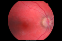
Acute Annular Outer Retinopathy for 3 Months - 25 YO Female Fundus Photograph510 x angesehen25-year-old female who, about three months ago, put on her sunglasses and noticed a smudge in the vision and at that point she noticed the right eye was hazy. She had a cold a week or two before the vision loss, but she didn’t have any fever. She has no focal neurological problems in her past history. VA 20/20 OU
|
|

Choroidal Neovascular Membrane - Multifocal Scars - PIC vs MFC vs OHS 510 x angesehen
|
|

Multifocal Choroiditis - Linear Scars - Schlaegel lines510 x angesehen66 year old woman with macular pucker right eye. Images show typical linear peripheral scars for MFC or POHS - this is unilateral.
|
|

Neuroretinitis - NOT cat scratch - 5 year old509 x angesehenPresented with nerve swelling, serous RD and uveitis. Ultimately responded to steroids and methotrexate. ANA is positive. All other extensive testing negative including MRI worried about MS
|
|
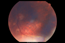
Sarcoidosis Multifocal Choroiditis509 x angesehen66 year old man with dense cataracts and recurrent uveitis. Images show multifocal choroidal granulomas from sarcoidosis more in the right eye than the left eye.
|
|

Punctate Inner Choroidopathy (PIC) Mild509 x angesehen24 year old female with high myopia. Noticing some distortion on grid. Vision 20/20 OU. Was followed frequently for several months with no progression and no CNVM.
|
|

Punctate Inner Choroidopathy (PIC) Mild509 x angesehen24 year old female with high myopia. Noticing some distortion on grid. Vision 20/20 OU. Was followed frequently for several months with no progression and no CNVM.
|
|

Choroidal Neovascular Membrane - Multifocal Scars - PIC vs MFC vs OHS 508 x angesehen
|
|

Birdshot Chorioretinitis - Fresh Presentation508 x angesehenUltawidefield ICG shows best the multifocal lesions. 61 year old woman with floaters for less than 6 months. HLA-A29 Positive
|
|

Multifocal Choroiditis Panuveitis - New Choroidal Neovascular Membrane Right Eye508 x angesehen74 year old female had vitrectomy elsewhere for possible ocular lymphoma - 5 years of shimmery vision changes then 3 weeks ago noticed vision loss right eye.
|
|

Multifocal Choroiditis Panuveitis with Subretinal Fibrosis Acute Phase508 x angesehen36 year old latin american female 3 weeks of vision loss and new headaches. MRI was normal. VA initially 20/32 OD; 20/20 OS Final vision was 20/63 OD; 20/20 OS
|
|

AZOOR - unilateral pseudo-retinitis pigmentosa508 x angesehen74 year old woman with unilateral vision loss 40 years ago.
|
|

AZOOR - unilateral pseudo-retinitis pigmentosa508 x angesehen74 year old woman with unilateral vision loss 40 years ago.
|
|

Multifocal Choroiditis and Subretinal Fibrosis with new Subretinal Neovascular Membrane508 x angesehen26 year old African American Girl Saw me at age 10 (16 years ago) with acute multifocal choroiditis and panuveitis which evolved into subretinal fibrosis. She was initially treated with 3 years of methotrexate. Vision ended up 20/20 in both eyes. Her eyes have been inactive since and she was lost to follow-up.
A week ago she noticed sudden decreased vision in the right eye. Within the past few weeks there are more floaters in the right eye.
VA OD: sc6’/200 PHNI
VA OS: sc20/16
|
|

Choroidal Neovascular Membrane - Multifocal Scars - PIC vs MFC vs OHS 507 x angesehen
|
|

Birdshot Chorioretinitis - Fresh Presentation507 x angesehenUltawidefield ICG shows best the multifocal lesions. 61 year old woman with floaters for less than 6 months. HLA-A29 Positive
|
|

Multifocal Choroiditis Panuveitis - New Choroidal Neovascular Membrane Right Eye507 x angesehen74 year old female had vitrectomy elsewhere for possible ocular lymphoma - 5 years of shimmery vision changes then 3 weeks ago noticed vision loss right eye.
|
|

506 x angesehen
|
|

Birdshot Chorioretinitis - Fresh Presentation506 x angesehenUltawidefield ICG shows best the multifocal lesions. 61 year old woman with floaters for less than 6 months. HLA-A29 Positive
|
|

Birdshot Chorioretinitis - Fresh Presentation506 x angesehenUltawidefield ICG shows best the multifocal lesions. 61 year old woman with floaters for less than 6 months. HLA-A29 Positive
|
|

Neuroretinitis - NOT cat scratch - 5 year old506 x angesehenPresented with nerve swelling, serous RD and uveitis. Ultimately responded to steroids and methotrexate. ANA is positive. All other extensive testing negative including MRI worried about MS
|
|
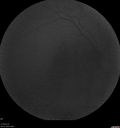
Sarcoidosis Multifocal Choroiditis506 x angesehen66 year old man with dense cataracts and recurrent uveitis. Images show multifocal choroidal granulomas from sarcoidosis more in the right eye than the left eye.
|
|

Multifocal Choroiditis Panuveitis with Subretinal Fibrosis Acute Phase506 x angesehen36 year old latin american female 3 weeks of vision loss and new headaches. MRI was normal. VA initially 20/32 OD; 20/20 OS Final vision was 20/63 OD; 20/20 OS
|
|

Birdshot Chorioretinitis - Fresh Presentation505 x angesehenUltawidefield ICG shows best the multifocal lesions. 61 year old woman with floaters for less than 6 months. HLA-A29 Positive
|
|

Birdshot Chorioretintis and PAMM505 x angesehenPatient presented with paracentral acute middle maculopathy as the presenting symptom of birdshot. This case was published. RETINAL CASES & BRIEF REPORTS 10:151–153, 2016.
|
|

Birdshot Chorioretinitis - Fresh Presentation504 x angesehenUltawidefield ICG shows best the multifocal lesions. 61 year old woman with floaters for less than 6 months. HLA-A29 Positive
|
|

Recurrent MEWDS Left Eye - Teenage Girl504 x angesehenPatient presented in 2014 (first images) 2 months following vision loss left eye. Vision went from 20/60 - 20/20. OCT showed outer retinal irregularities which normalized. Then 3 years later in 2017 she had full blown MEWDS. She presented acutely and all the images are typical. Both episodes where in the same time of year, about 3 months after her flu shot for sports.
|
|
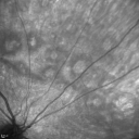
Acute Annular Outer Retinopathy for 3 Months - 25 YO Female InfraRed Image503 x angesehen25-year-old female who, about three months ago, put on her sunglasses and noticed a smudge in the vision and at that point she noticed the right eye was hazy. She had a cold a week or two before the vision loss, but she didn’t have any fever. She has no focal neurological problems in her past history. VA 20/20 OU
|
|

Choroidal Neovascular Membrane - Multifocal Scars - PIC vs MFC vs OHS 503 x angesehen
|
|

Birdshot Chorioretinitis - Fresh Presentation503 x angesehenUltawidefield ICG shows best the multifocal lesions. 61 year old woman with floaters for less than 6 months. HLA-A29 Positive
|
|

Birdshot Chorioretinitis - Fresh Presentation503 x angesehenUltawidefield ICG shows best the multifocal lesions. 61 year old woman with floaters for less than 6 months. HLA-A29 Positive
|
|

Recurrent MEWDS Left Eye - Teenage Girl503 x angesehenPatient presented in 2014 (first images) 2 months following vision loss left eye. Vision went from 20/60 - 20/20. OCT showed outer retinal irregularities which normalized. Then 3 years later in 2017 she had full blown MEWDS. She presented acutely and all the images are typical. Both episodes where in the same time of year, about 3 months after her flu shot for sports.
|
|

Recurrent MEWDS Left Eye - Teenage Girl503 x angesehenPatient presented in 2014 (first images) 2 months following vision loss left eye. Vision went from 20/60 - 20/20. OCT showed outer retinal irregularities which normalized. Then 3 years later in 2017 she had full blown MEWDS. She presented acutely and all the images are typical. Both episodes where in the same time of year, about 3 months after her flu shot for sports.
|
|

Multifocal Choroiditis Panuveitis with Subretinal Fibrosis Acute Phase503 x angesehen36 year old latin american female 3 weeks of vision loss and new headaches. MRI was normal. VA initially 20/32 OD; 20/20 OS Final vision was 20/63 OD; 20/20 OS
|
|

Choroidal Neovascular Membrane - Multifocal Scars - PIC vs MFC vs OHS 502 x angesehen
|
|

Unilateral Hemorrhagic Retinopathy502 x angesehenUnilateral Hemorrhagic Retinopathy
|
|

Birdshot Chorioretinitis - Fresh Presentation502 x angesehenUltawidefield ICG shows best the multifocal lesions. 61 year old woman with floaters for less than 6 months. HLA-A29 Positive
|
|

Birdshot Chorioretinitis - Fresh Presentation502 x angesehenUltawidefield ICG shows best the multifocal lesions. 61 year old woman with floaters for less than 6 months. HLA-A29 Positive
|
|

Recurrent MEWDS Left Eye - Teenage Girl502 x angesehenPatient presented in 2014 (first images) 2 months following vision loss left eye. Vision went from 20/60 - 20/20. OCT showed outer retinal irregularities which normalized. Then 3 years later in 2017 she had full blown MEWDS. She presented acutely and all the images are typical. Both episodes where in the same time of year, about 3 months after her flu shot for sports.
|
|

Multifocal Choroiditis Panuveitis with Subretinal Fibrosis Acute Phase502 x angesehen36 year old latin american female 3 weeks of vision loss and new headaches. MRI was normal. VA initially 20/32 OD; 20/20 OS Final vision was 20/63 OD; 20/20 OS
|
|

Birdshot Chorioretintis and PAMM502 x angesehenPatient presented with paracentral acute middle maculopathy as the presenting symptom of birdshot. This case was published. RETINAL CASES & BRIEF REPORTS 10:151–153, 2016.
|
|

Birdshot Chorioretintis and PAMM502 x angesehenPatient presented with paracentral acute middle maculopathy as the presenting symptom of birdshot. This case was published. RETINAL CASES & BRIEF REPORTS 10:151–153, 2016.
|
|

Multifocal Choroiditis and Subretinal Fibrosis with new Subretinal Neovascular Membrane502 x angesehen26 year old African American Girl Saw me at age 10 (16 years ago) with acute multifocal choroiditis and panuveitis which evolved into subretinal fibrosis. She was initially treated with 3 years of methotrexate. Vision ended up 20/20 in both eyes. Her eyes have been inactive since and she was lost to follow-up.
A week ago she noticed sudden decreased vision in the right eye. Within the past few weeks there are more floaters in the right eye.
VA OD: sc6’/200 PHNI
VA OS: sc20/16
|
|

Acute Annular Outer Retinopathy for 3 Months - 25 YO Female InfraRed Image501 x angesehen25-year-old female who, about three months ago, put on her sunglasses and noticed a smudge in the vision and at that point she noticed the right eye was hazy. She had a cold a week or two before the vision loss, but she didn’t have any fever. She has no focal neurological problems in her past history. VA 20/20 OU
|
|

Birdshot Chorioretinitis - Fresh Presentation501 x angesehenUltawidefield ICG shows best the multifocal lesions. 61 year old woman with floaters for less than 6 months. HLA-A29 Positive
|
|

Birdshot Chorioretinitis - Fresh Presentation501 x angesehenUltawidefield ICG shows best the multifocal lesions. 61 year old woman with floaters for less than 6 months. HLA-A29 Positive
|
|

Birdshot Chorioretinitis - Fresh Presentation501 x angesehenUltawidefield ICG shows best the multifocal lesions. 61 year old woman with floaters for less than 6 months. HLA-A29 Positive
|
|

Acute Annular Outer Retinopathy for 3 Months - 25 YO Female Fundus Photograph500 x angesehen25-year-old female who, about three months ago, put on her sunglasses and noticed a smudge in the vision and at that point she noticed the right eye was hazy. She had a cold a week or two before the vision loss, but she didn’t have any fever. She has no focal neurological problems in her past history. VA 20/20 OU
|
|

Choroidal Neovascular Membrane - Multifocal Scars - PIC vs MFC vs OHS 500 x angesehen
|
|

Recurrent MEWDS Left Eye - Teenage Girl500 x angesehenPatient presented in 2014 (first images) 2 months following vision loss left eye. Vision went from 20/60 - 20/20. OCT showed outer retinal irregularities which normalized. Then 3 years later in 2017 she had full blown MEWDS. She presented acutely and all the images are typical. Both episodes where in the same time of year, about 3 months after her flu shot for sports.
|
|

Recurrent MEWDS Left Eye - Teenage Girl500 x angesehenPatient presented in 2014 (first images) 2 months following vision loss left eye. Vision went from 20/60 - 20/20. OCT showed outer retinal irregularities which normalized. Then 3 years later in 2017 she had full blown MEWDS. She presented acutely and all the images are typical. Both episodes where in the same time of year, about 3 months after her flu shot for sports.
|
|

Recurrent MEWDS Left Eye - Teenage Girl500 x angesehenPatient presented in 2014 (first images) 2 months following vision loss left eye. Vision went from 20/60 - 20/20. OCT showed outer retinal irregularities which normalized. Then 3 years later in 2017 she had full blown MEWDS. She presented acutely and all the images are typical. Both episodes where in the same time of year, about 3 months after her flu shot for sports.
|
|
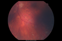
Sarcoidosis Multifocal Choroiditis500 x angesehen66 year old man with dense cataracts and recurrent uveitis. Images show multifocal choroidal granulomas from sarcoidosis more in the right eye than the left eye.
|
|

Multifocal Choroiditis Panuveitis with Subretinal Fibrosis Acute Phase500 x angesehen36 year old latin american female 3 weeks of vision loss and new headaches. MRI was normal. VA initially 20/32 OD; 20/20 OS Final vision was 20/63 OD; 20/20 OS
|
|

Punctate Inner Choroidopathy (PIC) Mild500 x angesehen24 year old female with high myopia. Noticing some distortion on grid. Vision 20/20 OU. Was followed frequently for several months with no progression and no CNVM.
|
|

Birdshot Chorioretinitis - Fresh Presentation498 x angesehenUltawidefield ICG shows best the multifocal lesions. 61 year old woman with floaters for less than 6 months. HLA-A29 Positive
|
|
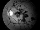
Unilateral Hemorrhagic Retinopathy497 x angesehenFrom 2012 ARVO ABSTRACT TITLED
Clinical Features Of Unilateral Hemorrhagic Retinopathy: A New Retinal Entity?
Conclusions: This unusual retinopathy with apparent spontaneous unilateral deep retinal hemorrhages remains unexplained and a new entity might be considered.
|
|

Multifocal Choroiditis Panuveitis - New Choroidal Neovascular Membrane Right Eye497 x angesehen74 year old female had vitrectomy elsewhere for possible ocular lymphoma - 5 years of shimmery vision changes then 3 weeks ago noticed vision loss right eye.
|
|

Multifocal Choroiditis Panuveitis with Subretinal Fibrosis Acute Phase497 x angesehen36 year old latin american female 3 weeks of vision loss and new headaches. MRI was normal. VA initially 20/32 OD; 20/20 OS Final vision was 20/63 OD; 20/20 OS
|
|

AZOOR - unilateral pseudo-retinitis pigmentosa497 x angesehen74 year old woman with unilateral vision loss 40 years ago.
|
|
| 1298 Dateien auf 13 Seite(n) |
 |
|
|
|
|
7 | |
|
|
|
|


