Most viewed - Choroidal Melanoma and Choroidal Nevus
|
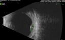
Melanosis Oculi and Choroidal Melanoma505 views88 year old female with 2.5 mm elevated choroidal melanoma and melanosis oculi. She also has geographic atrophy from AMD. The right eye is the better eye with 20/40 vision. After observation with growth, brachytherapy was done.
|
|

Choroidal Melanoma (Medium) Exudative Retinal Detachment 504 views87-year-old man noticed a new onset floater a few months ago. OD is 20/40, OS is 20/30
|
|

Pigmented Choroidal Mass Right Eye - Melanoma vs. Nevus vs. Unilateral Melanocytic Proliferation - 4 Month Follow-Up Images503 views71-year-old man his vision 4 month follow-up. Vision is 20/60 and patient sees flashing lights. Lesion has grown slightly in the superior half of the macula.
|
|
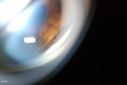
Ciliary Body Melanoma502 views71-year old male who was recently diagnosed with a large ciliary body melanoma that is pushing into the anterior chamber of the left eye. Patient is going to proceed with proton therapy.
|
|
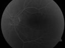
Pigmented Choroidal Mass Right Eye - Melanoma vs. Nevus vs. Unilateral Melanocytic Proliferation - 4 Month Follow-Up Images501 views71-year-old man his vision 4 month follow-up. Vision is 20/60 and patient sees flashing lights. Lesion has grown slightly in the superior half of the macula.
|
|
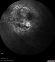
Medium Choroidal Melanoma Left Eye 499 views
|
|
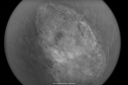
Choroidal Melanoma Medium498 views33-year-old man with a history of decreased in the right eye. He had blurring in the eye about a year ago. He reports that it improved spontaneously until it was “nearly resolvedâ€. He said he had some persistent distortion, but that over the past couple of weeks he has noticed this dark spot recurring in the right eye, the left eye is unaffected. He does not have any flashes, floaters or change in his peripheral visual acuity. 20/80 OD, 20/16 OS. No improvement with pinhole. IOP: 12 OD, 11 OS.
POSTERIOR SEGMENT EXAMINATION: Shows a clear view with no PVD in either eye. C/D ratio is 0.2 OU. Large retinal vessels are unremarkable. The macula of the right eye has a brown choroidal lesion with pigment overlying the surface and orange spots along its lateral surface. The left macula is normal.
B-SCAN: B-scan ecography of the ultrasound of the right eye shows a mass about 5 mm high with a moderate to low internal reflectivity on A-scan echogram. There is serous fluid inferiorly on the echogram
|
|
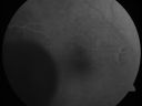
Choroidal Melanoma (Medium) Exudative Retinal Detachment 498 views87-year-old man noticed a new onset floater a few months ago. OD is 20/40, OS is 20/30
|
|
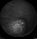
Melanosis Oculi and Choroidal Melanoma497 views88 year old female with 2.5 mm elevated choroidal melanoma and melanosis oculi. She also has geographic atrophy from AMD. The right eye is the better eye with 20/40 vision. After observation with growth, brachytherapy was done.
|
|
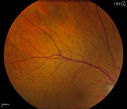
Uveal Choroidal Melanoma495 viewsPatient comes in for evaluation on a Choroidal Melanoma in the right eye. VA was 20/25 in both eyes. The melanoma is in the temporal aspect of the right eye. It measured at 0.7mm elevated after doing a BSCAN Ultrasound.
|
|
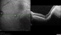
Medium Choroidal Melanoma Left Eye 495 views
|
|

Choroidal Nevus with Drusen494 views82 year old female with flat nevus for 3 years.
|
|
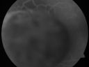
Choroidal Melanoma (Medium) Exudative Retinal Detachment 493 views87-year-old man noticed a new onset floater a few months ago. OD is 20/40, OS is 20/30
|
|
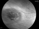
Uveal Choroidal Melanoma493 viewsPatient comes in for evaluation on a Choroidal Melanoma in the right eye. VA was 20/25 in both eyes. The melanoma is in the temporal aspect of the right eye. It measured at 0.7mm elevated after doing a BSCAN Ultrasound.
|
|
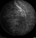
Melanosis Oculi and Choroidal Melanoma493 views88 year old female with 2.5 mm elevated choroidal melanoma and melanosis oculi. She also has geographic atrophy from AMD. The right eye is the better eye with 20/40 vision. After observation with growth, brachytherapy was done.
|
|

Pigmented Choroidal Mass Right Eye - Melanoma vs. Nevus vs. Unilateral Melanocytic Proliferation - 4 Month Follow-Up Images492 views71-year-old man his vision 4 month follow-up. Vision is 20/60 and patient sees flashing lights. Lesion has grown slightly in the superior half of the macula.
|
|
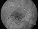
Pigmented Choroidal Mass Right Eye - Melanoma vs. Nevus vs. Unilateral Melanocytic Proliferation - Initial Visit492 views71-year-old man is vision has been bothering him for about a month. He does not notice one eye particularly worse than the other. OD is 20/40, OS is 20/25 - Initial Visit
|
|
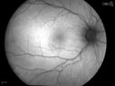
Uveal Choroidal Melanoma492 viewsPatient comes in for evaluation on a Choroidal Melanoma in the right eye. VA was 20/25 in both eyes. The melanoma is in the temporal aspect of the right eye. It measured at 0.7mm elevated after doing a BSCAN Ultrasound.
|
|
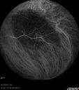
Medium Choroidal Melanoma Left Eye 492 views
|
|
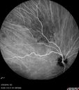
Amelanotic Choroidal Nevus - Multimodal Imaging492 views61 year old man with amelanotic choroidal nevus. Vision Normal - Stable over 4 years.
|
|
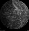
Melanosis Oculi and Choroidal Melanoma491 views88 year old female with 2.5 mm elevated choroidal melanoma and melanosis oculi. She also has geographic atrophy from AMD. The right eye is the better eye with 20/40 vision. After observation with growth, brachytherapy was done.
|
|

Choroidal Melanoma Medium490 views33-year-old man with a history of decreased in the right eye. He had blurring in the eye about a year ago. He reports that it improved spontaneously until it was “nearly resolvedâ€. He said he had some persistent distortion, but that over the past couple of weeks he has noticed this dark spot recurring in the right eye, the left eye is unaffected. He does not have any flashes, floaters or change in his peripheral visual acuity. 20/80 OD, 20/16 OS. No improvement with pinhole. IOP: 12 OD, 11 OS.
POSTERIOR SEGMENT EXAMINATION: Shows a clear view with no PVD in either eye. C/D ratio is 0.2 OU. Large retinal vessels are unremarkable. The macula of the right eye has a brown choroidal lesion with pigment overlying the surface and orange spots along its lateral surface. The left macula is normal.
B-SCAN: B-scan ecography of the ultrasound of the right eye shows a mass about 5 mm high with a moderate to low internal reflectivity on A-scan echogram. There is serous fluid inferiorly on the echogram
|
|

Pigmented Choroidal Mass Right Eye - Melanoma vs. Nevus vs. Unilateral Melanocytic Proliferation - Initial Visit490 views71-year-old man is vision has been bothering him for about a month. He does not notice one eye particularly worse than the other. OD is 20/40, OS is 20/25 - Initial Visit
|
|

Melanocytoma - Lightly Pigmented490 views42 year old female with very mild visual field and vision loss in the left eye: 20/20 OU
|
|

Atypical High Risk Choroidal Nevus vs Macular Melanoma489 views70 year old female with vision loss in the left eye for a few weeks. She was referred to a tumor center and being managed as an "atypical high risk choroidal nevus". The size is 6 mm x 6.5 mm x 1.6 mm (elevated).
Vision 20/40 - having serial avastin injections.
|
|

488 views
|
|
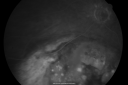
Choroidal Melanoma - Exudative Retinal Detachment 82 Year Old Man 488 views82-year-old man who had 3 falls 2 weeks ago. After the falls he started checking his vision and noticed there was a veil over the left eye, which he had seen for about two weeks in the superior visual field. OD 20/25, OS 20/40
(patient was medically never well enough for brachytherapy and died 6 months later from heart disease)
|
|
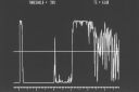
Choroidal Melanoma (Medium) Exudative Retinal Detachment 488 views87-year-old man noticed a new onset floater a few months ago. OD is 20/40, OS is 20/30
|
|
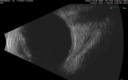
Choroidal Nevus with Drusen488 views82 year old female with flat nevus for 3 years.
|
|
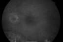
Choroidal Melanoma - Exudative Retinal Detachment 82 Year Old Man 487 views82-year-old man who had 3 falls 2 weeks ago. After the falls he started checking his vision and noticed there was a veil over the left eye, which he had seen for about two weeks in the superior visual field. OD 20/25, OS 20/40
(patient was medically never well enough for brachytherapy and died 6 months later from heart disease)
|
|

Choroidal Melanoma with Sub Retinal Fluid487 viewsFundus photography shows choroidal melanoma superiorily between 11-12-o'clock in the left eye. VA is 20/150 in the left eye.
|
|

486 views
|
|
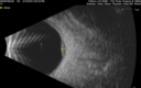
Atypical High Risk Choroidal Nevus vs Macular Melanoma486 views70 year old female with vision loss in the left eye for a few weeks. She was referred to a tumor center and being managed as an "atypical high risk choroidal nevus". The size is 6 mm x 6.5 mm x 1.6 mm (elevated).
Vision 20/40 - having serial avastin injections.
|
|
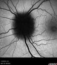
Melanocytoma - Lightly Pigmented485 views42 year old female with very mild visual field and vision loss in the left eye: 20/20 OU
|
|
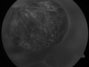
Choroidal Melanoma (Medium) Exudative Retinal Detachment 484 views87-year-old man noticed a new onset floater a few months ago. OD is 20/40, OS is 20/30
|
|
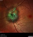
Melanocytoma - Lightly Pigmented484 views42 year old female with very mild visual field and vision loss in the left eye: 20/20 OU
|
|
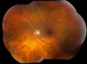
Multiple Uveal Choroidal Nevi483 viewsPatient referred for CHRPE which in this case has unusual appearance. VA is 20/20 in the both eyes. Distinguishing if there was any relation to polyposis. This is not CHRPE.
|
|
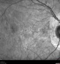
Melanosis Oculi and Choroidal Melanoma483 views88 year old female with 2.5 mm elevated choroidal melanoma and melanosis oculi. She also has geographic atrophy from AMD. The right eye is the better eye with 20/40 vision. After observation with growth, brachytherapy was done.
|
|
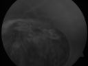
Choroidal Melanoma (Medium) Exudative Retinal Detachment 481 views87-year-old man noticed a new onset floater a few months ago. OD is 20/40, OS is 20/30
|
|

Melanocytoma - Lightly Pigmented481 views42 year old female with very mild visual field and vision loss in the left eye: 20/20 OU
|
|
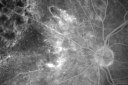
Radiation Retinopathy and Macular Pucker 4 years following Brachytherapy (Plaque) for Nasal Melanoma479 views67-year-old woman has a macular pucker in the left eye. She had radiation for a melanoma in that eye 4 years aog. She had cataract surgery 2 years ago and a posterior sub-Tenon Kenalog injection in 7 months ago. Her vision in the left eye is hazy and she has trouble seeing out of it. VA OS 20/70.
|
|
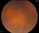
Uveal Choroidal Melanoma479 viewsPatient comes in for evaluation on a Choroidal Melanoma in the right eye. VA was 20/25 in both eyes. The melanoma is in the temporal aspect of the right eye. It measured at 0.7mm elevated after doing a BSCAN Ultrasound.
|
|
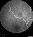
Amelanotic Choroidal Nevus - Multimodal Imaging479 views61 year old man with amelanotic choroidal nevus. Vision Normal - Stable over 4 years.
|
|
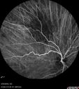
Amelanotic Choroidal Nevus - Multimodal Imaging479 views61 year old man with amelanotic choroidal nevus. Vision Normal - Stable over 4 years.
|
|

Amelanotic Choroidal Nevus478 views60 year old man. Nevus is stable over 6 years. Images are Photo, FAF, OCT, FA, ICG, and B-Scan
|
|

Melanocytoma - Lightly Pigmented476 views42 year old female with very mild visual field and vision loss in the left eye: 20/20 OU
|
|

Amelanotic Choroidal Nevus - Multimodal Imaging476 views61 year old man with amelanotic choroidal nevus. Vision Normal - Stable over 4 years.
|
|

Atypical High Risk Choroidal Nevus vs Macular Melanoma475 views70 year old female with vision loss in the left eye for a few weeks. She was referred to a tumor center and being managed as an "atypical high risk choroidal nevus". The size is 6 mm x 6.5 mm x 1.6 mm (elevated).
Vision 20/40 - having serial avastin injections.
|
|
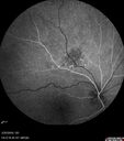
Amelanotic Choroidal Nevus - Multimodal Imaging475 views61 year old man with amelanotic choroidal nevus. Vision Normal - Stable over 4 years.
|
|

Large Choroidal Melanoma with Retinal Detachment474 viewsPatient notices a decline in her vision for the past 3-weeks in her right eye. VA was light perception in the right eye. Fundus photo shows a very large melanoma temporal to the optic nerve and retinal detachment inferiorly. Patient will under go proton beam therapy.
|
|
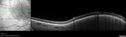
Amelanotic Choroidal Nevus - Multimodal Imaging474 views61 year old man with amelanotic choroidal nevus. Vision Normal - Stable over 4 years.
|
|
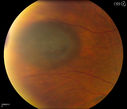
Uveal Choroidal Melanoma473 viewsPatient comes in for evaluation on a Choroidal Melanoma in the right eye. VA was 20/25 in both eyes. The melanoma is in the temporal aspect of the right eye. It measured at 0.7mm elevated after doing a BSCAN Ultrasound.
|
|
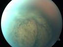
Choroidal Melanoma (Medium) Exudative Retinal Detachment 472 views87-year-old man noticed a new onset floater a few months ago. OD is 20/40, OS is 20/30
|
|

Choroidal Melanoma (Medium) Exudative Retinal Detachment 472 views87-year-old man noticed a new onset floater a few months ago. OD is 20/40, OS is 20/30
|
|
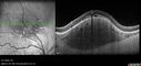
Amelanotic Choroidal Nevus472 views60 year old man. Nevus is stable over 6 years. Images are Photo, FAF, OCT, FA, ICG, and B-Scan
|
|
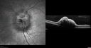
Melanocytoma - Lightly Pigmented471 views42 year old female with very mild visual field and vision loss in the left eye: 20/20 OU
|
|
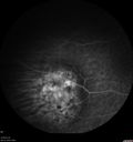
Melanosis Oculi and Choroidal Melanoma471 views88 year old female with 2.5 mm elevated choroidal melanoma and melanosis oculi. She also has geographic atrophy from AMD. The right eye is the better eye with 20/40 vision. After observation with growth, brachytherapy was done.
|
|

Atypical High Risk Choroidal Nevus vs Macular Melanoma471 views70 year old female with vision loss in the left eye for a few weeks. She was referred to a tumor center and being managed as an "atypical high risk choroidal nevus". The size is 6 mm x 6.5 mm x 1.6 mm (elevated).
Vision 20/40 - having serial avastin injections.
|
|

Multiple Uveal Choroidal Nevi469 viewsPatient referred for CHRPE which in this case has unusual appearance. VA is 20/20 in the both eyes. Distinguishing if there was any relation to polyposis. This is not CHRPE.
|
|
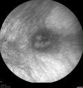
Choroidal Nevus with Drusen469 views82 year old female with flat nevus for 3 years.
|
|
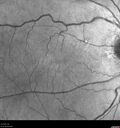
Amelanotic Choroidal Nevus - Multimodal Imaging468 views61 year old man with amelanotic choroidal nevus. Vision Normal - Stable over 4 years.
|
|

Amelanotic Choroidal Nevus - Multimodal Imaging467 views61 year old man with amelanotic choroidal nevus. Vision Normal - Stable over 4 years.
|
|
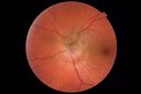
Melanocytoma - Lightly Pigmented466 views42 year old female with very mild visual field and vision loss in the left eye: 20/20 OU
|
|
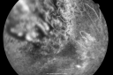
Radiation Retinopathy and Macular Pucker 4 years following Brachytherapy (Plaque) for Nasal Melanoma465 views67-year-old woman has a macular pucker in the left eye. She had radiation for a melanoma in that eye 4 years aog. She had cataract surgery 2 years ago and a posterior sub-Tenon Kenalog injection in 7 months ago. Her vision in the left eye is hazy and she has trouble seeing out of it. VA OS 20/70.
|
|

Large Choroidal Nevus465 viewsYoung male patient who comes in for follow up on choroidal nevus in the right eye. VA is 20/30 in the right eye. Fundus montage photo shows very large nevus branched out from the macula to the temporal aspect of the right eye. Patient will be followed up in 3-months.
|
|
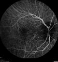
Melanosis Oculi and Choroidal Melanoma465 views88 year old female with 2.5 mm elevated choroidal melanoma and melanosis oculi. She also has geographic atrophy from AMD. The right eye is the better eye with 20/40 vision. After observation with growth, brachytherapy was done.
|
|
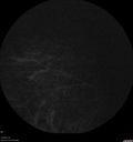
Melanosis Oculi and Choroidal Melanoma464 views88 year old female with 2.5 mm elevated choroidal melanoma and melanosis oculi. She also has geographic atrophy from AMD. The right eye is the better eye with 20/40 vision. After observation with growth, brachytherapy was done.
|
|
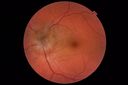
Melanocytoma - Lightly Pigmented463 views42 year old female with very mild visual field and vision loss in the left eye: 20/20 OU
|
|
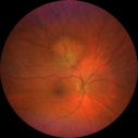
Amelanotic Choroidal Nevus - Multimodal Imaging463 views61 year old man with amelanotic choroidal nevus. Vision Normal - Stable over 4 years.
|
|
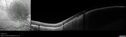
Amelanotic Choroidal Nevus - Multimodal Imaging463 views61 year old man with amelanotic choroidal nevus. Vision Normal - Stable over 4 years.
|
|
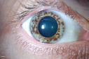
Multiple Uveal Choroidal Nevi462 viewsPatient referred for CHRPE which in this case has unusual appearance. VA is 20/20 in the both eyes. Distinguishing if there was any relation to polyposis. This is not CHRPE.
|
|
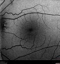
Amelanotic Choroidal Nevus - Multimodal Imaging462 views61 year old man with amelanotic choroidal nevus. Vision Normal - Stable over 4 years.
|
|
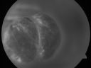
Choroidal Melanoma (Medium) Exudative Retinal Detachment 459 views87-year-old man noticed a new onset floater a few months ago. OD is 20/40, OS is 20/30
|
|
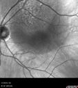
Atypical High Risk Choroidal Nevus vs Macular Melanoma458 views70 year old female with vision loss in the left eye for a few weeks. She was referred to a tumor center and being managed as an "atypical high risk choroidal nevus". The size is 6 mm x 6.5 mm x 1.6 mm (elevated).
Vision 20/40 - having serial avastin injections.
|
|

Choroidal Melanoma (Medium) Exudative Retinal Detachment 457 views87-year-old man noticed a new onset floater a few months ago. OD is 20/40, OS is 20/30
|
|
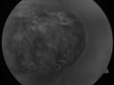
Choroidal Melanoma (Medium) Exudative Retinal Detachment 455 views87-year-old man noticed a new onset floater a few months ago. OD is 20/40, OS is 20/30
|
|

Melanocytoma - Lightly Pigmented455 views42 year old female with very mild visual field and vision loss in the left eye: 20/20 OU
|
|
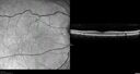
Amelanotic Choroidal Nevus - Multimodal Imaging455 views61 year old man with amelanotic choroidal nevus. Vision Normal - Stable over 4 years.
|
|
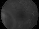
Choroidal Melanoma (Medium) Exudative Retinal Detachment 454 views87-year-old man noticed a new onset floater a few months ago. OD is 20/40, OS is 20/30
|
|

Choroidal Melanoma454 viewsYoung male patient notice a visual field defect in the right eye. Patient's VA was 20/60 with no improvement with pinhole. No history of cancer. Patient will be followed up in one week to discuss surgery.
|
|

Choroidal Nevus454 viewsPt comes in for 6-month follow up on a Choroidal Nevus in the left eye. Patient's VA was 20/60 in the left eye. There has been a slight enlargement in the past 6-months. Patient has a history of bladder cancer.
|
|
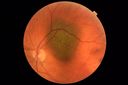
Atypical High Risk Choroidal Nevus vs Macular Melanoma454 views70 year old female with vision loss in the left eye for a few weeks. She was referred to a tumor center and being managed as an "atypical high risk choroidal nevus". The size is 6 mm x 6.5 mm x 1.6 mm (elevated).
Vision 20/40 - having serial avastin injections.
|
|

Amelanotic Choroidal Nevus454 views60 year old man. Nevus is stable over 6 years. Images are Photo, FAF, OCT, FA, ICG, and B-Scan
|
|
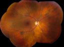
Multiple Uveal Choroidal Nevi453 viewsPatient referred for CHRPE which in this case has unusual appearance. VA is 20/20 in the both eyes. Distinguishing if there was any relation to polyposis. This is not CHRPE.
|
|

Recurrence of Melanoma after Transpupillary Thermotherapy452 views3 previous episodes of TTT. This recurrence occurred 1 year later
|
|
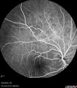
Amelanotic Choroidal Nevus - Multimodal Imaging452 views61 year old man with amelanotic choroidal nevus. Vision Normal - Stable over 4 years.
|
|
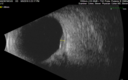
Amelanotic Choroidal Nevus - Multimodal Imaging450 views61 year old man with amelanotic choroidal nevus. Vision Normal - Stable over 4 years.
|
|
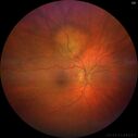
Amelanotic Choroidal Nevus450 views60 year old man. Nevus is stable over 6 years. Images are Photo, FAF, OCT, FA, ICG, and B-Scan
|
|

Melanocytoma - Lightly Pigmented449 views42 year old female with very mild visual field and vision loss in the left eye: 20/20 OU
|
|
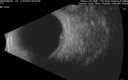
Atypical High Risk Choroidal Nevus vs Macular Melanoma448 views70 year old female with vision loss in the left eye for a few weeks. She was referred to a tumor center and being managed as an "atypical high risk choroidal nevus". The size is 6 mm x 6.5 mm x 1.6 mm (elevated).
Vision 20/40 - having serial avastin injections.
|
|
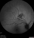
Amelanotic Choroidal Nevus - Multimodal Imaging446 views61 year old man with amelanotic choroidal nevus. Vision Normal - Stable over 4 years.
|
|
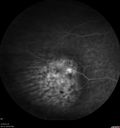
Melanosis Oculi and Choroidal Melanoma445 views88 year old female with 2.5 mm elevated choroidal melanoma and melanosis oculi. She also has geographic atrophy from AMD. The right eye is the better eye with 20/40 vision. After observation with growth, brachytherapy was done.
|
|
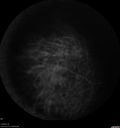
Melanosis Oculi and Choroidal Melanoma444 views88 year old female with 2.5 mm elevated choroidal melanoma and melanosis oculi. She also has geographic atrophy from AMD. The right eye is the better eye with 20/40 vision. After observation with growth, brachytherapy was done.
|
|
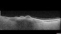
Choroidal Nevus with Drusen444 views82 year old female with flat nevus for 3 years.
|
|
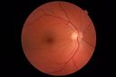
Melanocytoma - Lightly Pigmented443 views42 year old female with very mild visual field and vision loss in the left eye: 20/20 OU
|
|
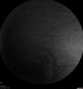
Melanosis Oculi and Choroidal Melanoma443 views88 year old female with 2.5 mm elevated choroidal melanoma and melanosis oculi. She also has geographic atrophy from AMD. The right eye is the better eye with 20/40 vision. After observation with growth, brachytherapy was done.
|
|
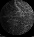
Melanosis Oculi and Choroidal Melanoma443 views88 year old female with 2.5 mm elevated choroidal melanoma and melanosis oculi. She also has geographic atrophy from AMD. The right eye is the better eye with 20/40 vision. After observation with growth, brachytherapy was done.
|
|
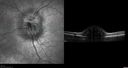
Melanocytoma - Lightly Pigmented441 views42 year old female with very mild visual field and vision loss in the left eye: 20/20 OU
|
|
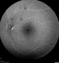
Amelanotic Choroidal Nevus - Multimodal Imaging440 views61 year old man with amelanotic choroidal nevus. Vision Normal - Stable over 4 years.
|
|
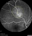
Amelanotic Choroidal Nevus - Multimodal Imaging438 views61 year old man with amelanotic choroidal nevus. Vision Normal - Stable over 4 years.
|
|
|
|


