Most viewed - Macular Degeneration - Age Related, Myopic, Choroidal Neovascular Membranes
|
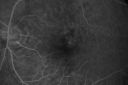
Hyperpigmentation of the Retinal Pigment Epithelium Right Eye (Treated wet AMD OS)502 views85-year-old man has wet age-related macular degeneration in the left eye treated for one year most recently one year ago. OD is 20/30, OS is 20/60
|
|

Polypoidal Choroidal Vasculopathy with Pulsating ICG polyp left eye502 views
|
|
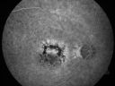
Patterned Pigment Dystrophy of the Fovea 79 YO Woman VA 20/20 Right 20/30 Left501 views
|
|
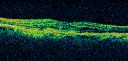
New Subfoveal Classic CNVM OS - Wet AMD - Macular Scar Congenital Toxoplasmosis Right Eye 501 views77-year-old woman as had poor vision in the right eye since birth from congenital toxoplasmosis of her macula in the right eye. Two to three weeks ago when she noticed vision loss in the left eye. VISUAL ACUITY: OD 2/200, OS 20/200.
|
|

Cuticular Drusen (Basal Laminar Drusen) and Bilateral Chronic Subfoveal Fluid (Serous Retinal Detachment) unresponsive to monthly Avastin - Fill ICG FA OD501 views66-year-old man with bilateral subfoveal fluid unresponsive to one year of monthly avastin therapy.
OD 20/160, Pinhole 20/80. OS 20/63, Pinhole 20/50. OCT scan shows subretinal fluid with normal choroidal thickness. Fluorescein angiogram shows cuticular/basal laminar drusen. ICG shows no foci of choriocapillaris leakage.
|
|

Cuticular Drusen (Basal Laminar Drusen) and Bilateral Chronic Subfoveal Fluid (Serous Retinal Detachment) unresponsive to monthly Avastin Early FA OD501 views66-year-old man with bilateral subfoveal fluid unresponsive to one year of monthly avastin therapy.
OD 20/160, Pinhole 20/80. OS 20/63, Pinhole 20/50. OCT scan shows subretinal fluid with normal choroidal thickness. Fluorescein angiogram shows cuticular/basal laminar drusen. ICG shows no foci of choriocapillaris leakage.
|
|

Adult Vitelliform Macular Dystrophy - Pattern Dystrophy501 views
|
|
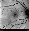
Confluent Soft Drusen with Subetinal Fluid - Right Eye501 viewsAsymptomatic 85 year old man. OCT shows pocket of subfoveal fluid in the center of the macula. This could be mechanical from the drusen preventing the retina from settling down. Testing did not reveal a CNVM and subsequent OCT 6 weeks later showed no change in structure.
|
|

Myopic CNVM - Wet - rx Avastin for 1 year500 views55-year-old woman OD is 20/40, OS is 20/20. There is a hyperpigmented disc diameter choroidal neovascular membrane with fluid touching the fovea.
|
|
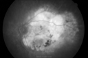
Disciform Macular Scar Both Eyes from Wet Macular Degeneration500 views86-year-old woman has disciform scars OD: 20/400; OS: 20/100
|
|
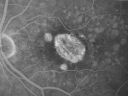
500 views
|
|
Angioid Streaks - Pseudoxanthoma Elasticum - Supressed CNVM500 views54-year-old woman had angioid streaks and macular degeneration in both eyes. She has had intravitreal Avastin in the right eye about every three months and her vision in that eye is good.
VISUAL ACUITY: Vision OD is 20/30, OS is 2/200
|
|
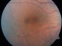
Pattern Dystrophy - Adult Vitelliform - Both Eyes500 views60-year-old woman noticing less distortion and less blind spots.
OD is 20/20, OS is 20/40
|
|
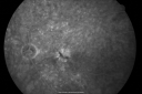
Small Retinal Pigment Epithelial Rip and Poor Vision (20/400)499 views80-year-old woman vision in the right eye has substantially declined.
OD: 20/400; OS: 20/50
|
|
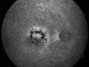
Patterned Pigment Dystrophy of the Fovea 79 YO Woman VA 20/20 Right 20/30 Left499 views
|
|
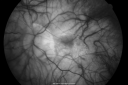
Myopic Traction Maculopathy - Vitreoretinal Traction - Possible Occult Neovascular Membrane499 views58-year-old woman was seen in the office on June 30, 2011. In 2002, her vision was 20/30. Subsequent to that she had her left eye made her distance eye, which was dominate. She had LASIK surgery on the right eye to make that her near eye and she was getting along well until about a year ago when she noticed substantial vision loss in the left eye only. VISUAL ACUITY: The near eye is 20/200, but she reads J2. OS 20/100- and J16.
|
|
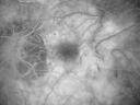
Myopic Traction Maculopathy - Vitreoretinal Traction - Possible Occult Neovascular Membrane499 views58-year-old woman was seen in the office on June 30, 2011. In 2002, her vision was 20/30. Subsequent to that she had her left eye made her distance eye, which was dominate. She had LASIK surgery on the right eye to make that her near eye and she was getting along well until about a year ago when she noticed substantial vision loss in the left eye only. VISUAL ACUITY: The near eye is 20/200, but she reads J2. OS 20/100- and J16.
|
|
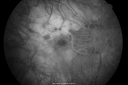
Myopic Traction Maculopathy - Vitreoretinal Traction - Possible Occult Neovascular Membrane499 views58-year-old woman was seen in the office on June 30, 2011. In 2002, her vision was 20/30. Subsequent to that she had her left eye made her distance eye, which was dominate. She had LASIK surgery on the right eye to make that her near eye and she was getting along well until about a year ago when she noticed substantial vision loss in the left eye only. VISUAL ACUITY: The near eye is 20/200, but she reads J2. OS 20/100- and J16.
|
|
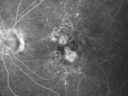
Non-Foveal Geographic Atrophy Both Eyes499 views81-year-old man has age-related macular degeneration in both eyes. Vision OD is 20/40, OS is 20/80.
|
|
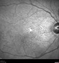
Adult Pseudovitelliform Macular Dystrophy499 views89 year old female states dark smudge on ceiling during night time came and went. Decreased vision in the left eye
VA OD: Dcc20/32
VA OS: Dcc20/63
|
|
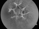
Angioid Streaks - New Extrafoveal Choroidal Neovascular Membrane Right Eye498 views61-year-old woman twelve days ago, saw a spot to the right of her central vision. OD is 20/25, OS is 20/25
Treated with one year of avastin with good result and no vision loss.
|
|
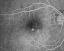
Macular Degeneration - Choroidal Laser Left eye - New Subfoveal CNVM left eye - Did well with Avastin498 views69-year-old woman new vision loss right eye and old macular scar left eye. OD 20/40, OS 20/200
|
|

New Subfoveal Classic CNVM OS - Wet AMD - Macular Scar Congenital Toxoplasmosis Right Eye 498 views77-year-old woman as had poor vision in the right eye since birth from congenital toxoplasmosis of her macula in the right eye. Two to three weeks ago when she noticed vision loss in the left eye. VISUAL ACUITY: OD 2/200, OS 20/200.
|
|
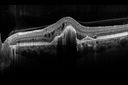
Presumed Ocular Histoplasmosis with new CNVM in right and better eye - VA 20/80 OD; 20/800 OS498 views
|
|
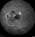
Asymptomatic wet AMD - Subfoveal CNM both eyes498 views82 year old woman - 20/25 OU. Images show occult subfoveal CNVM both eyes best seen on late ICG. The left eye also has probable polypoidal choroidal vasculopathy. No treatment was recommended on the first visit - follow-up at 1 month.
|
|

Small Retinal Pigment Epithelial Rip and Poor Vision (20/400)497 views80-year-old woman vision in the right eye has substantially declined.
OD: 20/400; OS: 20/50
|
|

Macular Scar Left Eye from Age-Related Macular Degeneration497 views81-year-old woman has a macular scar in the left eye and then unfortunately developed a central retinal artery occlusion in the right eye OD 20/100, OS 6/200.
|
|

Angioid Streaks and Macular Degeneration - CNVM OS - Band Keratopathy 497 views79-year-old man has wet age-related macular degeneration in the left eye, for which he is having Lucentis treatment. OD 20/40, Pinhole 20/32. OS 20/160, Pinhole 20/50. He has band keratopathy sparing the visual axis in both eyes. ICG shows angioid streaks both eye.
|
|
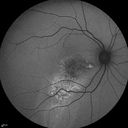
Serous Macular Detachment Both Eyes - Wet AMD vs. CSR - 62 Year Old Man - FAF497 views
|
|
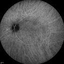
Cartwheel CNVM - Classic - wet AMD - evidence of possible old CSR - ICG is normal - shows nothing497 views75 y.o Decreased Vision right eye for one year. VA OD: sc20/125+2 OS 20/25. Autofluorescence and SD OCT show evidence of old serous retinal detachment superior to the fovea suggesting a history of CSR. The macular lesion on FA shows a classic cartwheel appearance
|
|

Asymptomatic wet AMD - Subfoveal CNM both eyes497 views82 year old woman - 20/25 OU. Images show occult subfoveal CNVM both eyes best seen on late ICG. The left eye also has probable polypoidal choroidal vasculopathy. No treatment was recommended on the first visit - follow-up at 1 month.
|
|

Small Retinal Pigment Epithelial Rip and Poor Vision (20/400)496 views80-year-old woman vision in the right eye has substantially declined.
OD: 20/400; OS: 20/50
|
|
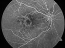
496 views
|
|

Hyperpigmentation of the Retinal Pigment Epithelium Right Eye (Treated wet AMD OS)496 views85-year-old man has wet age-related macular degeneration in the left eye treated for one year most recently one year ago. OD is 20/30, OS is 20/60
|
|
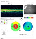
Juxtapapillary CNVM and Serous Macular Detachment Wet AMD Rx Laser495 views85-year-old man OD is 20/25, OS is 20/100. IOP: OD 21, OS 22.
OS: Vertical C/D ratio is 0.1. There is a posterior vitreous separation. There is a hemorrhagic pigment epithelial detachment on the superior pole of the optic nerve, extending a disc-and-a-half diameter off the nerve, with adjacent exudates. There is a serous macular detachment involving the center of the fovea.
VA improved from 20/100 to 20/50 in four months from laser.
|
|
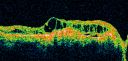
495 views
|
|

Angioid Streaks - Peau D'Orange - Pseudoxanthoma Elasticum - Asymptomatic Extrafoveal Choroidal Neovascular Membrane495 views60-year-old woman has pseudoxanthoma elasticum since 2008. Her sister has the same problem and lost vision in both eyes. She had a problem with blood vessel growth in the left eye for which she had injections of Avastin in August, September, and October of 2011 (5 months ago). Subsequent to that, the macula has been dry and you have just been watching her carefully. She is in Florida for a short time but will ultimately be spending nine months out of the year here. Both of her eyes have good vision at
|
|

Angioid Streaks - New Extrafoveal Choroidal Neovascular Membrane Right Eye494 views61-year-old woman twelve days ago, saw a spot to the right of her central vision. OD is 20/25, OS is 20/25
Treated with one year of avastin with good result and no vision loss.
|
|
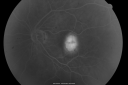
New Subfoveal Classic CNVM OS - Wet AMD - Macular Scar Congenital Toxoplasmosis Right Eye 493 views77-year-old woman as had poor vision in the right eye since birth from congenital toxoplasmosis of her macula in the right eye. Two to three weeks ago when she noticed vision loss in the left eye. VISUAL ACUITY: OD 2/200, OS 20/200.
|
|
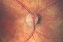
Wet AMD OD and Lamellar Macular hole OS492 viewsWet AMD in right and better eye failed extend to 2 months but was able to stop therapy once he started on systemic Avastin for cancer. The last image shows the macula dry 9 months without local therapy. The left eye has a lamellar macular hole.
|
|

Multifocal Pattern Dystrophy492 views51 year old with normal vision - images look a lot like fundus flavimaculatus
|
|
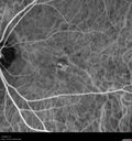
Confluent Soft Drusen with Subetinal Fluid - Right Eye492 viewsAsymptomatic 85 year old man. OCT shows pocket of subfoveal fluid in the center of the macula. This could be mechanical from the drusen preventing the retina from settling down. Testing did not reveal a CNVM and subsequent OCT 6 weeks later showed no change in structure.
|
|
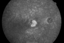
Small Retinal Pigment Epithelial Rip and Poor Vision (20/400)491 views80-year-old woman vision in the right eye has substantially declined.
OD: 20/400; OS: 20/50
|
|
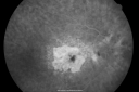
Concentric Geograophic Atrophy - Age-related Macular Degeneration - Charles Bonnet Syndrome - Bulls Eye491 views88-year-old woman has age-related with vision gradually declining over the last year. She sees spots in her vision, like red spots and dark spots and she even notices during the daytime, for example when she was looking at her minister, it looked like there were little flowering pots coming out of his head.
VISUAL ACUITY: OD 20/200, OS 20/400.
|
|
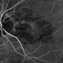
Angioid Streaks and Macular Degeneration - CNVM OS - Band Keratopathy 491 views79-year-old man has wet age-related macular degeneration in the left eye, for which he is having Lucentis treatment. OD 20/40, Pinhole 20/32. OS 20/160, Pinhole 20/50. He has band keratopathy sparing the visual axis in both eyes. ICG shows angioid streaks both eye.
|
|
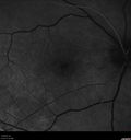
Confluent Soft Drusen with Subetinal Fluid - Right Eye491 viewsAsymptomatic 85 year old man. OCT shows pocket of subfoveal fluid in the center of the macula. This could be mechanical from the drusen preventing the retina from settling down. Testing did not reveal a CNVM and subsequent OCT 6 weeks later showed no change in structure.
|
|
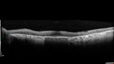
Concentric Geographic Atrophy491 views76 year old man Gradual vision loss
20/32 OD; 20/40 OS
No medicines, Non-smoker
Working and Driving
|
|
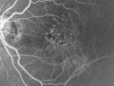
Basal Laminar (cuticular) Drusen both eyes 60 Year Old Woman490 views60-year-old woman with minimal vision loss. Vision OD is 20/30 OS 20/40
|
|

Serous Macular Detachment Both Eyes - Wet AMD vs. CSR - 62 Year Old Man - Color Photo490 views
|
|
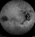
IPCV - idiopathic polypoidal choroidal vasculopathy - juxtapapillary CNVM with hemorrhage both eyes (also choroidal nevus right eye) - Subretinal hemorrhage both eyes490 views86 year old woman with shadow in vision OD and new hemmorhage OU. ICG angiogram
|
|
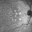
Dry AMD - Confluent Drusen Sparing Center of Macula which has Atrophy490 views65 year old woman, VA 20/40 OD; 20/80 OS. The center of the macula has few or no drusen with predominantly non-geographic atrophy
|
|
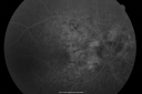
Angioid Streaks New Vision Change in Good Right Eye Treated with Lucentis489 views53-year-old man has angioid streaks. He has a macular scar in the left eye from choroidal neovasculari-zation that was treated with photodynamic laser 5-8 years ago. His vision in the right eye had been fine until about a week ago when he noticed temporally in the right eye an area that does not look normal and looks a little distorted.
VISUAL ACUITY: Vision OD is 20/30, OS is 20/400. Treatment was started within a month and patient responded well and is now 3 years out requiring every 3 month Lucentis
|
|
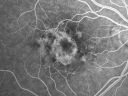
489 views
|
|
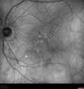
Reticular Macular Disease (Pseudo-drusen) Both Eyes - Occult CNVM and Macular Edema Right Eye 489 views
|
|
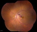
Macular Degeneration with Hemorrhage489 viewsPatient comes in with active leaking of AMD in the left eye. VA is 20/40 in the left eye. Patient was treated with Eylea and will return in 1-month for follow up.
|
|
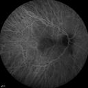
Cartwheel CNVM - Classic - wet AMD - evidence of possible old CSR - ICG is normal - shows nothing489 views75 y.o Decreased Vision right eye for one year. VA OD: sc20/125+2 OS 20/25. Autofluorescence and SD OCT show evidence of old serous retinal detachment superior to the fovea suggesting a history of CSR. The macular lesion on FA shows a classic cartwheel appearance
|
|

Polypoidal Choroidal Vasculopathy with Pulsating ICG polyp left eye489 views
|
|
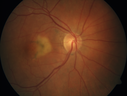
Pattern Dystrophy - Adult Vitellifrom (Best)489 views71 year old female - lesions nasal to the fovea in both eyes. (20/40 OU)
|
|

Angioid Streaks New Vision Change in Good Right Eye Treated with Lucentis488 views53-year-old man has angioid streaks. He has a macular scar in the left eye from choroidal neovasculari-zation that was treated with photodynamic laser 5-8 years ago. His vision in the right eye had been fine until about a week ago when he noticed temporally in the right eye an area that does not look normal and looks a little distorted.
VISUAL ACUITY: Vision OD is 20/30, OS is 20/400. Treatment was started within a month and patient responded well and is now 3 years out requiring every 3 month Lucentis
|
|
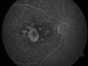
488 views
|
|
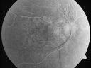
Angioid Streaks - Pseudoxanthoma Elasticum - Supressed CNVM488 views54-year-old woman had angioid streaks and macular degeneration in both eyes. She has had intravitreal Avastin in the right eye about every three months and her vision in that eye is good.
VISUAL ACUITY: Vision OD is 20/30, OS is 2/200
|
|
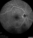
Asymptomatic Juxtapapillary Choroidal Neovascular Membrane - Reticular Macular Disease (Wet AMD) 488 views
|
|
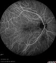
Asymptomatic Juxtapapillary Choroidal Neovascular Membrane - Reticular Macular Disease (Wet AMD) 488 views
|
|
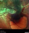
Idiopathic Polypoidal Choroidal Vasculopathy - False Color488 views
|
|
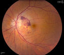
Macular Degeneration with Hemorrhage488 viewsPatient comes in with active leaking of AMD in the left eye. VA is 20/40 in the left eye. Patient was treated with Eylea and will return in 1-month for follow up.
|
|
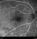
Confluent Soft Drusen with Subetinal Fluid - Right Eye488 viewsAsymptomatic 85 year old man. OCT shows pocket of subfoveal fluid in the center of the macula. This could be mechanical from the drusen preventing the retina from settling down. Testing did not reveal a CNVM and subsequent OCT 6 weeks later showed no change in structure.
|
|
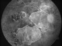
Myopic Macular Degeneration - Staphyloma - Recent Vision Loss Right Eye - Possible CNVM487 views83-year-old woman is a high myope with myopic macular degeneration. She had a retinal detachment repair in the right eye in 1968. I saw her in June of 2003. She had posterior colobomas and areas of pigment epithelial atrophy. She has noticed over the last six weeks substantial declining vision in the right eye. Prior to that, the vision in the two eyes was about the same. She uses Xalatan in the left eye and Alphagan in both eyes.
VISUAL ACUITY: Vision OD is 20/160, OS is 20/40
|
|
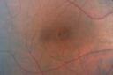
Pattern Dystrophy - Adult Vitelliform - Both Eyes486 views60-year-old woman noticing less distortion and less blind spots.
OD is 20/20, OS is 20/40
|
|
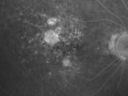
Non-Foveal Geographic Atrophy Both Eyes486 views81-year-old man has age-related macular degeneration in both eyes. Vision OD is 20/40, OS is 20/80.
|
|

Serous Macular Detachment Both Eyes - Wet AMD vs. CSR - 62 Year Old Man - Color Photo486 views
|
|

Presumed Ocular Histoplasmosis with new CNVM in right and better eye - VA 20/80 OD; 20/800 OS486 viewsIndocyanine Green Angiogram
|
|
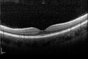
Cuticular Drusen - 26 year old female - Glomerulonephritis486 viewsVA 20/25 OU. Images are over 4 years
|
|
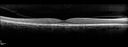
Multifocal Pattern Dystrophy486 views51 year old with normal vision - images look a lot like fundus flavimaculatus
|
|

Wet Macular Degeneration Left Eye with Gutter (atypical) and Geographic Atrophy Right Eye485 views80-year-old man has wet age-related macular degeneration in his left eye. He has an atypical angiogram and that there is a gutter under the choroidal neovascular membrane suggesting still leakage of subretinal fluid. OS 20/400.
|
|
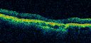
485 views
|
|
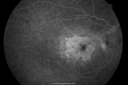
Concentric Geograophic Atrophy - Age-related Macular Degeneration - Charles Bonnet Syndrome - Bulls Eye485 views88-year-old woman has age-related with vision gradually declining over the last year. She sees spots in her vision, like red spots and dark spots and she even notices during the daytime, for example when she was looking at her minister, it looked like there were little flowering pots coming out of his head.
VISUAL ACUITY: OD 20/200, OS 20/400.
|
|
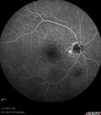
Asymptomatic Juxtapapillary Choroidal Neovascular Membrane - Reticular Macular Disease (Wet AMD) 485 views
|
|
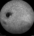
IPCV - idiopathic polypoidal choroidal vasculopathy - juxtapapillary CNVM with hemorrhage both eyes (also choroidal nevus right eye) - Subretinal hemorrhage both eyes485 views86 year old woman with shadow in vision OD and new hemmorhage OU. ICG shows no plaque
|
|
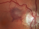
Juxtapapillary CNVM - Hemorrhage485 views83 year old female VA 20/200 improved to 20/32 within 2 months with Lucentis
|
|
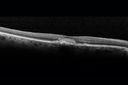
Adult Vitelliform Macular Dystrophy - Pattern Dystrophy484 viewsSD OCT shows material to be subretinal not sub-RPE as you would see in typical AMD
|
|
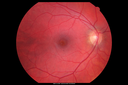
Cuticular Drusen - 26 year old female - Glomerulonephritis484 viewsVA 20/25 OU. Images are over 4 years
|
|
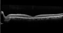
Confluent Soft Drusen with Subetinal Fluid - Right Eye484 viewsAsymptomatic 85 year old man. OCT shows pocket of subfoveal fluid in the center of the macula. This could be mechanical from the drusen preventing the retina from settling down. Testing did not reveal a CNVM and subsequent OCT 6 weeks later showed no change in structure.
|
|

Submacular Hemorrhage - wet AMD484 views84 year old man with 20/200 from submacular hemorrhage. Vision improved to 20/32 with only Avastin therapy (no surgical intervention). The initial FAF is interesting because parts of the blood hyper auto fluoresce.
|
|

Pigment Epithelial Detachment with Subretinal Fluid and Exudate 20/400 Vision483 views87-year-old woman has wet age-related macular degeneration in the left eye. She had treatment up north but there has been a gap in therapy and the vision in the left eye is poor. Vision OD is 20/25, OS is 20/400
|
|

Wet AMD controlled with Avastin 20/60 Vision483 views79-year-old woman has wet age-related macular degeneration in both eyes. The left eye had intravitreal injections of Avastin starting in May of 2009. Her vision remains good in the left eye and poor in the right eye.
VISUAL ACUITY: OD 8/200, OS 20/60.
|
|
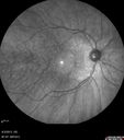
Idiopathic Polypoidal Choroidal Vasculopathy - IR483 views
|
|

Wet Macular Degeneration Left Eye with Gutter (atypical) and Geographic Atrophy Right Eye482 views80-year-old man has wet age-related macular degeneration in his left eye. He has an atypical angiogram and that there is a gutter under the choroidal neovascular membrane suggesting still leakage of subretinal fluid. OS 20/400.
|
|

Choriodal Neovascular Membrane - Subfoveal - Pseudo-Retinitis Pigmentosa - 50 years post Retinal Detachment Repair482 views86-year-old with a history of decreased vision in the right eye. The patient reports she has lost vision in the right eye over the past two months or so. She reports the vision was always compromised in that eye since retinal detachment surgery 50 years ago with a scleral buckler. Vision dropped from 20/50 to 20/200 right eye in 2 months.
|
|
482 views
|
|
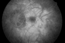
Myopic Traction Maculopathy - Vitreoretinal Traction - Possible Occult Neovascular Membrane482 views58-year-old woman was seen in the office on June 30, 2011. In 2002, her vision was 20/30. Subsequent to that she had her left eye made her distance eye, which was dominate. She had LASIK surgery on the right eye to make that her near eye and she was getting along well until about a year ago when she noticed substantial vision loss in the left eye only. VISUAL ACUITY: The near eye is 20/200, but she reads J2. OS 20/100- and J16.
|
|
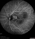
Asymptomatic Juxtapapillary Choroidal Neovascular Membrane - Reticular Macular Disease (Wet AMD) 482 views
|
|
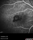
Presumed Ocular Histoplasmosis with new CNVM in right and better eye - VA 20/80 OD; 20/800 OS482 viewsFluorescein Angiogram
|
|
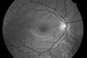
Cuticular Drusen - 26 year old female - Glomerulonephritis482 viewsVA 20/25 OU. Images are over 4 years
|
|
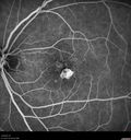
Confluent Soft Drusen with Subetinal Fluid - Right Eye482 viewsAsymptomatic 85 year old man. OCT shows pocket of subfoveal fluid in the center of the macula. This could be mechanical from the drusen preventing the retina from settling down. Testing did not reveal a CNVM and subsequent OCT 6 weeks later showed no change in structure.
|
|

Wet Macular Degeneration Left Eye with Gutter (atypical) and Geographic Atrophy Right Eye481 views80-year-old man has wet age-related macular degeneration in his left eye. He has an atypical angiogram and that there is a gutter under the choroidal neovascular membrane suggesting still leakage of subretinal fluid. OS 20/400.
|
|
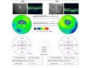
481 views
|
|
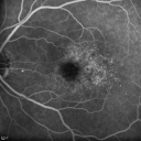
Cuticular Drusen (Basal Laminar Drusen) and Bilateral Chronic Subfoveal Fluid (Serous Retinal Detachment) unresponsive to monthly Avastin FA OS481 views66-year-old man with bilateral subfoveal fluid unresponsive to one year of monthly avastin therapy.
OD 20/160, Pinhole 20/80. OS 20/63, Pinhole 20/50. OCT scan shows subretinal fluid with normal choroidal thickness. Fluorescein angiogram shows cuticular/basal laminar drusen. ICG shows no foci of choriocapillaris leakage.
|
|
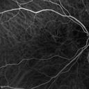
Angioid Streaks and Macular Degeneration - CNVM OS - Band Keratopathy 481 views79-year-old man has wet age-related macular degeneration in the left eye, for which he is having Lucentis treatment. OD 20/40, Pinhole 20/32. OS 20/160, Pinhole 20/50. He has band keratopathy sparing the visual axis in both eyes. ICG shows angioid streaks both eye.
|
|

IPCV - idiopathic polypoidal choroidal vasculopathy - juxtapapillary CNVM with hemorrhage both eyes (also choroidal nevus right eye) - Subretinal hemorrhage both eyes481 views86 year old woman with shadow in vision OD and new hemmorhage OU. OCT map shows progression of jpcnvm
|
|
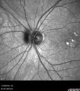
Confluent Soft Drusen with Subetinal Fluid - Right Eye481 viewsAsymptomatic 85 year old man. OCT shows pocket of subfoveal fluid in the center of the macula. This could be mechanical from the drusen preventing the retina from settling down. Testing did not reveal a CNVM and subsequent OCT 6 weeks later showed no change in structure.
|
|
| 2694 files on 27 page(s) |
 |
|
|
|
|
9 | |
|
|
|
|


