Most viewed - Macular Degeneration - Age Related, Myopic, Choroidal Neovascular Membranes
|

80 year old woman with Reticular Drusen (pseudodrusen) 20/30 Vision916 views80 year old woman with asteroid hyalosis and macular degeneration.
OPHTHALMIC EXAMINATION: Visual acuity with correction 20/30-3 OD and 20/30-2
|
|
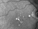
Reticular Macular Disease (Pseudo-drusen) Both Eyes - Wet AMD OS - Dry AMD OD InfraRed913 views84-year-old woman has wet age-related macular degeneration in the left eye and dry macular degeneration in the right eye. She takes the eye vitamins and her vision is stable since she was treated three months ago with Avastin. OD 20/50, OS 20/32
|
|

Geographic Atrophy 20/160 Vision911 views84-year-old woman has age-related macular degeneration in both eyes with foveal atrophy. She also has a central retinal vein occlusion
in the left eye for which she had intravitreal Kenalog and finally chorioretinal anastomosis laser a year
ago. VISUAL ACUITY: Vision OD is 20/160, OS is 7/200.
|
|

Subfoveal CNVM wet AMD color photo903 viewsShe has noticed decreased vision in the left eye. It is hard to say for how long, maybe a few months. She had her last eye exam about a year ago. She thoughts perhaps it was cataracts, but you saw a problem with the macula and suggested she come here for evaluation.
VISUAL ACUITY: Vision OD is 20/25, OS is 2/200
|
|
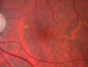
901 views
|
|

Myopic Degeneration - Staphyloma - Atrophy897 views53-year-old man was a -15.00 myope. He is aphakic. His left eye also had a retinal detachment repair and unfortunately the left eye does not see well. He is on Timolol in both eyes for glaucoma. VISUAL ACUITY: OD 20/50, OS: Faint light perception.
|
|
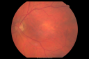
Cuticular Drusen (Basal Laminar Drusen) and Bilateral Chronic Subfoveal Fluid (Serous Retinal Detachment) unresponsive to monthly Avastin 894 views66-year-old man with bilateral subfoveal fluid unresponsive to one year of monthly avastin therapy.
OD 20/160, Pinhole 20/80. OS 20/63, Pinhole 20/50. OCT scan shows subretinal fluid with normal choroidal thickness. Fluorescein angiogram shows cuticular/basal laminar drusen. ICG shows no foci of choriocapillaris leakage.
|
|

Choroidal Neovascular Membrane - Optic Disc Drusen - Hemorrhage (Coumadin) 891 views82-year-old man with decreased vision in the left eye over about the last six to 12 months. He is also on Coumadin. OD 20/20, OS 20/80
|
|

Cuticular Drusen (Basal Laminar Drusen) and Bilateral Chronic Subfoveal Fluid (Serous Retinal Detachment) unresponsive to monthly Avastin 890 views66-year-old man with bilateral subfoveal fluid unresponsive to one year of monthly avastin therapy.
OD 20/160, Pinhole 20/80. OS 20/63, Pinhole 20/50. OCT scan shows subretinal fluid with normal choroidal thickness. Fluorescein angiogram shows cuticular/basal laminar drusen. ICG shows no foci of choriocapillaris leakage.
|
|
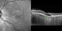
Cuticular Drusen (Basal Laminar Drusen) and Bilateral Chronic Subfoveal Fluid (Serous Retinal Detachment) unresponsive to monthly Avastin OCT OS880 views66-year-old man with bilateral subfoveal fluid unresponsive to one year of monthly avastin therapy.
OD 20/160, Pinhole 20/80. OS 20/63, Pinhole 20/50. OCT scan shows subretinal fluid with normal choroidal thickness. Fluorescein angiogram shows cuticular/basal laminar drusen. ICG shows no foci of choriocapillaris leakage.
|
|

Drusen Maculopathy - Confluent Subfoveal Soft Hydrophobic Drusen - AMD OU Red Free Image shows Confluent Drusen 876 views73-year-old woman. She is diabetic for thirteen years. She is also on Coumadin for her heart disease.
VISUAL ACUITY: OD 20/25, OS 20/50.
|
|

Cuticular Drusen (Basal Laminar Drusen) and Bilateral Chronic Subfoveal Fluid (Serous Retinal Detachment) unresponsive to monthly Avastin OCT OD863 views66-year-old man with bilateral subfoveal fluid unresponsive to one year of monthly avastin therapy.
OD 20/160, Pinhole 20/80. OS 20/63, Pinhole 20/50. OCT scan shows subretinal fluid with normal choroidal thickness. Fluorescein angiogram shows cuticular/basal laminar drusen. ICG shows no foci of choriocapillaris leakage.
|
|

Disciform Scar after Photodynamic Laser - Macular Hemorrhage and Vitrectomy862 views84-year-old man has age-related macular degeneration in both eyes, with a disciform scar in the right eye. He had a subfoveal hemorrhage in that eye treated with vitrectomy and displacement on May 3, 2006. He previous to that had photodynamic laser. These photos are from October 2010. VA is 1/200.
|
|
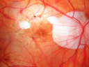
Myopic Traction Maculopathy - Vitreoretinal Traction - Possible Occult Neovascular Membrane860 views58-year-old woman was seen in the office on June 30, 2011. In 2002, her vision was 20/30. Subsequent to that she had her left eye made her distance eye, which was dominate. She had LASIK surgery on the right eye to make that her near eye and she was getting along well until about a year ago when she noticed substantial vision loss in the left eye only. VISUAL ACUITY: The near eye is 20/200, but she reads J2. OS 20/100- and J16.
|
|
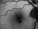
Reticular Macular Disease (Pseudo-drusen) Both Eyes - Wet AMD OS - Dry AMD OD FAF860 views84-year-old woman has wet age-related macular degeneration in the left eye and dry macular degeneration in the right eye. She takes the eye vitamins and her vision is stable since she was treated three months ago with Avastin. OD 20/50, OS 20/32
|
|
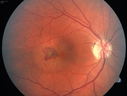
Presumed Ocular Histoplasmosis with new CNVM in right and better eye - VA 20/80 OD; 20/800 OS858 viewsNew subretinal hemorrhage in better eye
|
|

Myopic Macular Degeneration - Dry 856 views55-year-old woman has myopic macular degeneration in both eyes with vision and an Amsler grid change starting a few days ago in the left eye.
VISUAL ACUITY: OD: 20/60. PH: 20/25. OS: 20/200. PH: 20/30.
|
|
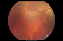
Equatorial Drusen Fundus Photo Right Eye856 views
|
|
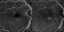
Reticular Macular Disease (Pseudo-drusen) Both Eyes - Wet AMD OS - Dry AMD OD FA and ICG early853 views84-year-old woman has wet age-related macular degeneration in the left eye and dry macular degeneration in the right eye. She takes the eye vitamins and her vision is stable since she was treated three months ago with Avastin. OD 20/50, OS 20/32
|
|
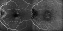
Reticular Macular Disease (Pseudo-drusen) Both Eyes - Wet AMD OS - Dry AMD OD FA and ICG late848 views84-year-old woman has wet age-related macular degeneration in the left eye and dry macular degeneration in the right eye. She takes the eye vitamins and her vision is stable since she was treated three months ago with Avastin. OD 20/50, OS 20/32
|
|

Reticular Macular Disease (Pseudo-drusen) Both Eyes - Wet AMD OS - Dry AMD OD FA and ICG late846 views84-year-old woman has wet age-related macular degeneration in the left eye and dry macular degeneration in the right eye. She takes the eye vitamins and her vision is stable since she was treated three months ago with Avastin. OD 20/50, OS 20/32
|
|
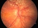
Basal Laminar Drusen - Cuticular Drusen - Equatorial Drusen - 70 Year Old Woman - Optic Nerve Photo Right Eye840 views70 year old woman has had macular drusen since her thirties and she said her sister has a similar problem. Recently the vision in the left eye has substantially declined. OD 20/16, OS 10/200.
|
|

Adult Vitelliform Macular Dystrophy - Pattern Dystrophy839 views69-year-old woman sees a dark spot centrally. When she looks at things she loses letters and numbers and is having more and more difficulty balancing her checkbook. OD is 20/80, OS is 20/40
|
|
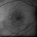
Cuticular Drusen (Basal Laminar Drusen) and Bilateral Chronic Subfoveal Fluid (Serous Retinal Detachment) unresponsive to monthly Avastin - Fundus Autofluorescence OD836 views66-year-old man with bilateral subfoveal fluid unresponsive to one year of monthly avastin therapy.
OD 20/160, Pinhole 20/80. OS 20/63, Pinhole 20/50. OCT scan shows subretinal fluid with normal choroidal thickness. Fluorescein angiogram shows cuticular/basal laminar drusen. ICG shows no foci of choriocapillaris leakage.
|
|

Myopic Degeneration - Staphyloma - Atrophy832 views53-year-old man was a -15.00 myope. He is aphakic. His left eye also had a retinal detachment repair and unfortunately the left eye does not see well. He is on Timolol in both eyes for glaucoma. VISUAL ACUITY: OD 20/50, OS: Faint light perception.
|
|
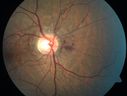
Angioid Streaks - Peau D'Orange - Pseudoxanthoma Elasticum - Asymptomatic Extrafoveal Choroidal Neovascular Membrane832 views60-year-old woman has pseudoxanthoma elasticum since 2008. Her sister has the same problem and lost vision in both eyes. She had a problem with blood vessel growth in the left eye for which she had injections of Avastin in August, September, and October of 2011 (5 months ago). Subsequent to that, the macula has been dry and you have just been watching her carefully. She is in Florida for a short time but will ultimately be spending nine months out of the year here. Both of her eyes have good vision at
|
|
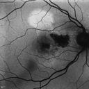
Cartwheel CNVM - Classic - wet AMD - evidence of possible old CSR - Autofluorescence shows brigh spot superior to subfoveal lesion832 views75 y.o Decreased Vision right eye for one year. VA OD: sc20/125+2 OS 20/25. Autofluorescence and SD OCT show evidence of old serous retinal detachment superior to the fovea suggesting a history of CSR. The macular lesion on FA shows a classic cartwheel appearance
|
|

Drusen Maculopathy - Confluent Subfoveal Soft Hydrophobic Drusen - OCT Line Scan Shows Thickening Under Retina with Retinal Thinning831 views73-year-old woman. She is diabetic for thirteen years. She is also on Coumadin for her heart disease.
VISUAL ACUITY: OD 20/25, OS 20/50.
|
|

Myopic Choroidal Neovascular Membrane 54 year old woman 20/20831 views54-year-old woman has had problems in her vision in the right eye for about the last three months. Inferotemporally she sees a gray area, which is distorted. OD is 20/20, OS is 20/20
|
|

Chorioretinal Scar with Radiating Chorioretinal Folds830 views79-year-old woman has wet age-related macular degeneration in both eyes. The left eye had intravitreal injections of Avastin starting in May of 2009. Her vision remains good in the left eye and poor in the right eye.
VISUAL ACUITY: OD 8/200, OS 20/60.
|
|

Classic Subfoveal Choroidal Neovascular Membrane - 20/400 Vision - 6 - 12 months old lesion828 views78-year-old woman six months or a year ago decreasing vision in the right eye. OD 20/400. Fluorescein angiography shows a classic subfoveal choroidal neovascular membrane in the right eye 3 disc-diameters across, which has brisk leakage. The left eye shows staining drusen.
|
|

Subfoveal CNVM wet AMD early fa (contrast enhanced with photoshop)825 viewsShe has noticed decreased vision in the left eye. It is hard to say for how long, maybe a few months. She had her last eye exam about a year ago. She thoughts perhaps it was cataracts, but you saw a problem with the macula and suggested she come here for evaluation.
VISUAL ACUITY: Vision OD is 20/25, OS is 2/200
|
|
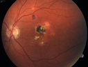
Presumed Ocular Histoplasmosis with new CNVM in right and better eye - VA 20/80 OD; 20/800 OS825 viewsMacular scar 20 years old
|
|

Angioid Streaks - Peau D'Orange - Pseudoxanthoma Elasticum - Asymptomatic Extrafoveal Choroidal Neovascular Membrane824 views60-year-old woman has pseudoxanthoma elasticum since 2008. Her sister has the same problem and lost vision in both eyes. She had a problem with blood vessel growth in the left eye for which she had injections of Avastin in August, September, and October of 2011 (5 months ago). Subsequent to that, the macula has been dry and you have just been watching her carefully. She is in Florida for a short time but will ultimately be spending nine months out of the year here. Both of her eyes have good vision at
|
|
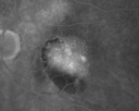
AMD - Wet - Fibrovascular Hemorrhagic PED - Occult CNVM823 views87-year-old woman for one month OS: 20/60 Fibrovascular PED and Hemorrhage
|
|

Choriodal Neovascular Membrane - Subfoveal - Pseudo-Retinitis Pigmentosa - 50 years post Retinal Detachment Repair823 views86-year-old with a history of decreased vision in the right eye. The patient reports she has lost vision in the right eye over the past two months or so. She reports the vision was always compromised in that eye since retinal detachment surgery 50 years ago with a scleral buckler. Vision dropped from 20/50 to 20/200 right eye in 2 months.
|
|
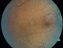
Angioid Streaks - Peau D'Orange - Pseudoxanthoma Elasticum - Asymptomatic Extrafoveal Choroidal Neovascular Membrane823 views60-year-old woman has pseudoxanthoma elasticum since 2008. Her sister has the same problem and lost vision in both eyes. She had a problem with blood vessel growth in the left eye for which she had injections of Avastin in August, September, and October of 2011 (5 months ago). Subsequent to that, the macula has been dry and you have just been watching her carefully. She is in Florida for a short time but will ultimately be spending nine months out of the year here. Both of her eyes have good vision at
|
|

Idiopathic CNVM in 6 year old girl823 viewsCNVM in OD of a 6 yr old girl
|
|

Drusen Maculopathy - Confluent Subfoveal Soft Hydrophobic Drusen - AMD OU- OCT Line Scan Shows Thickening Under Retina with Retinal Thinning821 views73-year-old woman. She is diabetic for thirteen years. She is also on Coumadin for her heart disease.
VISUAL ACUITY: OD 20/25, OS 20/50.
|
|
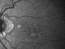
Reticular Macular Disease (Pseudo-drusen) Both Eyes - Wet AMD OS - Dry AMD OD InfraRed820 views84-year-old woman has wet age-related macular degeneration in the left eye and dry macular degeneration in the right eye. She takes the eye vitamins and her vision is stable since she was treated three months ago with Avastin. OD 20/50, OS 20/32
|
|
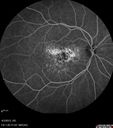
Idiopathic Polypoidal Choroidal Vasculopathy - Fluorescein Angiogram818 views
|
|
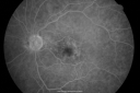
Drusen Maculopathy - Confluent Subfoveal Soft Hydrophobic Drusen - Mid Phase Fluorescein Angiogram Drusen Stain 815 views73-year-old woman. She is diabetic for thirteen years. She is also on Coumadin for her heart disease.
VISUAL ACUITY: OD 20/25, OS 20/50.
|
|

Juxtapapillary CNVM and Serous Macular Detachment Wet AMD Rx Laser813 views85-year-old man OD is 20/25, OS is 20/100. IOP: OD 21, OS 22.
OS: Vertical C/D ratio is 0.1. There is a posterior vitreous separation. There is a hemorrhagic pigment epithelial detachment on the superior pole of the optic nerve, extending a disc-and-a-half diameter off the nerve, with adjacent exudates. There is a serous macular detachment involving the center of the fovea.
VA improved from 20/100 to 20/50 in four months from laser.
|
|

Polypoidal Choroidal Vasculopathy with Pulsating ICG polyp left eye813 views
|
|

Retinal Angiomatous Proliferation - Type III choroidal neovascular membrane813 views83 year old female with wet AMD - 20/160 VA- Vision improved to 20/40 with 3 injections of anti-VEGF drugs. OCT shows break in RPE and FA and ICG show PED with hot spot on ICG
|
|
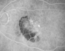
AMD - Wet - Fibrovascular Hemorrhagic PED - Occult CNVM812 views87-year-old woman for one month OS: 20/60 Fibrovascular PED and Hemorrhage
|
|
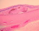
Macular Scar with Retinal Pigment Epithelial Hyperplasia - Hyperpigmentation - Wet Macular Degeneration809 viewsMacular Scar in age related macular degeneration with pathology showing retinal pigment epithelial hyperplasia at the edge of the scar. Histopathogy, microscopic slide, pathology.
|
|
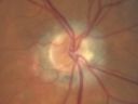
Choroidal Neovascular Membrane - Optic Disc Drusen - Hemorrhage (Coumadin) 806 views82-year-old man with decreased vision in the left eye over about the last six to 12 months. He is also on Coumadin. OD 20/20, OS 20/80
|
|
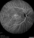
Asymptomatic Occult Subfoveal CNVM in Good Eye Visible on ICG803 views
|
|
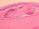
Macular Scar with Retinal Pigment Epithelial Hyperplasia - Hyperpigmentation - Wet Macular Degeneration802 viewsMacular Scar in age related macular degeneration with pathology showing retinal pigment epithelial hyperplasia at the edge of the scar. Histopathogy, microscopic slide, pathology.
|
|
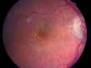
Reticular Macular Disease (Pseudo-drusen) Both Eyes - Wet AMD OS - Dry AMD OD 802 views84-year-old woman has wet age-related macular degeneration in the left eye and dry macular degeneration in the right eye. She takes the eye vitamins and her vision is stable since she was treated three months ago with Avastin. OD 20/50, OS 20/32
|
|

Serous Macular Detachment Both Eyes - Wet AMD vs. CSR - 62 Year Old Man - SD - OCT800 views
|
|
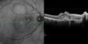
Cartwheel CNVM - Classic - wet AMD - evidence of possible old CSR797 views75 y.o Decreased Vision right eye for one year. VA OD: sc20/125+2 OS 20/25. Autofluorescence and SD OCT show evidence of old serous retinal detachment superior to the fovea suggesting a history of CSR. The macular lesion on FA shows a classic cartwheel appearance
|
|
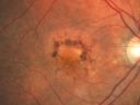
Patterned Pigment Dystrophy of the Fovea 79 YO Woman VA 20/20 Right 20/30 Left795 views
|
|

Confluent Soft Drusen with Subetinal Fluid - Right Eye795 viewsAsymptomatic 85 year old man. OCT shows pocket of subfoveal fluid in the center of the macula. This could be mechanical from the drusen preventing the retina from settling down. Testing did not reveal a CNVM and subsequent OCT 6 weeks later showed no change in structure.
|
|
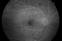
Drusen Maculopathy - Confluent Subfoveal Soft Hydrophobic Drusen - Early Fluorescein Angiogram Drusen do not yet Stain794 views73-year-old woman. She is diabetic for thirteen years. She is also on Coumadin for her heart disease.
VISUAL ACUITY: OD 20/25, OS 20/50.
|
|
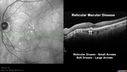
Reticular Macular Disease (Pseudo-drusen) Both Eyes - Occult CNVM and Macular Edema Right Eye 794 viewsSpectral Domain Optical Coherence Tomography
|
|

Hyperpigmentation of the Retinal Pigment Epithelium Right Eye (Treated wet AMD OS)789 views85-year-old man has wet age-related macular degeneration in the left eye treated for one year most recently one year ago. OD is 20/30, OS is 20/60
|
|

Basal Laminar Drusen - Cuticular Drusen - 19 Year Old with Lupus Nephritis788 views19-year-old. She has a history of developing severe systemic lupus erythematosus in 2001. She was in renal failure and had biopsy proven Class 2 lupus nephritis. She is currently on Plaquenil 400 mg for six years, as well as Prednisolone orally, every other day. OD 20/20, OS 20/25.
|
|

Juxtapapillary CNVM and Serous Macular Detachment Wet AMD Rx Laser788 views85-year-old man OD is 20/25, OS is 20/100. IOP: OD 21, OS 22.
OS: Vertical C/D ratio is 0.1. There is a posterior vitreous separation. There is a hemorrhagic pigment epithelial detachment on the superior pole of the optic nerve, extending a disc-and-a-half diameter off the nerve, with adjacent exudates. There is a serous macular detachment involving the center of the fovea.
VA improved from 20/100 to 20/50 in four months from laser.
|
|
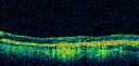
Vision Loss, Blood, and Cartwheel CNVM OD with no fluid on OCT Scan788 views86-year-old man has had changing vision. He says his vision has been gradually declining since I saw him in June. You saw a hemorrhage in the eye and suggested he comes in here for an evaluation. He is on Combigan and Travatan in the right eye for his glaucoma.
VISUAL ACUITY: Vision OD is 6/200 OS is 8/200 The macula in the right eye is wet even though interestingly the OCT does not show subretinal fluid.
|
|
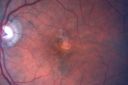
Hyperpigmentation of the Retinal Pigment Epithelium Right Eye (Treated wet AMD OS)788 views85-year-old man has wet age-related macular degeneration in the left eye treated for one year most recently one year ago. OD is 20/30, OS is 20/60
|
|
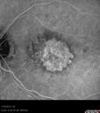
Wet AMD - New Small Hemorrhage Both Eyes - Mature CNVM visible on Early Indocyanine Green Angiogram786 views
|
|

Macular Scars - Laser (1980s) Ocular Histoplasmosis783 views70 year old man who had laser at Wills, Wilmer and the Mayo clinic before better therapies were available for ocular histoplasmosis related CNVM
|
|
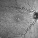
Juxtafoveal Retinal Telangiectasia (Telangiectasis) - MacTel - Striking Gray Ring in Macula781 views59-year-old woman decreasing vision in the right eye for a few years. She has had diabetes for five years. OD 20/100, Pinhole 20/40. OS 20/20
|
|

Reticular Macular Disease (Dry Macular Degeneration) - Spectral Domain Optical Coherence Tomography Line Scan781 views85 year old woman with 20/25 vision OU and reticular macular disease
|
|

Subfoveal CNVM wet AMD early fa full image779 viewsShe has noticed decreased vision in the left eye. It is hard to say for how long, maybe a few months. She had her last eye exam about a year ago. She thoughts perhaps it was cataracts, but you saw a problem with the macula and suggested she come here for evaluation.
VISUAL ACUITY: Vision OD is 20/25, OS is 2/200
|
|

Myopic Degeneration - Staphyloma - Atrophy779 views53-year-old man was a -15.00 myope. He is aphakic. His left eye also had a retinal detachment repair and unfortunately the left eye does not see well. He is on Timolol in both eyes for glaucoma. VISUAL ACUITY: OD 20/50, OS: Faint light perception.
|
|
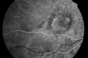
Classic Subfoveal Choroidal Neovascular Membrane - 20/400 Vision - 6 - 12 months old lesion779 views78-year-old woman six months or a year ago decreasing vision in the right eye. OD 20/400. Fluorescein angiography shows a classic subfoveal choroidal neovascular membrane in the right eye 3 disc-diameters across, which has brisk leakage. The left eye shows staining drusen.
|
|
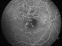
Calcified Drusen - Dry Age-related Macular Degeneration - Geographic Atrophy778 views87 Year old woman with dry age-related macular degeneration in both eyes.With both eyes open she is fine, but when she closes the left eye, things are wavier.
VISUAL ACUITY: Vision OD is 20/100, OS is 20/40.
|
|

Choriodal Neovascular Membrane - Subfoveal - Pseudo-Retinitis Pigmentosa - 50 years post Retinal Detachment Repair775 views86-year-old with a history of decreased vision in the right eye. The patient reports she has lost vision in the right eye over the past two months or so. She reports the vision was always compromised in that eye since retinal detachment surgery 50 years ago with a scleral buckler. Vision dropped from 20/50 to 20/200 right eye in 2 months.
|
|
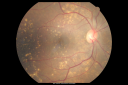
Drusen Maculopathy - Macular Degeneration (Dry) - 50 year old African-American Woman775 views50-year-old woman has drusen and maculopathy. VISUAL ACUITY: OD 20/30, OS 20/25.
|
|
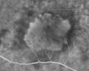
Classic Subfoveal Choroidal Neovascular Membrane - 20/400 Vision - 6 - 12 months old lesion774 views78-year-old woman six months or a year ago decreasing vision in the right eye. OD 20/400. Fluorescein angiography shows a classic subfoveal choroidal neovascular membrane in the right eye 3 disc-diameters across, which has brisk leakage. The left eye shows staining drusen.
|
|

Vision Loss, Blood, and Cartwheel CNVM OD with no fluid on OCT Scan774 views86-year-old man has had changing vision. He says his vision has been gradually declining since I saw him in June. You saw a hemorrhage in the eye and suggested he comes in here for an evaluation. He is on Combigan and Travatan in the right eye for his glaucoma.
VISUAL ACUITY: Vision OD is 6/200 OS is 8/200 The macula in the right eye is wet even though interestingly the OCT does not show subretinal fluid.
|
|

Reticular Macular Disease (Pseudo-drusen) Both Eyes - Wet AMD OS - Dry AMD OD FA and ICG early774 views84-year-old woman has wet age-related macular degeneration in the left eye and dry macular degeneration in the right eye. She takes the eye vitamins and her vision is stable since she was treated three months ago with Avastin. OD 20/50, OS 20/32
|
|

Subfoveal CNVM wet AMD late fa full image771 viewsShe has noticed decreased vision in the left eye. It is hard to say for how long, maybe a few months. She had her last eye exam about a year ago. She thoughts perhaps it was cataracts, but you saw a problem with the macula and suggested she come here for evaluation.
VISUAL ACUITY: Vision OD is 20/25, OS is 2/200
|
|
767 views
|
|

Subfoveal CNVM wet AMD fellow eye765 viewsShe has noticed decreased vision in the left eye. It is hard to say for how long, maybe a few months. She had her last eye exam about a year ago. She thoughts perhaps it was cataracts, but you saw a problem with the macula and suggested she come here for evaluation.
VISUAL ACUITY: Vision OD is 20/25, OS is 2/200
|
|
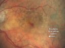
Pigment Epithelial Detachment with Subretinal Fluid and Exudate 20/400 Vision763 views87-year-old woman has wet age-related macular degeneration in the left eye. She had treatment up north but there has been a gap in therapy and the vision in the left eye is poor. Vision OD is 20/25, OS is 20/400
|
|
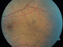
Angioid Streaks - Peau D'Orange - Pseudoxanthoma Elasticum - Asymptomatic Extrafoveal Choroidal Neovascular Membrane762 views60-year-old woman has pseudoxanthoma elasticum since 2008. Her sister has the same problem and lost vision in both eyes. She had a problem with blood vessel growth in the left eye for which she had injections of Avastin in August, September, and October of 2011 (5 months ago). Subsequent to that, the macula has been dry and you have just been watching her carefully. She is in Florida for a short time but will ultimately be spending nine months out of the year here. Both of her eyes have good vision at
|
|
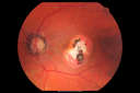
Macular Scars - Laser (1980s) Ocular Histoplasmosis761 views70 year old man who had laser at Wills, Wilmer and the Mayo clinic before better therapies were available for ocular histoplasmosis related CNVM
|
|

Drusen Maculopathy - Confluent Subfoveal Soft Hydrophobic Drusen - Early Fluorescein Shows no Staining of Drusen758 views73-year-old woman. She is diabetic for thirteen years. She is also on Coumadin for her heart disease.
VISUAL ACUITY: OD 20/25, OS 20/50.
|
|
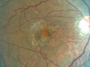
Basal Laminar (cuticular) Drusen both eyes 60 Year Old Woman758 views60-year-old woman with minimal vision loss. Vision OD is 20/30 OS 20/40
|
|
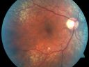
Equatorial Drusen756 views76-year-old man with equatorial drusen
VISUAL ACUITY: Vision OD is 20/40, OS is 20/25.
|
|

Reticular Macular Disease (Dry Macular Degeneration) - Color Photo756 views85 year old woman with 20/25 vision OU and reticular macular disease
|
|

Confluent Soft Drusen with Subetinal Fluid - Right Eye756 viewsAsymptomatic 85 year old man. OCT shows pocket of subfoveal fluid in the center of the macula. This could be mechanical from the drusen preventing the retina from settling down. Testing did not reveal a CNVM and subsequent OCT 6 weeks later showed no change in structure.
|
|
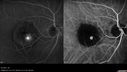
Retinal Angiomatous Proliferation - Type III choroidal neovascular membrane755 views83 year old female with wet AMD - 20/160 VA- Vision improved to 20/40 with 3 injections of anti-VEGF drugs. OCT shows break in RPE and FA and ICG show PED with hot spot on ICG
|
|
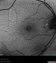
Asymptomatic Juxtapapillary Choroidal Neovascular Membrane - Reticular Macular Disease (Wet AMD) - FAF754 views
|
|
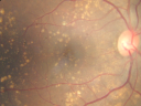
Drusen Maculopathy - Macular Degeneration (Dry) - 50 year old African-American Woman750 views50-year-old woman has drusen and maculopathy. VISUAL ACUITY: OD 20/30, OS 20/25.
|
|

Angioid Streaks and Macular Degeneration - CNVM OS - Band Keratopathy 750 views79-year-old man has wet age-related macular degeneration in the left eye, for which he is having Lucentis treatment. OD 20/40, Pinhole 20/32. OS 20/160, Pinhole 20/50. He has band keratopathy sparing the visual axis in both eyes. ICG shows angioid streaks both eye.
|
|
749 views
|
|
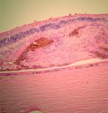
Macular Scar with Retinal Pigment Epithelial Hyperplasia - Hyperpigmentation - Wet Macular Degeneration748 viewsMacular Scar in age related macular degeneration with pathology showing retinal pigment epithelial hyperplasia at the edge of the scar. Histopathogy, microscopic slide, pathology.
|
|
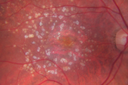
Calcified Macular Drusen - Age Related Macular Degeneration - Dry746 views74-year-old woman
OD 20/25, OS 20/32
|
|
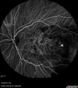
Idiopathic Polypoidal Choroidal Vasculopathy - Indocyanine Green Angiogram - Polyp Visible Left Eye. Branching Network Both Eye744 views
|
|
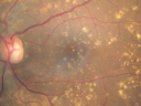
Drusen Maculopathy - Macular Degeneration (Dry) - 50 year old African-American Woman743 views50-year-old woman has drusen and maculopathy. VISUAL ACUITY: OD 20/30, OS 20/25.
|
|

Serous Macular Detachment Both Eyes - Wet AMD vs. CSR - 62 Year Old Man - SD - OCT742 views
|
|
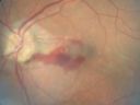
Choroidal Neovascular Membrane - Optic Disc Drusen - Hemorrhage (Coumadin) 738 views82-year-old man with decreased vision in the left eye over about the last six to 12 months. He is also on Coumadin. OD 20/20, OS 20/80
|
|

Angioid Streaks - Peau D'Orange - Pseudoxanthoma Elasticum - Asymptomatic Extrafoveal Choroidal Neovascular Membrane738 views60-year-old woman has pseudoxanthoma elasticum since 2008. Her sister has the same problem and lost vision in both eyes. She had a problem with blood vessel growth in the left eye for which she had injections of Avastin in August, September, and October of 2011 (5 months ago). Subsequent to that, the macula has been dry and you have just been watching her carefully. She is in Florida for a short time but will ultimately be spending nine months out of the year here. Both of her eyes have good vision at
|
|

Idiopathic Polypoidal Choroidal Vasculopathy - False Color738 views
|
|

Choriodal Neovascular Membrane - Subfoveal - Pseudo-Retinitis Pigmentosa - 50 years post Retinal Detachment Repair737 views86-year-old with a history of decreased vision in the right eye. The patient reports she has lost vision in the right eye over the past two months or so. She reports the vision was always compromised in that eye since retinal detachment surgery 50 years ago with a scleral buckler. Vision dropped from 20/50 to 20/200 right eye in 2 months.
|
|
| 2694 files on 27 page(s) |
2 |  |
|
|
|
|
|
|
|
|
|


