Les plus populaires - Age-Related Macular Degeneration - Wet
|
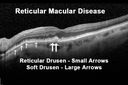
Reticular Macular Disease (Pseudo-drusen) Both Eyes - Occult CNVM and Macular Edema Right Eye vu 1305 foisSpectral Domain Optical Coherence Tomography
|
|
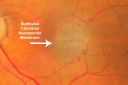
New Subfoveal Classic CNVM OS - Wet AMD - Macular Scar Congenital Toxoplasmosis Right Eye vu 1265 fois77-year-old woman as had poor vision in the right eye since birth from congenital toxoplasmosis of her macula in the right eye. Two to three weeks ago when she noticed vision loss in the left eye. VISUAL ACUITY: OD 2/200, OS 20/200.
|
|

Retinal Hemorrhage vu 1237 foisFundus photography shows central retinal hemorrhage. Patient was treated with Avastin Injection and will be followed up in 4-weeks.
|
|
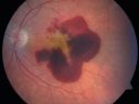
Wet AMD Fresh Submacular Hemorrhage - Displaced with Vitrectomy and Subretinal TPAvu 1232 fois85-year-old woman has wet age-related macular degeneration in her better eye and she was reading with it until two days ago when she had sudden severe vision loss in the eye. Her last injection with Avastin 2 weeks ago 20/160 (Pre-Surgical Displacement)
|
|

AMD - Wet - Fibrovascular Hemorrhagic PED - Occult CNVMvu 1195 fois87-year-old woman for one month OS: 20/60 Fibrovascular PED and Hemorrhage
|
|

Macular Degeneration with Hemorrhagevu 1158 foisPatient comes in for follow up exam for wet AMD. Fundus photography reveals retinal hemorrhage temporally to the macula. Patient was not treated. Wait to see if the blood will absorb on its own.
|
|

Hyperpigmentation of Retinal Pigment Epithlium in Dry AMD (hyperplasia)vu 1118 fois87-year-old woman has wet age-related macular degeneration in the left eye. She had treatment up north but there has been a gap in therapy and the vision in the left eye is poor. Vision OD is 20/25, OS is 20/400
|
|

AMD - Wet - Fibrovascular Hemorrhagic PED - Occult CNVMvu 1092 fois87-year-old woman for one month OS: 20/60 Fibrovascular PED and Hemorrhage
|
|

New Subfoveal Classic CNVM OS - Wet AMD - Macular Scar Congenital Toxoplasmosis Right Eye vu 1090 fois77-year-old woman as had poor vision in the right eye since birth from congenital toxoplasmosis of her macula in the right eye. Two to three weeks ago when she noticed vision loss in the left eye. VISUAL ACUITY: OD 2/200, OS 20/200.
|
|

AMD - Wet - Fibrovascular Hemorrhagic PED - Occult CNVMvu 1069 fois87-year-old woman for one month OS: 20/60 Fibrovascular PED and Hemorrhage
|
|
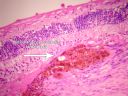
Macular Scar with Retinal Pigment Epithelial Hyperplasia - Hyperpigmentation - Wet Macular Degenerationvu 1049 foisMacular Scar in age related macular degeneration with pathology showing retinal pigment epithelial hyperplasia at the edge of the scar. Histopathogy, microscopic slide, pathology.
|
|

Hyperpigmentation of Retinal Pigment Epithlium in Dry AMD (hyperplasia)vu 1048 fois87-year-old woman has wet age-related macular degeneration in the left eye. She had treatment up north but there has been a gap in therapy and the vision in the left eye is poor. Vision OD is 20/25, OS is 20/400
|
|

Large serous PEDvu 1036 fois63-year-old female with acute onset large retinal pigment epithelium detachment from age-related macular degeneration
|
|

Subfoveal CNVM wet AMD oct line scanvu 998 foisShe has noticed decreased vision in the left eye. It is hard to say for how long, maybe a few months. She had her last eye exam about a year ago. She thoughts perhaps it was cataracts, but you saw a problem with the macula and suggested she come here for evaluation.
VISUAL ACUITY: Vision OD is 20/25, OS is 2/200
|
|

Cartwheel CNVM - Classic - wet AMD - evidence of possible old CSR - Subretinal hemorrhage next to subfoveal lesionvu 989 fois75 y.o Decreased Vision right eye for one year. VA OD: sc20/125+2 OS 20/25. Autofluorescence and SD OCT show evidence of old serous retinal detachment superior to the fovea suggesting a history of CSR. The macular lesion on FA shows a classic cartwheel appearance
|
|

AMD - Wet - Fibrovascular Hemorrhagic PED - Occult CNVMvu 976 fois87-year-old woman for one month OS: 20/60 Fibrovascular PED and Hemorrhage
|
|

Wet AMD Fresh Submacular Hemorrhage - Displaced with Vitrectomy and Subretinal TPAvu 952 fois85-year-old woman has wet age-related macular degeneration in her better eye and she was reading with it until two days ago when she had sudden severe vision loss in the eye. Post-surgical displacment vision was 20/120.
|
|

Disciform Scar after Photodynamic Laser - Macular Hemorrhage and Vitrectomyvu 927 fois84-year-old man has age-related macular degeneration in both eyes, with a disciform scar in the right eye. He had a subfoveal hemorrhage in that eye treated with vitrectomy and displacement on May 3, 2006. He previous to that had photodynamic laser. These photos are from October 2010. VA is 1/200.
|
|

Subfoveal CNVM wet AMD color photovu 903 foisShe has noticed decreased vision in the left eye. It is hard to say for how long, maybe a few months. She had her last eye exam about a year ago. She thoughts perhaps it was cataracts, but you saw a problem with the macula and suggested she come here for evaluation.
VISUAL ACUITY: Vision OD is 20/25, OS is 2/200
|
|

Disciform Scar after Photodynamic Laser - Macular Hemorrhage and Vitrectomyvu 863 fois84-year-old man has age-related macular degeneration in both eyes, with a disciform scar in the right eye. He had a subfoveal hemorrhage in that eye treated with vitrectomy and displacement on May 3, 2006. He previous to that had photodynamic laser. These photos are from October 2010. VA is 1/200.
|
|
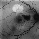
Cartwheel CNVM - Classic - wet AMD - evidence of possible old CSR - Autofluorescence shows brigh spot superior to subfoveal lesionvu 832 fois75 y.o Decreased Vision right eye for one year. VA OD: sc20/125+2 OS 20/25. Autofluorescence and SD OCT show evidence of old serous retinal detachment superior to the fovea suggesting a history of CSR. The macular lesion on FA shows a classic cartwheel appearance
|
|

Chorioretinal Scar with Radiating Chorioretinal Foldsvu 830 fois79-year-old woman has wet age-related macular degeneration in both eyes. The left eye had intravitreal injections of Avastin starting in May of 2009. Her vision remains good in the left eye and poor in the right eye.
VISUAL ACUITY: OD 8/200, OS 20/60.
|
|

Classic Subfoveal Choroidal Neovascular Membrane - 20/400 Vision - 6 - 12 months old lesionvu 828 fois78-year-old woman six months or a year ago decreasing vision in the right eye. OD 20/400. Fluorescein angiography shows a classic subfoveal choroidal neovascular membrane in the right eye 3 disc-diameters across, which has brisk leakage. The left eye shows staining drusen.
|
|
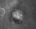
AMD - Wet - Fibrovascular Hemorrhagic PED - Occult CNVMvu 823 fois87-year-old woman for one month OS: 20/60 Fibrovascular PED and Hemorrhage
|
|
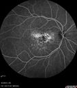
Idiopathic Polypoidal Choroidal Vasculopathy - Fluorescein Angiogramvu 818 fois
|
|

Juxtapapillary CNVM and Serous Macular Detachment Wet AMD Rx Laservu 814 fois85-year-old man OD is 20/25, OS is 20/100. IOP: OD 21, OS 22.
OS: Vertical C/D ratio is 0.1. There is a posterior vitreous separation. There is a hemorrhagic pigment epithelial detachment on the superior pole of the optic nerve, extending a disc-and-a-half diameter off the nerve, with adjacent exudates. There is a serous macular detachment involving the center of the fovea.
VA improved from 20/100 to 20/50 in four months from laser.
|
|

Retinal Angiomatous Proliferation - Type III choroidal neovascular membranevu 814 fois83 year old female with wet AMD - 20/160 VA- Vision improved to 20/40 with 3 injections of anti-VEGF drugs. OCT shows break in RPE and FA and ICG show PED with hot spot on ICG
|
|

Polypoidal Choroidal Vasculopathy with Pulsating ICG polyp left eyevu 813 fois
|
|
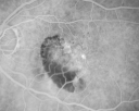
AMD - Wet - Fibrovascular Hemorrhagic PED - Occult CNVMvu 812 fois87-year-old woman for one month OS: 20/60 Fibrovascular PED and Hemorrhage
|
|
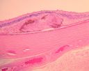
Macular Scar with Retinal Pigment Epithelial Hyperplasia - Hyperpigmentation - Wet Macular Degenerationvu 809 foisMacular Scar in age related macular degeneration with pathology showing retinal pigment epithelial hyperplasia at the edge of the scar. Histopathogy, microscopic slide, pathology.
|
|
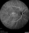
Asymptomatic Occult Subfoveal CNVM in Good Eye Visible on ICGvu 803 fois
|
|
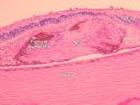
Macular Scar with Retinal Pigment Epithelial Hyperplasia - Hyperpigmentation - Wet Macular Degenerationvu 802 foisMacular Scar in age related macular degeneration with pathology showing retinal pigment epithelial hyperplasia at the edge of the scar. Histopathogy, microscopic slide, pathology.
|
|

Serous Macular Detachment Both Eyes - Wet AMD vs. CSR - 62 Year Old Man - SD - OCTvu 801 fois
|
|
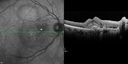
Cartwheel CNVM - Classic - wet AMD - evidence of possible old CSRvu 797 fois75 y.o Decreased Vision right eye for one year. VA OD: sc20/125+2 OS 20/25. Autofluorescence and SD OCT show evidence of old serous retinal detachment superior to the fovea suggesting a history of CSR. The macular lesion on FA shows a classic cartwheel appearance
|
|
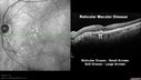
Reticular Macular Disease (Pseudo-drusen) Both Eyes - Occult CNVM and Macular Edema Right Eye vu 794 foisSpectral Domain Optical Coherence Tomography
|
|

Juxtapapillary CNVM and Serous Macular Detachment Wet AMD Rx Laservu 789 fois85-year-old man OD is 20/25, OS is 20/100. IOP: OD 21, OS 22.
OS: Vertical C/D ratio is 0.1. There is a posterior vitreous separation. There is a hemorrhagic pigment epithelial detachment on the superior pole of the optic nerve, extending a disc-and-a-half diameter off the nerve, with adjacent exudates. There is a serous macular detachment involving the center of the fovea.
VA improved from 20/100 to 20/50 in four months from laser.
|
|
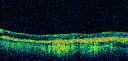
Vision Loss, Blood, and Cartwheel CNVM OD with no fluid on OCT Scanvu 788 fois86-year-old man has had changing vision. He says his vision has been gradually declining since I saw him in June. You saw a hemorrhage in the eye and suggested he comes in here for an evaluation. He is on Combigan and Travatan in the right eye for his glaucoma.
VISUAL ACUITY: Vision OD is 6/200 OS is 8/200 The macula in the right eye is wet even though interestingly the OCT does not show subretinal fluid.
|
|
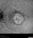
Wet AMD - New Small Hemorrhage Both Eyes - Mature CNVM visible on Early Indocyanine Green Angiogramvu 786 fois
|
|

Subfoveal CNVM wet AMD early fa full imagevu 779 foisShe has noticed decreased vision in the left eye. It is hard to say for how long, maybe a few months. She had her last eye exam about a year ago. She thoughts perhaps it was cataracts, but you saw a problem with the macula and suggested she come here for evaluation.
VISUAL ACUITY: Vision OD is 20/25, OS is 2/200
|
|
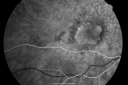
Classic Subfoveal Choroidal Neovascular Membrane - 20/400 Vision - 6 - 12 months old lesionvu 779 fois78-year-old woman six months or a year ago decreasing vision in the right eye. OD 20/400. Fluorescein angiography shows a classic subfoveal choroidal neovascular membrane in the right eye 3 disc-diameters across, which has brisk leakage. The left eye shows staining drusen.
|
|
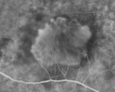
Classic Subfoveal Choroidal Neovascular Membrane - 20/400 Vision - 6 - 12 months old lesionvu 775 fois78-year-old woman six months or a year ago decreasing vision in the right eye. OD 20/400. Fluorescein angiography shows a classic subfoveal choroidal neovascular membrane in the right eye 3 disc-diameters across, which has brisk leakage. The left eye shows staining drusen.
|
|

Vision Loss, Blood, and Cartwheel CNVM OD with no fluid on OCT Scanvu 774 fois86-year-old man has had changing vision. He says his vision has been gradually declining since I saw him in June. You saw a hemorrhage in the eye and suggested he comes in here for an evaluation. He is on Combigan and Travatan in the right eye for his glaucoma.
VISUAL ACUITY: Vision OD is 6/200 OS is 8/200 The macula in the right eye is wet even though interestingly the OCT does not show subretinal fluid.
|
|

Subfoveal CNVM wet AMD late fa full imagevu 771 foisShe has noticed decreased vision in the left eye. It is hard to say for how long, maybe a few months. She had her last eye exam about a year ago. She thoughts perhaps it was cataracts, but you saw a problem with the macula and suggested she come here for evaluation.
VISUAL ACUITY: Vision OD is 20/25, OS is 2/200
|
|

Subfoveal CNVM wet AMD fellow eyevu 766 foisShe has noticed decreased vision in the left eye. It is hard to say for how long, maybe a few months. She had her last eye exam about a year ago. She thoughts perhaps it was cataracts, but you saw a problem with the macula and suggested she come here for evaluation.
VISUAL ACUITY: Vision OD is 20/25, OS is 2/200
|
|
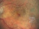
Pigment Epithelial Detachment with Subretinal Fluid and Exudate 20/400 Visionvu 763 fois87-year-old woman has wet age-related macular degeneration in the left eye. She had treatment up north but there has been a gap in therapy and the vision in the left eye is poor. Vision OD is 20/25, OS is 20/400
|
|
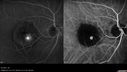
Retinal Angiomatous Proliferation - Type III choroidal neovascular membranevu 756 fois83 year old female with wet AMD - 20/160 VA- Vision improved to 20/40 with 3 injections of anti-VEGF drugs. OCT shows break in RPE and FA and ICG show PED with hot spot on ICG
|
|
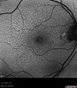
Asymptomatic Juxtapapillary Choroidal Neovascular Membrane - Reticular Macular Disease (Wet AMD) - FAFvu 754 fois
|
|
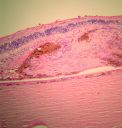
Macular Scar with Retinal Pigment Epithelial Hyperplasia - Hyperpigmentation - Wet Macular Degenerationvu 750 foisMacular Scar in age related macular degeneration with pathology showing retinal pigment epithelial hyperplasia at the edge of the scar. Histopathogy, microscopic slide, pathology.
|
|
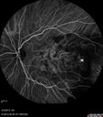
Idiopathic Polypoidal Choroidal Vasculopathy - Indocyanine Green Angiogram - Polyp Visible Left Eye. Branching Network Both Eyevu 744 fois
|
|

Serous Macular Detachment Both Eyes - Wet AMD vs. CSR - 62 Year Old Man - SD - OCTvu 742 fois
|
|

Idiopathic Polypoidal Choroidal Vasculopathy - False Colorvu 741 fois
|
|
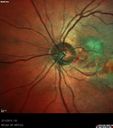
Asymptomatic Juxtapapillary Choroidal Neovascular Membrane - Reticular Macular Disease (Wet AMD) - Heidelberg Colorvu 737 fois
|
|
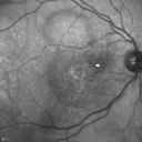
Cartwheel CNVM - Classic - wet AMD - evidence of possible old CSRvu 737 fois75 y.o Decreased Vision right eye for one year. VA OD: sc20/125+2 OS 20/25. Autofluorescence and SD OCT show evidence of old serous retinal detachment superior to the fovea suggesting a history of CSR. The macular lesion on FA shows a classic cartwheel appearance
|
|
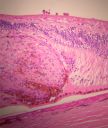
Macular Scar with Retinal Pigment Epithelial Hyperplasia - Hyperpigmentation - Wet Macular Degenerationvu 736 foisMacular Scar in age related macular degeneration with pathology showing retinal pigment epithelial hyperplasia at the edge of the scar. Histopathogy, microscopic slide, pathology.
|
|
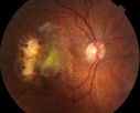
vu 736 fois87 year old male seen for neovascular macular degeneration. Patient had huge disciform scar and lipid.
|
|
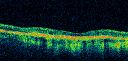
Vision Loss, Blood, and Cartwheel CNVM OD with no fluid on OCT Scanvu 735 fois86-year-old man has had changing vision. He says his vision has been gradually declining since I saw him in June. You saw a hemorrhage in the eye and suggested he comes in here for an evaluation. He is on Combigan and Travatan in the right eye for his glaucoma.
VISUAL ACUITY: Vision OD is 6/200 OS is 8/200 The macula in the right eye is wet even though interestingly the OCT does not show subretinal fluid.
|
|
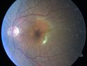
Wet Age-related Macular Degeneration (AMD) Radiating Chorioretinal Folds from Contracting Macular Scarvu 734 fois93-year-old man has wet age-related macular degeneration in his left eye with a subfoveal pigment epithelial detachment.
Vision OS is 20/40. There are striking radiating chorioretinal folds emanating off of the scar, which suggests it is contracting.
|
|
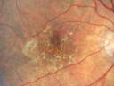
Hyperpigmentation of Retinal Pigment Epithlium in Dry AMD (hyperplasia)vu 728 fois87-year-old woman has wet age-related macular degeneration in the left eye. She had treatment up north but there has been a gap in therapy and the vision in the left eye is poor. Vision OD is 20/25, OS is 20/400
|
|
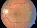
Cartwheel CNVM - Classic - wet AMD - evidence of possible old CSR - Fellow eye has hydrophilic Drusenvu 725 fois75 y.o Decreased Vision right eye for one year. VA OD: sc20/125+2 OS 20/25. Autofluorescence and SD OCT show evidence of old serous retinal detachment superior to the fovea suggesting a history of CSR. The macular lesion on FA shows a classic cartwheel appearance
|
|
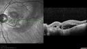
Asymptomatic Juxtapapillary Choroidal Neovascular Membrane - Reticular Macular Disease (Wet AMD) vu 723 fois
|
|
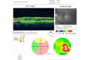
New Subfoveal Classic CNVM OS - Wet AMD - Macular Scar Congenital Toxoplasmosis Right Eye vu 721 fois77-year-old woman as had poor vision in the right eye since birth from congenital toxoplasmosis of her macula in the right eye. Two to three weeks ago when she noticed vision loss in the left eye. VISUAL ACUITY: OD 2/200, OS 20/200.
|
|
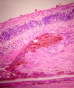
Macular Scar with Retinal Pigment Epithelial Hyperplasia - Hyperpigmentation - Wet Macular Degenerationvu 719 foisMacular Scar in age related macular degeneration with pathology showing retinal pigment epithelial hyperplasia at the edge of the scar. Histopathogy, microscopic slide, pathology.
|
|
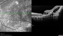
Idiopathic Polypoidal Choroidal Vasculopathy - SD-OCTvu 715 fois
|
|
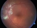
Wet Macular Degeneration with Juxtapapillary Neovascular Membrane and Choroidal Laser Treatmentvu 712 fois87-year-old woman has wet age-related macular degeneration in the left eye with an extrafoveal choroidal neovascular membrane OD is 20/25, OS is 20/25
|
|
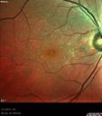
Asymptomatic Juxtapapillary Choroidal Neovascular Membrane - Reticular Macular Disease (Wet AMD) - Heidelberg Colorvu 712 fois
|
|
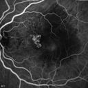
Polypoidal Choroidal Vasculopathy with Pulsating ICG polyp left eyevu 712 fois
|
|
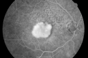
Classic Subfoveal Choroidal Neovascular Membrane - 20/400 Vision - 6 - 12 months old lesionvu 701 fois78-year-old woman six months or a year ago decreasing vision in the right eye. OD 20/400. Fluorescein angiography shows a classic subfoveal choroidal neovascular membrane in the right eye 3 disc-diameters across, which has brisk leakage. The left eye shows staining drusen.
|
|
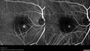
Retinal Angiomatous Proliferation - Type III choroidal neovascular membranevu 701 fois83 year old female with wet AMD - 20/160 VA- Vision improved to 20/40 with 3 injections of anti-VEGF drugs. OCT shows break in RPE and FA and ICG show PED with hot spot on ICG
|
|
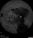
Idiopathic Polypoidal Choroidal Vasculopathy - Indocyanine Green Angiogram - Polyp Visible Left Eye. Branching Network Both Eyevu 700 fois
|
|

Wet AMD 62 year old male smoker vu 696 fois62-year-old man has wet age-related macular degeneration in the right eye. He has been undergoing treatment with Lucentis for that. Then his left eye developed hazy vision a few weeks ago while he was overseas and now his vision is hazy in both eyes. OD 20/40, OS 20/30. FA shows a classic subfoveal choroidal neovascular membrane in the left eye, which is leaking and in the right eye, which is dry. He is going to stop smoking today. FU: VA 20/30 OU with 2 years of every 6 weeks bilateral avastin!
|
|
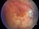
Vision Loss, Blood, and Cartwheel CNVM OD with no fluid on OCT Scanvu 696 fois86-year-old man has had changing vision. He says his vision has been gradually declining since I saw him in June. You saw a hemorrhage in the eye and suggested he comes in here for an evaluation. He is on Combigan and Travatan in the right eye for his glaucoma.
VISUAL ACUITY: Vision OD is 6/200 OS is 8/200 The macula in the right eye is wet even though interestingly the OCT does not show subretinal fluid.
|
|
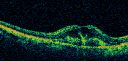
Classic Subfoveal Choroidal Neovascular Membrane - 20/400 Vision - 6 - 12 months old lesionvu 694 fois78-year-old woman six months or a year ago decreasing vision in the right eye. OD 20/400. Fluorescein angiography shows a classic subfoveal choroidal neovascular membrane in the right eye 3 disc-diameters across, which has brisk leakage. The left eye shows staining drusen.
|
|

Retinal Pigment Epithlial Rip and Good Vision (20/40)vu 689 fois82-year-old woman has age-related macular degeneration in the left eye. She developed a pigment epithelial rip in the macula and remarkably her vision is improved with 2 years of treatment. She has had no injections for one year. OS 20/40. (fellow eye has a disciform scar)
|
|

Pigment Epithelial Detachment with Subretinal Fluid and Exudate 20/400 Visionvu 688 fois87-year-old woman has wet age-related macular degeneration in the left eye. She had treatment up north but there has been a gap in therapy and the vision in the left eye is poor. Vision OD is 20/25, OS is 20/400
|
|

Wet AMD Fresh Submacular Hemorrhage - Displaced with Vitrectomy and Subretinal TPAvu 686 fois85-year-old woman has wet age-related macular degeneration in her better eye and she was reading with it until two days ago when she had sudden severe vision loss in the eye. Post-surgical displacment vision was 20/120.
|
|
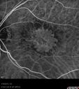
Wet AMD - New Small Hemorrhage Both Eyes - Mature CNVM visible on Early Indocyanine Green Angiogramvu 685 fois
|
|
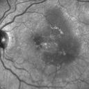
Polypoidal Choroidal Vasculopathy with Pulsating ICG polyp left eyevu 685 fois
|
|

Idiopathic Polypoidal Choroidal Vasculopathy - Indocyanine Green Angiogram - Polyp Visible Left Eye. Branching Network Both Eyevu 684 fois
|
|

Polypoidal Choroidal Vasculopathy with Pulsating ICG polyp left eyevu 684 fois
|
|
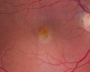
Macular Degeneration - Choroidal Laser Left eye - New Subfoveal CNVM left eye - Did well with Avastinvu 683 fois69-year-old woman new vision loss right eye and old macular scar left eye. OD 20/40, OS 20/200
|
|

Subfoveal CNVM wet AMD fellow eye oct line scanvu 678 foisShe has noticed decreased vision in the left eye. It is hard to say for how long, maybe a few months. She had her last eye exam about a year ago. She thoughts perhaps it was cataracts, but you saw a problem with the macula and suggested she come here for evaluation.
VISUAL ACUITY: Vision OD is 20/25, OS is 2/200
|
|
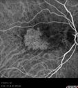
Wet AMD - New Small Hemorrhage Both Eyes - Mature CNVM visible on Early Indocyanine Green Angiogramvu 678 fois
|
|

Polypoidal Choroidal Vasculopathy with Pulsating ICG polyp left eyevu 678 fois
|
|
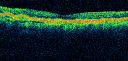
Hyperpigmentation of Retinal Pigment Epithlium in Dry AMD (hyperplasia)vu 677 fois87-year-old woman has wet age-related macular degeneration in the left eye. She had treatment up north but there has been a gap in therapy and the vision in the left eye is poor. Vision OD is 20/25, OS is 20/400
|
|
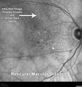
Reticular Macular Disease (Pseudo-drusen) Both Eyes - Occult CNVM and Macular Edema Right Eye vu 676 foisInfra-Red
|
|
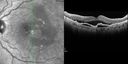
Polypoidal Choroidal Vasculopathy with Pulsating ICG polyp left eyevu 674 fois
|
|
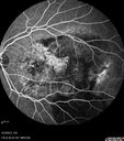
Idiopathic Polypoidal Choroidal Vasculopathy - Fluorescein Angiogramvu 671 fois
|
|
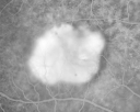
Classic Subfoveal Choroidal Neovascular Membrane - 20/400 Vision - 6 - 12 months old lesionvu 668 fois78-year-old woman six months or a year ago decreasing vision in the right eye. OD 20/400. Fluorescein angiography shows a classic subfoveal choroidal neovascular membrane in the right eye 3 disc-diameters across, which has brisk leakage. The left eye shows staining drusen.
|
|
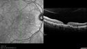
Idiopathic Polypoidal Choroidal Vasculopathy - SD-OCTvu 668 fois
|
|
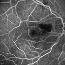
Cartwheel CNVM - Classic - wet AMD - evidence of possible old CSR - FA shows cartwheel shaped CNVMvu 667 fois75 y.o Decreased Vision right eye for one year. VA OD: sc20/125+2 OS 20/25. Autofluorescence and SD OCT show evidence of old serous retinal detachment superior to the fovea suggesting a history of CSR. The macular lesion on FA shows a classic cartwheel appearance
|
|
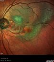
Asymptomatic Juxtapapillary Choroidal Neovascular Membrane - Reticular Macular Disease (Wet AMD) - Heidelberg Colorvu 665 fois
|
|
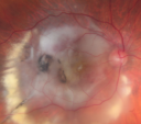
Disciform Macular Scar Both Eyes from Wet Macular Degenerationvu 660 fois86-year-old woman has disciform scars OD: 20/400; OS: 20/100
|
|
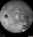
Idiopathic Polypoidal Choroidal Vasculopathy - IRvu 660 fois
|
|
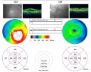
Classic Subfoveal Choroidal Neovascular Membrane - 20/400 Vision - 6 - 12 months old lesionvu 657 fois78-year-old woman six months or a year ago decreasing vision in the right eye. OD 20/400. Fluorescein angiography shows a classic subfoveal choroidal neovascular membrane in the right eye 3 disc-diameters across, which has brisk leakage. The left eye shows staining drusen.
|
|
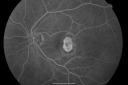
New Subfoveal Classic CNVM OS - Wet AMD - Macular Scar Congenital Toxoplasmosis Right Eye vu 657 fois77-year-old woman as had poor vision in the right eye since birth from congenital toxoplasmosis of her macula in the right eye. Two to three weeks ago when she noticed vision loss in the left eye. VISUAL ACUITY: OD 2/200, OS 20/200.
|
|
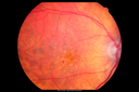
Reticular Macular Disease (Pseudo-drusen) Both Eyes - Occult CNVM and Macular Edema Right Eye vu 657 foisColor Fundus Photo
|
|
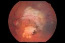
Disciform Macular Scar Both Eyes from Wet Macular Degenerationvu 656 fois86-year-old woman has disciform scars OD: 20/400; OS: 20/100
|
|
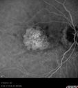
Wet AMD - New Small Hemorrhage Both Eyes - Mature CNVM visible on Early Indocyanine Green Angiogramvu 656 fois
|
|
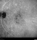
Reticular Macular Disease (Pseudo-drusen) Both Eyes - Occult CNVM and Macular Edema Right Eyevu 655 foisIndocyanine Green Angiogram
|
|
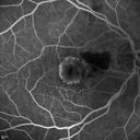
Cartwheel CNVM - Classic - wet AMD - evidence of possible old CSR - FA shows cartwheel shaped CNVMvu 655 fois75 y.o Decreased Vision right eye for one year. VA OD: sc20/125+2 OS 20/25. Autofluorescence and SD OCT show evidence of old serous retinal detachment superior to the fovea suggesting a history of CSR. The macular lesion on FA shows a classic cartwheel appearance
|
|
| 1175 photos sur 12 page(s) |
1 |
 |
|
|
|
|
|
|
|
|
|


