Most viewed - Retina Vascular Occlusions
|

CRVO 25 year old man - Heterozygous Factor V Leiden528 views20/100 initial vision. Improved to 20/30 with Lucentis which continued for 3 years, treat and extend, and stopped.
|
|
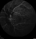
Central Retinal Vein Occlusion with Macular Edema528 views74 year old female The vision in the left eye has gotten worse over the last 3 weeks.
VA OD: Dcc20/25
VA OS: DccCF 3ft
IOP: TP: OD:16 OS:14
Vision improved to 20/40 in 3 months with monthly Aflibercept
|
|

Branch Retinal Arterial Occlusion 20/20 Vision - Symptoms for 1 Day527 views75-year-old man was seen in the office on 4/6/2010. He noticed a sudden vision change in the right eye yesterday morning. 20/20 Vision both Eyes
|
|

Branch Retinal Vein Occlusion with NVE (Neovascularization Elsewhere)527 views
|
|
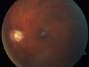
527 views
|
|

Branch Retinal Artery Occlusion527 viewsAcute Inferotemporal Occlusion Right Eye - FA shows delayed circulation, OCT shows edema of superficial layers of retina
|
|
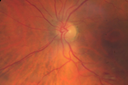
Branch Retinal Vein Occlusion527 views84 year old man - 2 months vision loss - VA 20/40 - 20/20 with one injection of Eylea
|
|
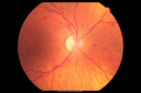
CRVO with Paracentral Acute Middle Maculopath - PAMM527 views56 year old woman with sudden vision loss, hemorrhages in all four quadrants. FA shows good retinal circulation. The OCT shows PAMM lesions in the affected eye. Vision did improve on second visit from 20/160 to 20/60 in about a month
|
|
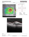
Venous Macroaneurysm 6 year old CRVO - treated with focal laser527 views93 year old female with CRVO in the right eye 6 years ago. The visual potential is 20/100 and has recently subjectively and objectively declined. VA is 20/200 and she has new onset macular edema from a venous macroaneurysm that formed at a collateral vessel. This responded nicely to a single laser spot.
|
|
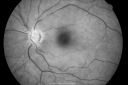
526 views
|
|
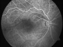
Branch Retinal Arterial Occlusion 20/20 Vision - Symptoms for 1 Day526 views75-year-old man was seen in the office on 4/6/2010. He noticed a sudden vision change in the right eye yesterday morning. 20/20 Vision both Eyes
|
|
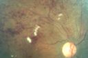
Branch Retinal Vein Occlusion with Cotton Wool Spot 526 views70-year-old man with blurred vision in the right eye for about one or two months. OD is 20/80, OS is 20/25.
|
|

526 views
|
|
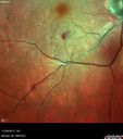
526 views
|
|
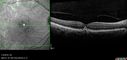
Holenhorst Plaque - Small and asymptomatic526 views72 year old woman with mild dry ARMD. VA 20/40 OU. Asymptomatic plaque noted in the right eye.
|
|
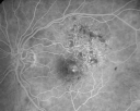
New BRVO Left Eye Old BRVO Right Eye - Left Eye Shows Twig Superior Vein Occlusion with Edema525 views90-year-old man decreased vision maybe one year since cataract surgery he is no longer able to read. OD 20/200, OS 20/200
|
|

CVO525 viewsmid phase (3min) fa of a 27 year old male with crvo os VA 20/20
|
|
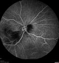
Branch Retinal Artery Occlusion525 viewsAcute Inferotemporal Occlusion Right Eye - FA shows delayed circulation, OCT shows edema of superficial layers of retina
|
|
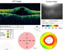
524 views
|
|
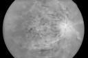
524 views49-year-old woman has central retinal vein occlusion right eye with fluctuating vision for a few month now with OD 20/100. IOP: OD 20
|
|
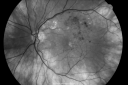
New BRVO Left Eye Old BRVO Right Eye 524 views90-year-old man decreased vision maybe one year since cataract surgery he is no longer able to read. OD 20/200, OS 20/200
|
|
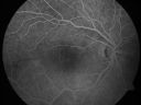
Branch Retinal Arterial Occlusion 20/20 Vision - Symptoms for 1 Day524 views75-year-old man was seen in the office on 4/6/2010. He noticed a sudden vision change in the right eye yesterday morning. 20/20 Vision both Eyes
|
|
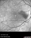
Cardiac Catheterization induced Cilioretinal Artery Occlusion - Paracentral Acute Middle Maculopathy524 views69 year old female with sudden vision loss on completing cardiac catheterization. The SD OCT shows a PAMM lesion and the lesion looks gray on color imaging. There is delayed filling of the cilioretinal artery.
|
|

Twig Old Macular Branch Retinal Vein Occlusion - Venous Venous Collaterals524 views88 year old man with 20/25 vision in his left and only eye. There is vascular remodeling consistent with an old twig BRVO.
|
|
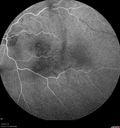
Branch Retinal Vein Occlusion523 views84 year old man - 2 months vision loss - VA 20/40 - 20/20 with one injection of Eylea
|
|
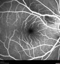
Bilateral Branch Retinal Artery Occlusion - Intraarterial (hollenhorst) plaques visible523 views67 year old with acute visual field loss in each eye. Vision is 20/25 right eye 20/32 left eye. Images show intra-arterial plaques in each eye. The right eye on the optic nerve and the left eye in a branch retinal artery.
|
|

Branch Retinal Arterial Occlusion Right Eye - Atheromata - Both Eyes523 views83 year old woman with no symptoms - VA 20/40 in both eyes. Right eye has ST BRAO with silver wiring. Left eye has superotemporal atheromata
|
|

Non-ischemic Central Retinal Vein Occlusion with Macular Edema523 views62 year old man The vision in the right eye started to decline about 10 days ago. Prior to that the two eyes were about the same.
VA OD: Dcc20/125 PH20/50-2
VA OS: Dcc20/20
IOP: TP: OD:21 OS:21
|
|

CRVO522 views58 year old male with present CRVO OU. Visual Acuity 20/30 OD 20/200 OS. Pt. recently underwent surgery for cancer of the bladder and has other blood and hemoglobin issues. Pt sent for further blood work and see a neurologist.
|
|
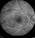
CRVO with Paracentral Acute Middle Maculopath - PAMM522 views56 year old woman with sudden vision loss, hemorrhages in all four quadrants. FA shows good retinal circulation. The OCT shows PAMM lesions in the affected eye. Vision did improve on second visit from 20/160 to 20/60 in about a month
|
|
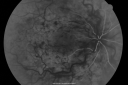
521 views49-year-old woman has central retinal vein occlusion right eye with fluctuating vision for a few month now with OD 20/100. IOP: OD 20
|
|
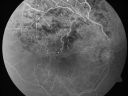
Branch Retinal Vein Occlusion with Cotton Wool Spot 521 views70-year-old man with blurred vision in the right eye for about one or two months. OD is 20/80, OS is 20/25.
|
|
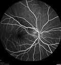
Bilateral Branch Retinal Artery Occlusion - Intraarterial (hollenhorst) plaques visible521 views67 year old with acute visual field loss in each eye. Vision is 20/25 right eye 20/32 left eye. Images show intra-arterial plaques in each eye. The right eye on the optic nerve and the left eye in a branch retinal artery.
|
|
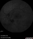
Branch Retinal Vein Occlusion (BRVO) no macular edema - cotton wool spot521 views85 year old man with asymptomatic BRVO - his vision stayed great without needing any treatment.
|
|

Non-ischemic Central Retinal Vein Occlusion with Macular Edema521 views62 year old man The vision in the right eye started to decline about 10 days ago. Prior to that the two eyes were about the same.
VA OD: Dcc20/125 PH20/50-2
VA OS: Dcc20/20
IOP: TP: OD:21 OS:21
|
|
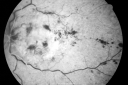
Central Retinal Vein Occlusion - New- and old Macular Scar from AMD520 views87-year-old woman with longstaning macular scars and new vision loss left eye for 2 months. OD: 20/200, OS 20/400.
|
|
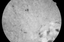
Central Retinal Vein Occlusion - New- and old Macular Scar from AMD520 views87-year-old woman with longstaning macular scars and new vision loss left eye for 2 months. OD: 20/200, OS 20/400.
|
|

Central Retinal Artery Occlusion less than 24 hours - 69 year old man VA light perception520 views
|
|

Branch Retinal Vein Occlusion with Cotton Wool Spot 520 views70-year-old man with blurred vision in the right eye for about one or two months. OD is 20/80, OS is 20/25.
|
|
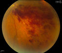
Branch Retinal Vein Occlusion520 viewsPatient's VA was 20/40, right eye. Fundus exam shows branch retinal vein occlusion nasally to the optic nerve in the right eye.
|
|
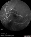
Ischemic BRVO with non-perfusion (nonperfusion) - recovered from severe vitreous hemorrhage520 views
|
|
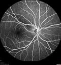
Bilateral Branch Retinal Artery Occlusion - Intraarterial (hollenhorst) plaques visible520 views67 year old with acute visual field loss in each eye. Vision is 20/25 right eye 20/32 left eye. Images show intra-arterial plaques in each eye. The right eye on the optic nerve and the left eye in a branch retinal artery.
|
|
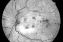
Central Retinal Vein Occlusion - New- and old Macular Scar from AMD519 views87-year-old woman with longstaning macular scars and new vision loss left eye for 2 months. OD: 20/200, OS 20/400.
|
|
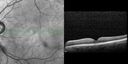
Branch Retinal Vein Occlusion with worse macular edema from hypertension - SD-OCT shows Edema Right Eye519 views
|
|
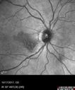
Cardiac Catheterization induced Cilioretinal Artery Occlusion - Paracentral Acute Middle Maculopathy519 views69 year old female with sudden vision loss on completing cardiac catheterization. The SD OCT shows a PAMM lesion and the lesion looks gray on color imaging. There is delayed filling of the cilioretinal artery.
|
|
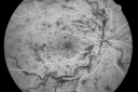
518 views49-year-old woman has central retinal vein occlusion right eye with fluctuating vision for a few month now with OD 20/100. IOP: OD 20
|
|

Branch Retinal Arterial Occlusion 20/20 Vision - Symptoms for 1 Day518 views75-year-old man was seen in the office on 4/6/2010. He noticed a sudden vision change in the right eye yesterday morning. 20/20 Vision both Eyes
|
|
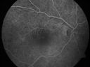
Branch Retinal Arterial Occlusion 20/20 Vision - Symptoms for 1 Day518 views75-year-old man was seen in the office on 4/6/2010. He noticed a sudden vision change in the right eye yesterday morning. 20/20 Vision both Eyes
|
|

Macular Branch Retinal Vein Occlusion and Adult Vitelliform Dystrophy518 views72-year-old woman 2 months post ECCE with poor vision since surgery especially left eye.
VISUAL ACUITY: OD 20/50, OS 20/200
|
|
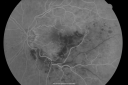
Macular Branch Retinal Vein Occlusion and Adult Vitelliform Dystrophy518 views72-year-old woman 2 months post ECCE with poor vision since surgery especially left eye.
VISUAL ACUITY: OD 20/50, OS 20/200
|
|

Macular Branch Retinal Vein Occlusion and Adult Vitelliform Dystrophy518 views72-year-old woman 2 months post ECCE with poor vision since surgery especially left eye.
VISUAL ACUITY: OD 20/50, OS 20/200
|
|

Branch Retinal Vein Occlusion with Cotton Wool Spot 518 views70-year-old man with blurred vision in the right eye for about one or two months. OD is 20/80, OS is 20/25.
|
|
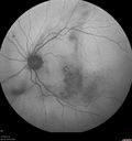
Branch Retinal Vein Occlusion518 views84 year old man - 2 months vision loss - VA 20/40 - 20/20 with one injection of Eylea
|
|
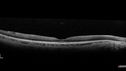
Branch Retinal Artery Occlusion - No Plaque518 views74 year old man with 2 days of vision loss. Vision OS is 20/200 but improved to 20/25 in 1 month. He has dysfibrinogen anemia.
|
|

Chronic BRAO with Hemorrhages - Sclerosed Vessel518 views95 year old female with blurred vision in both eyes in for a checkup. History of perpapillary CNVM in the left eye 2 years prior not treated. History of stroke with multiple emboli 1 year ago.
Vision 20/32 OD, 20/40 OS
|
|
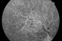
517 views49-year-old woman has central retinal vein occlusion right eye with fluctuating vision for a few month now with OD 20/100. IOP: OD 20
|
|
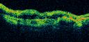
Central Retinal Vein Occlusion - New- and old Macular Scar from AMD517 views87-year-old woman with longstaning macular scars and new vision loss left eye for 2 months. OD: 20/200, OS 20/400.
|
|
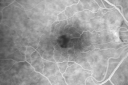
Pattern Dystrophy - Adult Vitelliform Macular Degeneration517 views72-year-old woman 2 months post ECCE with poor vision since surgery especially left eye.
VISUAL ACUITY: OD 20/50, OS 20/200
|
|
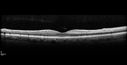
Bilateral Branch Retinal Artery Occlusion - Intraarterial (hollenhorst) plaques visible517 views67 year old with acute visual field loss in each eye. Vision is 20/25 right eye 20/32 left eye. Images show intra-arterial plaques in each eye. The right eye on the optic nerve and the left eye in a branch retinal artery.
|
|

CRVO with Paracentral Acute Middle Maculopath - PAMM517 views56 year old woman with sudden vision loss, hemorrhages in all four quadrants. FA shows good retinal circulation. The OCT shows PAMM lesions in the affected eye. Vision did improve on second visit from 20/160 to 20/60 in about a month
|
|
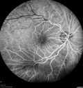
Branch Retinal Arterial Occlusion Right Eye - Atheromata - Both Eyes517 views83 year old woman with no symptoms - VA 20/40 in both eyes. Right eye has ST BRAO with silver wiring. Left eye has superotemporal atheromata
|
|

Branch Retinal Arterial Occlusion Right Eye - Atheromata - Both Eyes517 views83 year old woman with no symptoms - VA 20/40 in both eyes. Right eye has ST BRAO with silver wiring. Left eye has superotemporal atheromata
|
|
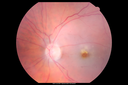
Central Retinal Artery Occlusion then Neovascular Glaucoma517 views81 year old male with sudden vision loss. 2 months later developed NVG
|
|

Branch Retinal Arterial Occlusion 20/20 Vision - Symptoms for 1 Day516 views75-year-old man was seen in the office on 4/6/2010. He noticed a sudden vision change in the right eye yesterday morning. 20/20 Vision both Eyes
|
|

Mtehylenetetrahydrofolate reductase c677T homozygous CRVO pregnant woman (MTHFR)516 viewsVision improved from 20/50 to 20/16 in one month following ozurdex injections. 38 year old woman with only one eye. The left eye was removed as a child for retinoblastoma.
|
|

Branch Retinal Vein Occlusion516 views84 year old man - 2 months vision loss - VA 20/40 - 20/20 with one injection of Eylea
|
|
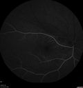
Bilateral Branch Retinal Artery Occlusion - Intraarterial (hollenhorst) plaques visible516 views67 year old with acute visual field loss in each eye. Vision is 20/25 right eye 20/32 left eye. Images show intra-arterial plaques in each eye. The right eye on the optic nerve and the left eye in a branch retinal artery.
|
|
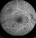
CRVO with Paracentral Acute Middle Maculopath - PAMM516 views56 year old woman with sudden vision loss, hemorrhages in all four quadrants. FA shows good retinal circulation. The OCT shows PAMM lesions in the affected eye. Vision did improve on second visit from 20/160 to 20/60 in about a month
|
|

Subtle Branch Retinal Artery Occlusion516 views68 year old man with shadow in the left eye yesterday. IR and OCT show subtle retinal swelling. FA is normal
|
|

Acute Branch Retinal Artery Occlusion Left Eye515 viewsThis 84 year old woman had an old BRVO in the right eye and saw a shadow for 2 days in the left eye. She has a superotemporal BRAO. The photos show retinal whitening, the OCT shows inner retinal edema and the FA shows poor circulation.
|
|
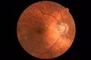
Branch Retinal Artery Occlusion - No Plaque515 views74 year old man with 2 days of vision loss. Vision OS is 20/200 but improved to 20/25 in 1 month. He has dysfibrinogen anemia.
|
|
514 views49-year-old woman has central retinal vein occlusion right eye with fluctuating vision for a few month now with OD 20/100. IOP: OD 20
|
|
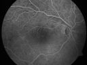
Branch Retinal Arterial Occlusion 20/20 Vision - Symptoms for 1 Day514 views75-year-old man was seen in the office on 4/6/2010. He noticed a sudden vision change in the right eye yesterday morning. 20/20 Vision both Eyes
|
|
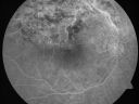
Branch Retinal Vein Occlusion with Cotton Wool Spot 514 views70-year-old man with blurred vision in the right eye for about one or two months. OD is 20/80, OS is 20/25.
|
|

Vasular Anomalies514 viewsThis patient shows vasculopathy seen after an old vein occlusion.
|
|

Acute Branch Retinal Artery Occlusion Left Eye514 viewsThis 84 year old woman had an old BRVO in the right eye and saw a shadow for 2 days in the left eye. She has a superotemporal BRAO. The photos show retinal whitening, the OCT shows inner retinal edema and the FA shows poor circulation.
|
|

Branch Retinal Vein Occlusion514 views84 year old man - 2 months vision loss - VA 20/40 - 20/20 with one injection of Eylea
|
|
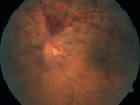
Hemicentral Vein Occlusion Left Eye514 viewsVision improved from 20/100 - 2 to 20/32 with 5 monthly Avastin treatments.
|
|
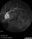
Hemicentral Vein Occlusion Left Eye514 viewsVision improved from 20/100 - 2 to 20/32 with 5 monthly Avastin treatments.
|
|

Hemicentral Vein Occlusion Left Eye514 viewsVision improved from 20/100 - 2 to 20/32 with 5 monthly Avastin treatments.
|
|
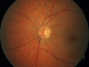
Acute Central Retinal Artery Occlusion514 views1 day of vision loss 79 year old man to HM. NVG developed 2 months later.
|
|
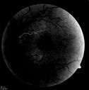
Corresponding FA513 viewsCorresponding FA for Cilioretinal artery occlusion with early venous stasis retinopathy - early
|
|
Embolic CRAO Late513 views
|
|

Normal513 views
|
|
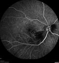
Branch Retinal Artery Occlusion513 viewsAcute Inferotemporal Occlusion Right Eye - FA shows delayed circulation, OCT shows edema of superficial layers of retina
|
|
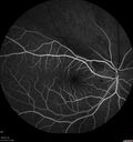
Bilateral Branch Retinal Artery Occlusion - Intraarterial (hollenhorst) plaques visible513 views67 year old with acute visual field loss in each eye. Vision is 20/25 right eye 20/32 left eye. Images show intra-arterial plaques in each eye. The right eye on the optic nerve and the left eye in a branch retinal artery.
|
|
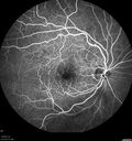
Vascular Remodeling Bilateral BRVO513 views85 year old female 20/25 OU with BRVO 7 years ago in both eyes. She has a change in vision. The FA shows extensive collaterallization and vascular remodeling in each macula.
|
|
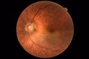
Branch Retinal Artery Occlusion - No Plaque513 views74 year old man with 2 days of vision loss. Vision OS is 20/200 but improved to 20/25 in 1 month. He has dysfibrinogen anemia.
|
|

Subtle Branch Retinal Artery Occlusion513 views68 year old man with shadow in the left eye yesterday. IR and OCT show subtle retinal swelling. FA is normal
|
|
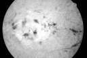
Central Retinal Vein Occlusion - New- and old Macular Scar from AMD512 views87-year-old woman with longstaning macular scars and new vision loss left eye for 2 months. OD: 20/200, OS 20/400.
|
|
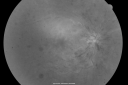
Central Retinal Vein Occlusion Recurrent Edema 6 weeks after Lucentis - Nonperfusion Temporally512 views66-year-old woman has a central retinal vein occlusion in the right eye with macular edema. She had intravitreal Lucentis treatment six weeks ago and she has noticed over the last week or two her vision is declining.
VISUAL ACUITY: OD 20/70, OS 20/30.
|
|
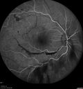
Branch Retinal Vein Occlusion with NVE (Neovascularization Elsewhere)512 views
|
|

Acute Branch Retinal Artery Occlusion Left Eye512 viewsThis 84 year old woman had an old BRVO in the right eye and saw a shadow for 2 days in the left eye. She has a superotemporal BRAO. The photos show retinal whitening, the OCT shows inner retinal edema and the FA shows poor circulation.
|
|
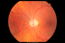
CRVO with Paracentral Acute Middle Maculopath - PAMM512 views56 year old woman with sudden vision loss, hemorrhages in all four quadrants. FA shows good retinal circulation. The OCT shows PAMM lesions in the affected eye. Vision did improve on second visit from 20/160 to 20/60 in about a month
|
|
512 views
|
|

Vascular Remodeling Bilateral BRVO512 views85 year old female 20/25 OU with BRVO 7 years ago in both eyes. She has a change in vision. The FA shows extensive collaterallization and vascular remodeling in each macula.
|
|
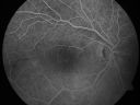
Branch Retinal Arterial Occlusion 20/20 Vision - Symptoms for 1 Day511 views75-year-old man was seen in the office on 4/6/2010. He noticed a sudden vision change in the right eye yesterday morning. 20/20 Vision both Eyes
|
|
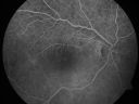
Branch Retinal Arterial Occlusion 20/20 Vision - Symptoms for 1 Day511 views75-year-old man was seen in the office on 4/6/2010. He noticed a sudden vision change in the right eye yesterday morning. 20/20 Vision both Eyes
|
|
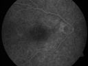
Branch Retinal Arterial Occlusion 20/20 Vision - Symptoms for 1 Day511 views75-year-old man was seen in the office on 4/6/2010. He noticed a sudden vision change in the right eye yesterday morning. 20/20 Vision both Eyes
|
|

Macular Branch Retinal Vein Occlusion and Adult Vitelliform Dystrophy511 views72-year-old woman 2 months post ECCE with poor vision since surgery especially left eye.
VISUAL ACUITY: OD 20/50, OS 20/200
|
|
| 1845 files on 19 page(s) |
 |
|
|
|
|
7 | |
|
|
|
|


