Most viewed - Retina Vascular Occlusions
|
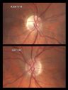
Sectoral Optic Atrophy from an old Branch Retinal Artery Occlusion 825 views74-year-old woman had cataract surgery 3 months ago. Her vision in the right eye has been abnormal since. She notices a shadow in the inferior vision in the right eye. OD is 20/60, OS is 20/50
|
|

Central Retinal Artery Occlusion Left Eye 2 Weeks Old - Still Non-Perfusion824 views85-year-old woman who had sudden severevision loss in the left eye two weeks ago. she can see a little more centrally and much more in the periphery, since about the last four days.
VISUAL ACUITY: OD 20/30, OS: Hand motion
|
|

Central Retinal Vein Occlusion and Laser Induced Chorioretinal Anastomosis824 views84-year-old woman has age-related macular degeneration in both eyes with foveal atrophy. She also has a central retinal vein occlusion
in the left eye for which she had intravitreal Kenalog and finally chorioretinal anastomosis laser a year
ago. VISUAL ACUITY: Vision OD is 20/160, OS is 7/200
|
|

Macular Branch Retinal Vein Occlusion and Adult Vitelliform Dystrophy823 views72-year-old woman 2 months post ECCE with poor vision since surgery especially left eye.
VISUAL ACUITY: OD 20/50, OS 20/200
|
|

brvo cme with small serous macular detachment 820 views70 year old woman with 20/80 vision from a branch retinal vein occlusion and macular edema. She has had laser treatment and intravitreal steroids. Her vision with further laser improved to 20/50.
|
|
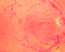
Central Retinal Vein Occlusion and Cystoid Macular Edema - Acute - Vision 20/50 817 views68-year-old man six weeks of vision loss right eye from CRVO and CME, vision right eye 20/50
|
|
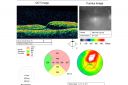
Twig Branch Retinal Vein Occlusion - Recurrent Macular Edema with extended interval Lucentis Therapy after first 6 monthly Injections817 views39-year-old man has a macular branch retinal vein occlusion with macular edema. He had an excellent response to Lucentis for his first 6 treatments. He was last here 6 weeks ago and his vision seems OK to him. OD 20/40.
|
|

Central Retinal Artery Occlusion Left Eye 2 Weeks Old - Still Non-Perfusion816 views85-year-old woman who had sudden severevision loss in the left eye two weeks ago. she can see a little more centrally and much more in the periphery, since about the last four days.
VISUAL ACUITY: OD 20/30, OS: Hand motion
|
|
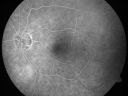
Cilioretinal artery occlusion and Ophthalmic Artery Occlusion with Relative Sparing of the Central Retinal Artery816 views80-year-old woman who says she was looking in the mirror, covered her right eye and noticed she could not see well out of the left eye. That just happened yesterday. Her last eye check was a few years ago. She also had a carotid endarterectomy on the right side in 2006 shortly after coronary bypass surgery. OD is 20/40, OS is 20/200.
|
|

New BRVO Left Eye Old BRVO Right Eye - Left Eye Shows Twig Superior Vein Occlusion with Edema812 views90-year-old man decreased vision maybe one year since cataract surgery he is no longer able to read. OD 20/200, OS 20/200
|
|

Cilioretinal artery occlusion and Ophthalmic Artery Occlusion with Relative Sparing of the Central Retinal Artery808 views80-year-old woman who says she was looking in the mirror, covered her right eye and noticed she could not see well out of the left eye. That just happened yesterday. Her last eye check was a few years ago. She also had a carotid endarterectomy on the right side in 2006 shortly after coronary bypass surgery. OD is 20/40, OS is 20/200.
|
|

Macular Branch Retinal Vein Occlusions (2) Left Eye - No Macular Edema (yet) 806 views77-year-old woman with two macular branch retinal vein occlusions in the left eye OD 20/25
|
|

Hemicentral Retinal Vein Occlusion Vision 20/60 800 views84-year-old man has a hemicentral retinal vein occlusion in the right eye. His vision has declined since then.
VISUAL ACUITY: Vision OD is 20/60.
|
|

New BRVO Left Eye Old BRVO Right Eye - Right Eye Showing Bulls Eye Atrophy from Chronic CME and Non-Perfusion Infero-temporally799 views90-year-old man decreased vision maybe one year since cataract surgery he is no longer able to read. OD 20/200, OS 20/200
|
|

Branch Retinal Vein Occlusion with Macular Edema797 viewsCentral foveal thickness came to normal after laser and intravitreal Kenalog
|
|

Central Retinal Artery Occlusion Left Eye 2 Weeks Old - Still Non-Perfusion797 views85-year-old woman who had sudden severevision loss in the left eye two weeks ago. she can see a little more centrally and much more in the periphery, since about the last four days.
VISUAL ACUITY: OD 20/30, OS: Hand motion
|
|

Central Retinal Artery Occlusion Left Eye 2 Weeks Old - Still Non-Perfusion795 views85-year-old woman who had sudden severevision loss in the left eye two weeks ago. she can see a little more centrally and much more in the periphery, since about the last four days.
VISUAL ACUITY: OD 20/30, OS: Hand motion
|
|

Central Retinal Vein Occlusion Recurrent Edema 6 weeks after Lucentis - Nonperfusion Temporally795 views66-year-old woman has a central retinal vein occlusion in the right eye with macular edema. She had intravitreal Lucentis treatment six weeks ago and she has noticed over the last week or two her vision is declining.
VISUAL ACUITY: OD 20/70, OS 20/30.
|
|
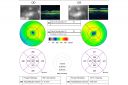
Macular Branch Retinal Vein Occlusions (2) Left Eye - No Macular Edema (yet) 788 views77-year-old woman with two macular branch retinal vein occlusions in the left eye OD 20/25
|
|

Pathology - Central Retinal Artery Occlusion - 6 weeks old - Inner Retinal Atrophy786 viewspatient died about 6 to 8 weeks after the artery occlusion, and he had no history of glaucoma. The inner retina atrophy, which looks very similar to that caused by COAG, is due to recent infarct.
|
|
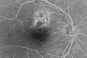
Twig Branch Retinal Vein Occlusion - Recurrent Macular Edema with extended interval Lucentis Therapy after first 6 monthly Injections785 views39-year-old man has a macular branch retinal vein occlusion with macular edema. He had an excellent response to Lucentis for his first 6 treatments. He was last here 6 weeks ago and his vision seems OK to him. OD 20/40.
|
|
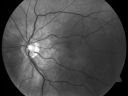
Cilioretinal artery occlusion and Ophthalmic Artery Occlusion with Relative Sparing of the Central Retinal Artery785 views80-year-old woman who says she was looking in the mirror, covered her right eye and noticed she could not see well out of the left eye. That just happened yesterday. Her last eye check was a few years ago. She also had a carotid endarterectomy on the right side in 2006 shortly after coronary bypass surgery. OD is 20/40, OS is 20/200.
|
|

785 views
|
|
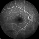
Central Retinal Artery Occlusion with Cilioretinal Sparing - Acute785 views 77-year-old man was doing fine until midnight of last night when he noticed sudden severe vision loss in the left eye. VISUAL ACUITY: OD 20/20, OS 2/200.
|
|
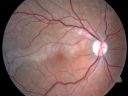
Branch Retinal Arterial Occlusion 20/20 Vision - Symptoms for 1 Day784 views
|
|
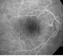
Old BRAO twig with plaque783 views81-year-old woman Her left eye is amblyopic. Her right eye was doing fine until about last Sunday, which is five days ago. She had a change in vision where for five minutes she saw a shadow in her side vision out of the right eye. She describes it as being more up into the left. It lasted for just a brief time and then went away. She has had atrial fibrillation since January of 2008. She is on Coumadin for that. She is diabetic for eleven years. OD is 20/25, OS is 20/60. IOP: OD 15, OS 18.
|
|
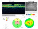
Branch Retinal Arterial Occlusion 20/20 Vision - Symptoms for 1 Day779 views75-year-old man was seen in the office on 4/6/2010. He noticed a sudden vision change in the right eye yesterday morning. 20/20 Vision both Eyes
|
|
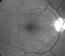
Old BRAO twig with plaque776 views81-year-old woman Her left eye is amblyopic. Her right eye was doing fine until about last Sunday, which is five days ago. She had a change in vision where for five minutes she saw a shadow in her side vision out of the right eye. She describes it as being more up into the left. It lasted for just a brief time and then went away. She has had atrial fibrillation since January of 2008. She is on Coumadin for that. She is diabetic for eleven years. OD is 20/25, OS is 20/60. IOP: OD 15, OS 18.
|
|
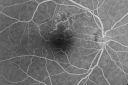
Twig Branch Retinal Vein Occlusion - Recurrent Macular Edema with extended interval Lucentis Therapy after first 6 monthly Injections776 views39-year-old man has a macular branch retinal vein occlusion with macular edema. He had an excellent response to Lucentis for his first 6 treatments. He was last here 6 weeks ago and his vision seems OK to him. OD 20/40.
|
|

Central Retinal Vein Occlusion and Laser Induced Chorioretinal Anastomosis775 views84-year-old woman has age-related macular degeneration in both eyes with foveal atrophy. She also has a central retinal vein occlusion
in the left eye for which she had intravitreal Kenalog and finally chorioretinal anastomosis laser a year
ago. VISUAL ACUITY: Vision OD is 20/160, OS is 7/200
|
|
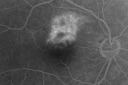
Twig Branch Retinal Vein Occlusion - Recurrent Macular Edema with extended interval Lucentis Therapy after first 6 monthly Injections774 views39-year-old man has a macular branch retinal vein occlusion with macular edema. He had an excellent response to Lucentis for his first 6 treatments. He was last here 6 weeks ago and his vision seems OK to him. OD 20/40.
|
|

branch retinal vein occlusion - fluorescein angiogram771 views70 year old woman with 20/80 vision from a branch retinal vein occlusion and macular edema. She has had laser treatment and intravitreal steroids. Her vision with further laser improved to 20/50.
|
|
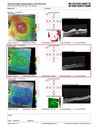
Central Retinal Artery Occlusion - MAP770 viewsProgression from acute to atrophy - decreasing retinal thickenss
|
|

Branch Retinal Vein Occlusion (BRVO) no macular edema - cotton wool spot770 views85 year old man with asymptomatic BRVO - his vision stayed great without needing any treatment.
|
|

CRVO Shunt Vessels Right Eye - Optociliary Shunt Vessels770 viewsPrior CRVO right eye treated with injections
|
|

CRVO with Paracentral Acute Middle Maculopath - PAMM769 views56 year old woman with sudden vision loss, hemorrhages in all four quadrants. FA shows good retinal circulation. The OCT shows PAMM lesions in the affected eye. Vision did improve on second visit from 20/160 to 20/60 in about a month
|
|

New BRVO Left Eye Old BRVO Right Eye - Right Eye Showing Bulls Eye Atrophy from Chronic CME and Non-Perfusion Infero-temporally768 views90-year-old man decreased vision maybe one year since cataract surgery he is no longer able to read. OD 20/200, OS 20/200
|
|

Central Retinal Vein Occlusion and Cystoid Macular Edema - Acute - Vision 20/50 766 views68-year-old man six weeks of vision loss right eye from CRVO and CME, vision right eye 20/50
|
|
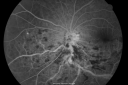
Hemi Central Retinal Vein Occlusion 35 year old non-compliant man764 views35-year-old man hazy vision in his left eye for about the last three days. OD 20/20, OS 20/30
|
|
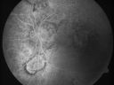
Central Retinal Vein Occlusion and Laser Induced Chorioretinal Anastomosis764 views84-year-old woman has age-related macular degeneration in both eyes with foveal atrophy. She also has a central retinal vein occlusion
in the left eye for which she had intravitreal Kenalog and finally chorioretinal anastomosis laser a year
ago. VISUAL ACUITY: Vision OD is 20/160, OS is 7/200
|
|
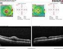
Branch Retinal Vein Occlusion with worse macular edema from hypertension - SD-OCT shows Edema Right Eye762 views
|
|
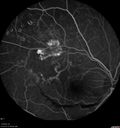
Branch Retinal Vein Occlusion with NVE (Neovascularization Elsewhere)761 viewsLate Leakage on FA - NV is adjacent to non-perfusion
|
|

Hemi Central Retinal Vein Occlusion 35 year old non-compliant man759 views35-year-old man hazy vision in his left eye for about the last three days. OD 20/20, OS 20/30
|
|

759 views
|
|

BRVO with collaterals and cystoid macular edema758 views66 Year Old Man with has had a branch retinal vein occlusion in the right eye with vision loss for the last few months. OD is 20/40, OS is 20/20. IOP: OD 18, OS 19. There is a superotemporal macular branch retinal vein occlusion with edema, hemorrhage, and exudate.
OCT SCAN: The right eye shows edema with a central foveal thickness of 354 microns FA: shows remodeling of the superotemporal vasculature. There are already venous-venous collaterals, suggesting the vein occlusion may be a little more than a few months old. There are multiple microaneurysms superotemporal to the fovea, and there is leakage in the late frames involving the fovea.
|
|

CRVO with Paracentral Acute Middle Maculopath - PAMM755 views56 year old woman with sudden vision loss, hemorrhages in all four quadrants. FA shows good retinal circulation. The OCT shows PAMM lesions in the affected eye. Vision did improve on second visit from 20/160 to 20/60 in about a month
|
|

Acute Branch Retinal Artery Occlusion Left Eye754 viewsThis 84 year old woman had an old BRVO in the right eye and saw a shadow for 2 days in the left eye. She has a superotemporal BRAO. The photos show retinal whitening, the OCT shows inner retinal edema and the FA shows poor circulation.
|
|

New BRVO Left Eye Old BRVO Right Eye - Left Eye Shows Twig Superior Vein Occlusion with Edema748 views90-year-old man decreased vision maybe one year since cataract surgery he is no longer able to read. OD 20/200, OS 20/200
|
|
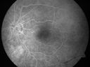
Cilioretinal artery occlusion and Ophthalmic Artery Occlusion with Relative Sparing of the Central Retinal Artery748 views80-year-old woman who says she was looking in the mirror, covered her right eye and noticed she could not see well out of the left eye. That just happened yesterday. Her last eye check was a few years ago. She also had a carotid endarterectomy on the right side in 2006 shortly after coronary bypass surgery. OD is 20/40, OS is 20/200.
|
|
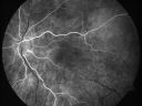
Cilioretinal artery occlusion and Ophthalmic Artery Occlusion with Relative Sparing of the Central Retinal Artery747 views80-year-old woman who says she was looking in the mirror, covered her right eye and noticed she could not see well out of the left eye. That just happened yesterday. Her last eye check was a few years ago. She also had a carotid endarterectomy on the right side in 2006 shortly after coronary bypass surgery. OD is 20/40, OS is 20/200.
|
|

Branch Retinal Vein Occlusion with NVE (Neovascularization Elsewhere)747 views
|
|
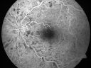
Non-Ischemic Central Retinal Vein Occlusion in Young Man (30 years) - Many Cotton Wool Spots746 views30-year-old man with a history of a central retinal vein occlusion for which he was first examined on 2 months ago and his vision then was 4/200. He responded nicely to two Avastin treatments. OD is 20/20, OS is 20/60
|
|

Twig Old Macular Branch Retinal Vein Occlusion - Venous Venous Collaterals744 views88 year old man with 20/25 vision in his left and only eye. There is vascular remodeling consistent with an old twig BRVO.
|
|

Sectoral Optic Atrophy from an old Branch Retinal Artery Occlusion 743 views74-year-old woman had cataract surgery 3 months ago. Her vision in the right eye has been abnormal since. She notices a shadow in the inferior vision in the right eye. OD is 20/60, OS is 20/50
|
|
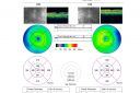
Cilioretinal artery occlusion and Ophthalmic Artery Occlusion with Relative Sparing of the Central Retinal Artery743 views80-year-old woman who says she was looking in the mirror, covered her right eye and noticed she could not see well out of the left eye. That just happened yesterday. Her last eye check was a few years ago. She also had a carotid endarterectomy on the right side in 2006 shortly after coronary bypass surgery. OD is 20/40, OS is 20/200.
|
|

Branch Retinal Vein Occlusion with NVE (Neovascularization Elsewhere)742 viewsPre and post laser therapy - NV has partially regressed
|
|

Central Retinal Artery Occlusion Left Eye 2 Weeks Old - Still Non-Perfusion741 views85-year-old woman who had sudden severevision loss in the left eye two weeks ago. she can see a little more centrally and much more in the periphery, since about the last four days.
VISUAL ACUITY: OD 20/30, OS: Hand motion
|
|

CRVO (retinal thrombophlebitis)741 views42 yr old female
20/25 VA
Labs normal
|
|

Central Retinal Artery Occlusion less than 24 hours - 69 year old man VA light perception739 views
|
|

Macular Branch Retinal Vein Occlusions (2) Left Eye - No Macular Edema (yet) 739 views77-year-old woman with two macular branch retinal vein occlusions in the left eye OD 20/25
|
|

Non-Ischemic Central Retinal Vein Occlusion in Young Man (30 years) - Many Cotton Wool Spots738 views30-year-old man with a history of a central retinal vein occlusion for which he was first examined on 2 months ago and his vision then was 4/200. He responded nicely to two Avastin treatments. OD is 20/20, OS is 20/60
|
|
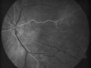
Cilioretinal artery occlusion and Ophthalmic Artery Occlusion with Relative Sparing of the Central Retinal Artery737 views80-year-old woman who says she was looking in the mirror, covered her right eye and noticed she could not see well out of the left eye. That just happened yesterday. Her last eye check was a few years ago. She also had a carotid endarterectomy on the right side in 2006 shortly after coronary bypass surgery. OD is 20/40, OS is 20/200.
|
|

Montage of Hemi-Central Retinal Artery Occlusion737 views
|
|
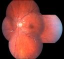
Non-Ischemic Central Retinal Vein Occlusion in Young Man (30 years) - Many Cotton Wool Spots736 views30-year-old man with a history of a central retinal vein occlusion for which he was first examined on 2 months ago and his vision then was 4/200. He responded nicely to two Avastin treatments. OD is 20/20, OS is 20/60
|
|

Sclerosed vein left eye736 views
|
|

Branch Retinal Vein Occlusion (BRVO) no macular edema - cotton wool spot735 views85 year old man with asymptomatic BRVO - his vision stayed great without needing any treatment.
|
|
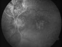
Central Retinal Vein Occlusion and Laser Induced Chorioretinal Anastomosis728 views84-year-old woman has age-related macular degeneration in both eyes with foveal atrophy. She also has a central retinal vein occlusion
in the left eye for which she had intravitreal Kenalog and finally chorioretinal anastomosis laser a year
ago. VISUAL ACUITY: Vision OD is 20/160, OS is 7/200
|
|

BRVO with collaterals and cystoid macular edema727 views66 Year Old Man with has had a branch retinal vein occlusion in the right eye with vision loss for the last few months. OD is 20/40, OS is 20/20. IOP: OD 18, OS 19. There is a superotemporal macular branch retinal vein occlusion with edema, hemorrhage, and exudate.
OCT SCAN: The right eye shows edema with a central foveal thickness of 354 microns FA: shows remodeling of the superotemporal vasculature. There are already venous-venous collaterals, suggesting the vein occlusion may be a little more than a few months old. There are multiple microaneurysms superotemporal to the fovea, and there is leakage in the late frames involving the fovea.
|
|
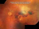
Central Retinal Vein Occlusion and Tractional Retinal Detachment for 3 Years (Patient is Diabetic)727 views67-year-old woman has severe vision loss in the left eye for 3 years OD 20/32, OS 3/200
|
|
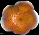
Stasis Retinopathy, Central Retinal Vein Occlusion726 viewsYoung male who complains of decreased vision in the left eye. VA is 20/20 in the left eye. Fundus photo shows mild central retinal vein occlusion in the left eye.
|
|

Central Retinal Vein Occlusion and Laser Induced Chorioretinal Anastomosis725 views84-year-old woman has age-related macular degeneration in both eyes with foveal atrophy. She also has a central retinal vein occlusion
in the left eye for which she had intravitreal Kenalog and finally chorioretinal anastomosis laser a year
ago. VISUAL ACUITY: Vision OD is 20/160, OS is 7/200
|
|

Hemi Central Retinal Vein Occlusion 35 year old non-compliant man724 views35-year-old man hazy vision in his left eye for about the last three days. OD 20/20, OS 20/30
|
|
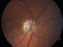
Sectoral Optic Atrophy from an old Branch Retinal Artery Occlusion 724 views74-year-old woman had cataract surgery 3 months ago. Her vision in the right eye has been abnormal since. She notices a shadow in the inferior vision in the right eye. OD is 20/60, OS is 20/50
|
|
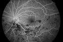
Hemi Central Retinal Vein Occlusion 35 year old non-compliant man723 views35-year-old man hazy vision in his left eye for about the last three days. OD 20/20, OS 20/30
|
|
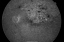
branch retinal vein occlusion - fluorescein angiogram720 views70 year old woman with 20/80 vision from a branch retinal vein occlusion and macular edema. She has had laser treatment and intravitreal steroids. Her vision with further laser improved to 20/50.
|
|

Sectoral Optic Atrophy from an old Branch Retinal Artery Occlusion 720 views74-year-old woman had cataract surgery 3 months ago. Her vision in the right eye has been abnormal since. She notices a shadow in the inferior vision in the right eye. OD is 20/60, OS is 20/50
|
|

Branch Retinal Vein Occlusion with NVE (Neovascularization Elsewhere)720 views
|
|

Hemicentral Retinal Vein Occlusion Vision 20/60 717 views84-year-old man has a hemicentral retinal vein occlusion in the right eye. His vision has declined since then.
VISUAL ACUITY: Vision OD is 20/60.
|
|

Central Retinal Vein Occlusion and Cystoid Macular Edema - Acute - Vision 20/50 716 views68-year-old man six weeks of vision loss right eye from CRVO and CME, vision right eye 20/50
|
|

Branch Retinal Vein Occlusion with NVE (Neovascularization Elsewhere)712 views
|
|
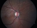
Branch Retinal Arterial Occlusion 20/20 Vision - Symptoms for 1 Day711 views
|
|
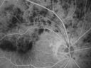
Hemicentral Retinal Vein Occlusion Vision 20/60 711 views84-year-old man has a hemicentral retinal vein occlusion in the right eye. His vision has declined since then.
VISUAL ACUITY: Vision OD is 20/60.
|
|
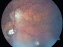
Central Retinal Vein Occlusion and Laser Induced Chorioretinal Anastomosis711 views84-year-old woman has age-related macular degeneration in both eyes with foveal atrophy. She also has a central retinal vein occlusion
in the left eye for which she had intravitreal Kenalog and finally chorioretinal anastomosis laser a year
ago. VISUAL ACUITY: Vision OD is 20/160, OS is 7/200
|
|

Twig Branch Retinal Vein Occlusion - Recurrent Macular Edema with extended interval Lucentis Therapy after first 6 monthly Injections710 views39-year-old man has a macular branch retinal vein occlusion with macular edema. He had an excellent response to Lucentis for his first 6 treatments. He was last here 6 weeks ago and his vision seems OK to him. OD 20/40.
|
|
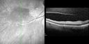
BRAO and PAMM710 views
|
|

Non-Ischemic Central Retinal Vein Occlusion in Young Man (30 years) - Many Cotton Wool Spots709 views30-year-old man with a history of a central retinal vein occlusion for which he was first examined on 2 months ago and his vision then was 4/200. He responded nicely to two Avastin treatments. OD is 20/20, OS is 20/60
|
|

Central Retinal Artery Occlusion Left Eye 2 Weeks Old - Still Non-Perfusion708 views85-year-old woman who had sudden severevision loss in the left eye two weeks ago. she can see a little more centrally and much more in the periphery, since about the last four days.
VISUAL ACUITY: OD 20/30, OS: Hand motion
|
|

Cilioretinal artery occlusion708 viewsCilioretinal artery occlusion with early venous stasis retinopathy
|
|

Central Retinal Artery Occlusion Left Eye 2 Weeks Old - Still Non-Perfusion707 views85-year-old woman who had sudden severevision loss in the left eye two weeks ago. she can see a little more centrally and much more in the periphery, since about the last four days.
VISUAL ACUITY: OD 20/30, OS: Hand motion
|
|
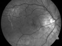
Cilioretinal artery occlusion and Ophthalmic Artery Occlusion with Relative Sparing of the Central Retinal Artery707 views80-year-old woman who says she was looking in the mirror, covered her right eye and noticed she could not see well out of the left eye. That just happened yesterday. Her last eye check was a few years ago. She also had a carotid endarterectomy on the right side in 2006 shortly after coronary bypass surgery. OD is 20/40, OS is 20/200.
|
|

Macular Branch Retinal Vein Occlusions (2) Left Eye - No Macular Edema (yet) 706 views77-year-old woman with two macular branch retinal vein occlusions in the left eye OD 20/25
|
|

Montage of Hemi-Central Retinal Artery Occlusion706 views
|
|

Macular Branch Retinal Vein Occlusions (2) Left Eye - No Macular Edema (yet) 705 views77-year-old woman with two macular branch retinal vein occlusions in the left eye OD 20/25
|
|

branch retinal vein occlusion - fluorescein angiogram702 views70 year old woman with 20/80 vision from a branch retinal vein occlusion and macular edema. She has had laser treatment and intravitreal steroids. Her vision with further laser improved to 20/50.
|
|

Branch Retinal Vein Occlusion with Macular Edema702 viewsCentral foveal thickness came to normal after laser and intravitreal Kenalog
|
|
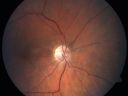
Sectoral Optic Atrophy from an old Branch Retinal Artery Occlusion 702 views74-year-old woman had cataract surgery 3 months ago. Her vision in the right eye has been abnormal since. She notices a shadow in the inferior vision in the right eye. OD is 20/60, OS is 20/50
|
|
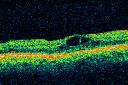
Twig Branch Retinal Vein Occlusion - Recurrent Macular Edema with extended interval Lucentis Therapy after first 6 monthly Injections702 views39-year-old man has a macular branch retinal vein occlusion with macular edema. He had an excellent response to Lucentis for his first 6 treatments. He was last here 6 weeks ago and his vision seems OK to him. OD 20/40.
|
|
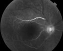
702 views60 year old male with LP vision due to extensive blood flow loss.
|
|

Central Retinal Artery Occlusion with Cilioretinal Sparing - Acute701 views 77-year-old man was doing fine until midnight of last night when he noticed sudden severe vision loss in the left eye. VISUAL ACUITY: OD 20/20, OS 2/200.
|
|
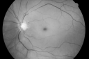
Central Retinal Artery Occlusion less than 24 hours - 69 year old man VA light perception700 views
|
|
| 1845 files on 19 page(s) |
2 |  |
|
|
|
|
|
|
|
|
|


