Most viewed - Retina Vascular Occlusions
|
Central Retinal Artery Occlusion then Neovascular Glaucoma437 views81 year old male with sudden vision loss. 2 months later developed NVG
|
|
Central Retinal Vein Occlusion - No macular edema - Pre-foveal opercula437 views62 year old female with 20/20 vision. She had been seeing a spot out of the left eye for 4 years and then noticed splotches in her vision. She never devleoped edema or NV
|
|

Central Retinal Vein Occlusion with Macular Edema437 views74 year old female The vision in the left eye has gotten worse over the last 3 weeks.
VA OD: Dcc20/25
VA OS: DccCF 3ft
IOP: TP: OD:16 OS:14
Vision improved to 20/40 in 3 months with monthly Aflibercept
|
|
437 views
|
|
CRVO 25 year old man - Heterozygous Factor V Leiden436 views20/100 initial vision. Improved to 20/30 with Lucentis which continued for 3 years, treat and extend, and stopped.
|
|
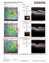
Venous Macroaneurysm 6 year old CRVO - treated with focal laser436 views93 year old female with CRVO in the right eye 6 years ago. The visual potential is 20/100 and has recently subjectively and objectively declined. VA is 20/200 and she has new onset macular edema from a venous macroaneurysm that formed at a collateral vessel. This responded nicely to a single laser spot.
|
|
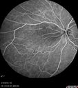
Cilioretinal Artery Occlusion - Nasal (not macular)436 views77 year old female There is a shadow in the top half of her temporal vision in the left eye that came on suddenly yesterday morning. It has not changed since then and the right eye is OK. She has had sinus headaches. She had no focal neurological changes. Â
VA OD: Dcc20/32
VA OS: Dcc20/16-1
IOP: TP: OD:18 OS:19Â
|
|
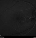
Cilioretinal Artery Occlusion - Good vision436 views67 year old female - 5 days ago she was getting ready for bed and closed one eye (she had worked for an optometrist for 20 years) and she noticed a spot in the vision in the right eye and came in. She has no focal neurological problems. She once in a while gets some weakness. She has a neurologist and there are some brain lesions. Father had stroke at age 52.
VA OD: Dcc20/20 NccJ1
VA OS: Dcc20/16-2 NccJ1-2
|
|

Branch retinal vein occlusion with macular edema436 views71 year old man Starting about 2 weeks ago the left eye was blurred. The right eye is seeing well.
VA OD: sc20/25+2 NccJ1+
VA OS: sc20/63+2 PH20/50-2 Ncc20/400
IOP: TP: OD:19 OS:16
|
|

Elevated Episcleral Venous Pressure and Retinal Hemorrhages in both eyes436 views78 year old female with chronic red eye. The eyes are red all the time now. She is on Xeralto because of a history of blood clots in her lungs (2005 about). She has not had problems since. She has had floaters for many years. Ever since her eyes got funny she sees more floaters in both eyes. She had a brain scan over 6 months ago which was OK. She also had double vision when this first started which went away. She could not drive for a few months.
VA OD: Dcc20/32-1
VA OS: Dcc20/125
IOP: TP: OD:17 OS:16
Neuroimaging did not show a CC fistula
|
|
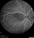
Cilioretinal Artery Occlusion - Nasal (not macular)435 views77 year old female There is a shadow in the top half of her temporal vision in the left eye that came on suddenly yesterday morning. It has not changed since then and the right eye is OK. She has had sinus headaches. She had no focal neurological changes. Â
VA OD: Dcc20/32
VA OS: Dcc20/16-1
IOP: TP: OD:18 OS:19Â
|
|
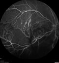
Branch Retinal Vein Occlusion - Both Eyes - Edema OD and NVE OS - Severe Non-perfusion Both Eyes435 views83 year old man with vision loss for 3 months not sure which eye. VA 20/100 OD and 20/50 OS. Tried avastin once without much improvement in the right eye and then lost to follow-up. FA in the left eye shows leaking NVE
|
|

CVO434 viewslate FA (5min) optos photo of a 27 year old male with crvo os VA 20/20-
|
|
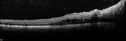
Mtehylenetetrahydrofolate reductase c677T homozygous CRVO pregnant woman (MTHFR)434 viewsVision improved from 20/50 to 20/16 in one month following ozurdex injections. 38 year old woman with only one eye. The left eye was removed as a child for retinoblastoma.
|
|

Mtehylenetetrahydrofolate reductase c677T homozygous CRVO pregnant woman (MTHFR)434 viewsVision improved from 20/50 to 20/16 in one month following ozurdex injections. 38 year old woman with only one eye. The left eye was removed as a child for retinoblastoma.
|
|

CRVO433 views58 year old male with present CRVO OU. Visual Acuity 20/30 OD 20/200 OS. Pt. recently underwent surgery for cancer of the bladder and has other blood and hemoglobin issues. Pt sent for further blood work and see a neurologist.
|
|
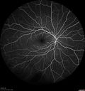
CRVO 25 year old man - Heterozygous Factor V Leiden432 views20/100 initial vision. Improved to 20/30 with Lucentis which continued for 3 years, treat and extend, and stopped.
|
|
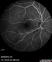
Central Retinal Vein Occlusion - No macular edema - Pre-foveal opercula432 views62 year old female with 20/20 vision. She had been seeing a spot out of the left eye for 4 years and then noticed splotches in her vision. She never devleoped edema or NV
|
|
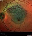
Branch Retinal Vein Occlusion with Edema - Failed avastin (bevacizumab) and did well with Eylea (Aflibercept)432 views67 year old female The vision in the left eye was good until about 4 months ago. Prior to that it was OK. The vision in the right eye is fine. She is not sure if she has had strokes from studies taken in December. She was, at that time, sent to the ER for high blood pressure. Â
VA OD: Dcc20/40 OS: 20/100
Four treatments with Avastin she was still 20/100 with edema. With two Eylea treatments she improved to 20/32 and has remained dry and with good vision
|
|
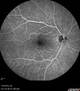
Cilioretinal Artery Occlusion - Nasal (not macular)432 views77 year old female There is a shadow in the top half of her temporal vision in the left eye that came on suddenly yesterday morning. It has not changed since then and the right eye is OK. She has had sinus headaches. She had no focal neurological changes. Â
VA OD: Dcc20/32
VA OS: Dcc20/16-1
IOP: TP: OD:18 OS:19Â
|
|
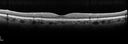
Cardiac Catheterization induced Cilioretinal Artery Occlusion - Paracentral Acute Middle Maculopathy431 views69 year old female with sudden vision loss on completing cardiac catheterization. The SD OCT shows a PAMM lesion and the lesion looks gray on color imaging. There is delayed filling of the cilioretinal artery.
|
|
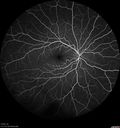
CRVO 25 year old man - Heterozygous Factor V Leiden431 views20/100 initial vision. Improved to 20/30 with Lucentis which continued for 3 years, treat and extend, and stopped.
|
|
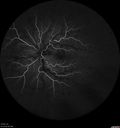
CRVO 25 year old man - Heterozygous Factor V Leiden431 views20/100 initial vision. Improved to 20/30 with Lucentis which continued for 3 years, treat and extend, and stopped.
|
|

Central Retinal Vein Occlusion with Macular Edema431 views74 year old female The vision in the left eye has gotten worse over the last 3 weeks.
VA OD: Dcc20/25
VA OS: DccCF 3ft
IOP: TP: OD:16 OS:14
Vision improved to 20/40 in 3 months with monthly Aflibercept
|
|
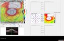
Non-ischemic Central Retinal Vein Occlusion with Macular Edema431 views62 year old man The vision in the right eye started to decline about 10 days ago. Prior to that the two eyes were about the same.
VA OD: Dcc20/125 PH20/50-2
VA OS: Dcc20/20
IOP: TP: OD:21 OS:21
|
|

Branch Retinal Vein Occlusion - Both Eyes - Edema OD and NVE OS - Severe Non-perfusion Both Eyes431 views83 year old man with vision loss for 3 months not sure which eye. VA 20/100 OD and 20/50 OS. Tried avastin once without much improvement in the right eye and then lost to follow-up. FA in the left eye shows leaking NVE
|
|

Elevated Episcleral Venous Pressure and Retinal Hemorrhages in both eyes431 views78 year old female with chronic red eye. The eyes are red all the time now. She is on Xeralto because of a history of blood clots in her lungs (2005 about). She has not had problems since. She has had floaters for many years. Ever since her eyes got funny she sees more floaters in both eyes. She had a brain scan over 6 months ago which was OK. She also had double vision when this first started which went away. She could not drive for a few months.
VA OD: Dcc20/32-1
VA OS: Dcc20/125
IOP: TP: OD:17 OS:16
Neuroimaging did not show a CC fistula
|
|
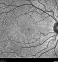
CRVO 25 year old man - Heterozygous Factor V Leiden430 views20/100 initial vision. Improved to 20/30 with Lucentis which continued for 3 years, treat and extend, and stopped.
|
|
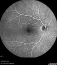
Cilioretinal Artery Occlusion - Nasal (not macular)430 views77 year old female There is a shadow in the top half of her temporal vision in the left eye that came on suddenly yesterday morning. It has not changed since then and the right eye is OK. She has had sinus headaches. She had no focal neurological changes. Â
VA OD: Dcc20/32
VA OS: Dcc20/16-1
IOP: TP: OD:18 OS:19Â
|
|
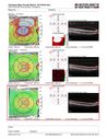
Non-ischemic Central Retinal Vein Occlusion with Macular Edema430 views62 year old man The vision in the right eye started to decline about 10 days ago. Prior to that the two eyes were about the same.
VA OD: Dcc20/125 PH20/50-2
VA OS: Dcc20/20
IOP: TP: OD:21 OS:21
|
|
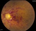
CRVO, Left Eye429 viewsPatient comes in with central retinal vein occlusion in the left eye. VA was count fingers in the left eye. Fundus photo shows scattered hemorrhages with cotton wool spots. Patient was treated with Avastin in the left eye.
|
|

Ischemic CRVO OD and Arterial Macroaneurysm OS429 views83 year old female post PPV, Laser, Ahmed valve for CRVO with vitreous heme and NVG in the right eye. VA was 20/160 3 months following vitrectomy. Left eye has MA which was not treated and involuted post-heme
|
|
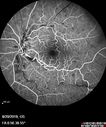
Central Retinal Vein Occlusion - No macular edema - Pre-foveal opercula429 views62 year old female with 20/20 vision. She had been seeing a spot out of the left eye for 4 years and then noticed splotches in her vision. She never devleoped edema or NV
|
|

Chronic BRAO with Hemorrhages - Sclerosed Vessel429 views95 year old female with blurred vision in both eyes in for a checkup. History of perpapillary CNVM in the left eye 2 years prior not treated. History of stroke with multiple emboli 1 year ago.
Vision 20/32 OD, 20/40 OS
|
|

Subtle Branch Retinal Artery Occlusion429 views68 year old man with shadow in the left eye yesterday. IR and OCT show subtle retinal swelling. FA is normal
|
|
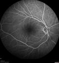
Acute Branch Retinal Vein Occlusion - 20/200429 views66 year old female with 3 weeks of vision loss in the left eye. VA 20/20 OD 20/200 OS
|
|

Branch retinal vein occlusion with macular edema429 views71 year old man Starting about 2 weeks ago the left eye was blurred. The right eye is seeing well.
VA OD: sc20/25+2 NccJ1+
VA OS: sc20/63+2 PH20/50-2 Ncc20/400
IOP: TP: OD:19 OS:16
|
|

Central Retinal Artery Occlusion then Neovascular Glaucoma428 views81 year old male with sudden vision loss. 2 months later developed NVG
|
|

Central Retinal Vein Occlusion - No macular edema - Pre-foveal opercula428 views62 year old female with 20/20 vision. She had been seeing a spot out of the left eye for 4 years and then noticed splotches in her vision. She never devleoped edema or NV
|
|
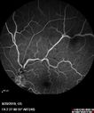
Central Retinal Vein Occlusion - No macular edema - Pre-foveal opercula428 views62 year old female with 20/20 vision. She had been seeing a spot out of the left eye for 4 years and then noticed splotches in her vision. She never devleoped edema or NV
|
|
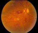
BRVO Inferiorly with Retinal Hemorrhages in the Left Eye427 viewsPatient comes in with complaint of "spot in vision", sort of a cloudy haze in the left eye. Patient's VA was 20/100-1 with no improvement with pinhole in the left eye. Fundus photos show multiple retinal hemorrhages scattered with a branch retinal vein occlusion inferiorily. Patient is also diabetic.
|
|
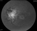
Central Retinal Vein Occlusion with Macular Edema427 viewsPatient notices decreased vision in the left eye. VA is 20/60, left eye. Fundus and Fluorescence Angiogram shows CRVO in the left eye with scattered hemorrhages throughout the retina.
|
|
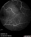
Central Retinal Vein Occlusion - No macular edema - Pre-foveal opercula427 views62 year old female with 20/20 vision. She had been seeing a spot out of the left eye for 4 years and then noticed splotches in her vision. She never devleoped edema or NV
|
|
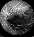
Acute Branch Retinal Vein Occlusion - 20/200427 views66 year old female with 3 weeks of vision loss in the left eye. VA 20/20 OD 20/200 OS
|
|
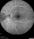
Cilioretinal Artery Occlusion - Nasal (not macular)427 views77 year old female There is a shadow in the top half of her temporal vision in the left eye that came on suddenly yesterday morning. It has not changed since then and the right eye is OK. She has had sinus headaches. She had no focal neurological changes. Â
VA OD: Dcc20/32
VA OS: Dcc20/16-1
IOP: TP: OD:18 OS:19Â
|
|
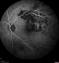
Branch retinal vein occlusion with macular edema427 views71 year old man Starting about 2 weeks ago the left eye was blurred. The right eye is seeing well.
VA OD: sc20/25+2 NccJ1+
VA OS: sc20/63+2 PH20/50-2 Ncc20/400
IOP: TP: OD:19 OS:16
|
|
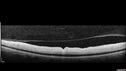
Central Retinal Artery Occlusion427 views80 Year old man Sudden severe vision loss about 2 weeks ago. Today he can see light for the first time. The right eye is OK. His neuroimaging showed a brain stroke along with the eye stroke. He was stepping in the shower and the vision was suddenly worse. He went, later, to the ER and had a full evaluation. VA 20/25 right eye, LP left eye
|
|

Cardiac Catheterization induced Cilioretinal Artery Occlusion - Paracentral Acute Middle Maculopathy426 views69 year old female with sudden vision loss on completing cardiac catheterization. The SD OCT shows a PAMM lesion and the lesion looks gray on color imaging. There is delayed filling of the cilioretinal artery.
|
|
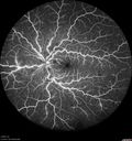
CRVO 25 year old man - Heterozygous Factor V Leiden426 views20/100 initial vision. Improved to 20/30 with Lucentis which continued for 3 years, treat and extend, and stopped.
|
|
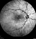
426 views
|
|

Branch Retinal Artery Occlusion - No Plaque425 views74 year old man with 2 days of vision loss. Vision OS is 20/200 but improved to 20/25 in 1 month. He has dysfibrinogen anemia.
|
|
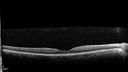
Acute Branch Retinal Vein Occlusion - 20/200425 views66 year old female with 3 weeks of vision loss in the left eye. VA 20/20 OD 20/200 OS
|
|
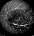
Acute Branch Retinal Vein Occlusion - 20/200425 views66 year old female with 3 weeks of vision loss in the left eye. VA 20/20 OD 20/200 OS
|
|

Cilioretinal Artery Occlusion - Nasal (not macular)425 views77 year old female There is a shadow in the top half of her temporal vision in the left eye that came on suddenly yesterday morning. It has not changed since then and the right eye is OK. She has had sinus headaches. She had no focal neurological changes. Â
VA OD: Dcc20/32
VA OS: Dcc20/16-1
IOP: TP: OD:18 OS:19Â
|
|
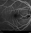
Cilioretinal Artery Occlusion - Good vision425 views67 year old female - 5 days ago she was getting ready for bed and closed one eye (she had worked for an optometrist for 20 years) and she noticed a spot in the vision in the right eye and came in. She has no focal neurological problems. She once in a while gets some weakness. She has a neurologist and there are some brain lesions. Father had stroke at age 52.
VA OD: Dcc20/20 NccJ1
VA OS: Dcc20/16-2 NccJ1-2
|
|

Branch Retinal Vein Occlusion - Both Eyes - Edema OD and NVE OS - Severe Non-perfusion Both Eyes425 views83 year old man with vision loss for 3 months not sure which eye. VA 20/100 OD and 20/50 OS. Tried avastin once without much improvement in the right eye and then lost to follow-up. FA in the left eye shows leaking NVE
|
|

Branch Retinal Vein Occlusion with Neovascularization Elsewhere and Vitreous Hemorrhage425 views57 year old man New floaters in the left eye about a month ago. Now they are getting better. PMHx A-Fib on Pradaxa
VA 20/32 OD, 20/40 OS
1+ NS OU
BP 137/77, 200LBs, 5’8â€
PRP laser was done
|
|

Combined Artery and Vein Occlusion424 viewsPatient comes in with decreased vision in both eyes. VA 20/200-OD, 20/60-OS. Fundus exam shows great amount of macular edema due to artery and vein occlusions. There is some neovascularization on the optic nerve in the right eye. Patient was treated with Eylea Injection in the left eye today and will return for Eylea Injection in the right eye.
|
|
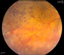
Central Retinal Vein Occlusion with Macular Edema and Retinal Hemorrhages 424 viewsFemale patient of 86-years of age presents a central retinal vein occlusion with macular edema in the right eye. Patient's VA was 20/70 in the right eye. Proceeded with laser treatment to the right eye.
|
|

Cilioretinal Artery Occlusion - Nasal (not macular)424 views77 year old female There is a shadow in the top half of her temporal vision in the left eye that came on suddenly yesterday morning. It has not changed since then and the right eye is OK. She has had sinus headaches. She had no focal neurological changes. Â
VA OD: Dcc20/32
VA OS: Dcc20/16-1
IOP: TP: OD:18 OS:19Â
|
|
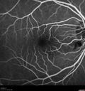
Cilioretinal Artery Occlusion - Good vision424 views67 year old female - 5 days ago she was getting ready for bed and closed one eye (she had worked for an optometrist for 20 years) and she noticed a spot in the vision in the right eye and came in. She has no focal neurological problems. She once in a while gets some weakness. She has a neurologist and there are some brain lesions. Father had stroke at age 52.
VA OD: Dcc20/20 NccJ1
VA OS: Dcc20/16-2 NccJ1-2
|
|
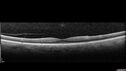
Central Retinal Artery Occlusion424 views80 Year old man Sudden severe vision loss about 2 weeks ago. Today he can see light for the first time. The right eye is OK. His neuroimaging showed a brain stroke along with the eye stroke. He was stepping in the shower and the vision was suddenly worse. He went, later, to the ER and had a full evaluation. VA 20/25 right eye, LP left eye
|
|

23.7 seconds423 viewsCorresponding FA for Cilioretinal artery occlusion with early venous stasis retinopathy
|
|
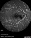
Cardiac Catheterization induced Cilioretinal Artery Occlusion - Paracentral Acute Middle Maculopathy423 views69 year old female with sudden vision loss on completing cardiac catheterization. The SD OCT shows a PAMM lesion and the lesion looks gray on color imaging. There is delayed filling of the cilioretinal artery.
|
|
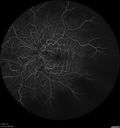
CRVO 25 year old man - Heterozygous Factor V Leiden423 views20/100 initial vision. Improved to 20/30 with Lucentis which continued for 3 years, treat and extend, and stopped.
|
|
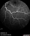
Central Retinal Vein Occlusion - No macular edema - Pre-foveal opercula423 views62 year old female with 20/20 vision. She had been seeing a spot out of the left eye for 4 years and then noticed splotches in her vision. She never devleoped edema or NV
|
|

Branch retinal vein occlusion with macular edema423 views71 year old man Starting about 2 weeks ago the left eye was blurred. The right eye is seeing well.
VA OD: sc20/25+2 NccJ1+
VA OS: sc20/63+2 PH20/50-2 Ncc20/400
IOP: TP: OD:19 OS:16
|
|
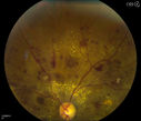
Combined Artery and Vein Occlusion422 viewsPatient comes in with decreased vision in both eyes. VA 20/200-OD, 20/60-OS. Fundus exam shows great amount of macular edema due to artery and vein occlusions. There is some neovascularization on the optic nerve in the right eye. Patient was treated with Eylea Injection in the left eye today and will return for Eylea Injection in the right eye.
|
|
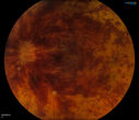
Retinal Vein Occlusion422 viewsPatient comes in with loss of vision, left eye. VA is count fingers in the left eye. Fundus photos show large vein occlusion with macular edema in the left eye. Patient proceeded with Avastin Injection..
|
|

Branch retinal vein occlusion with macular edema422 views71 year old man Starting about 2 weeks ago the left eye was blurred. The right eye is seeing well.
VA OD: sc20/25+2 NccJ1+
VA OS: sc20/63+2 PH20/50-2 Ncc20/400
IOP: TP: OD:19 OS:16
|
|
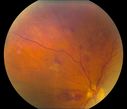
BRVO Inferiorly with Retinal Hemorrhages in the Left Eye421 viewsPatient comes in with complaint of "spot in vision", sort of a cloudy haze in the left eye. Patient's VA was 20/100-1 with no improvement with pinhole in the left eye. Fundus photos show multiple retinal hemorrhages scattered with a branch retinal vein occlusion inferiorily. Patient is also diabetic.
|
|

Chronic BRAO with Hemorrhages - Sclerosed Vessel421 views95 year old female with blurred vision in both eyes in for a checkup. History of perpapillary CNVM in the left eye 2 years prior not treated. History of stroke with multiple emboli 1 year ago.
Vision 20/32 OD, 20/40 OS
|
|
421 views
|
|

Branch Retinal Vein Occlusion with Neovascularization Elsewhere and Vitreous Hemorrhage421 views57 year old man New floaters in the left eye about a month ago. Now they are getting better. PMHx A-Fib on Pradaxa
VA 20/32 OD, 20/40 OS
1+ NS OU
BP 137/77, 200LBs, 5’8â€
PRP laser was done
|
|
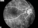
CRAO420 viewsCRAO in OD Tranisit Phase of FA
|
|
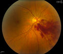
Branch Retinal Vein Occlusion420 viewsPatient's VA was 20/40, right eye. Fundus exam shows branch retinal vein occlusion nasally to the optic nerve in the right eye.
|
|
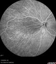
Cilioretinal Artery Occlusion - Nasal (not macular)420 views77 year old female There is a shadow in the top half of her temporal vision in the left eye that came on suddenly yesterday morning. It has not changed since then and the right eye is OK. She has had sinus headaches. She had no focal neurological changes. Â
VA OD: Dcc20/32
VA OS: Dcc20/16-1
IOP: TP: OD:18 OS:19Â
|
|
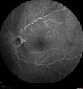
Non-ischemic Central Retinal Vein Occlusion with Macular Edema420 views62 year old man The vision in the right eye started to decline about 10 days ago. Prior to that the two eyes were about the same.
VA OD: Dcc20/125 PH20/50-2
VA OS: Dcc20/20
IOP: TP: OD:21 OS:21
|
|
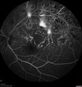
Branch Retinal Vein Occlusion - Both Eyes - Edema OD and NVE OS - Severe Non-perfusion Both Eyes420 views83 year old man with vision loss for 3 months not sure which eye. VA 20/100 OD and 20/50 OS. Tried avastin once without much improvement in the right eye and then lost to follow-up. FA in the left eye shows leaking NVE
|
|
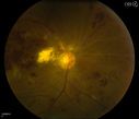
Combined Artery and Vein Occlusion419 viewsPatient comes in with decreased vision in both eyes. VA 20/200-OD, 20/60-OS. Fundus exam shows great amount of macular edema due to artery and vein occlusions. There is some neovascularization on the optic nerve in the right eye. Patient was treated with Eylea Injection in the left eye today and will return for Eylea Injection in the right eye.
|
|
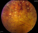
Central Retinal Vein Occlusion with Macular Edema and Retinal Hemorrhages 419 viewsFemale patient of 86-years of age presents a central retinal vein occlusion with macular edema in the right eye. Patient's VA was 20/70 in the right eye. Proceeded with laser treatment to the right eye.
|
|
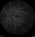
CRVO 25 year old man - Heterozygous Factor V Leiden419 views20/100 initial vision. Improved to 20/30 with Lucentis which continued for 3 years, treat and extend, and stopped.
|
|
419 views
|
|

419 views
|
|
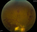
Combined Artery and Vein Occlusion418 viewsPatient comes in with decreased vision in both eyes. VA 20/200-OD, 20/60-OS. Fundus exam shows great amount of macular edema due to artery and vein occlusions. There is some neovascularization on the optic nerve in the right eye. Patient was treated with Eylea Injection in the left eye today and will return for Eylea Injection in the right eye.
|
|

Central Retinal Vein Occlusion - No macular edema - Pre-foveal opercula417 views62 year old female with 20/20 vision. She had been seeing a spot out of the left eye for 4 years and then noticed splotches in her vision. She never devleoped edema or NV
|
|

Central Retinal Vein Occlusion - No macular edema - Pre-foveal opercula417 views62 year old female with 20/20 vision. She had been seeing a spot out of the left eye for 4 years and then noticed splotches in her vision. She never devleoped edema or NV
|
|
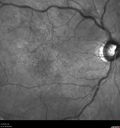
417 views
|
|
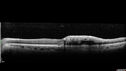
Branch retinal vein occlusion with macular edema417 views71 year old man Starting about 2 weeks ago the left eye was blurred. The right eye is seeing well.
VA OD: sc20/25+2 NccJ1+
VA OS: sc20/63+2 PH20/50-2 Ncc20/400
IOP: TP: OD:19 OS:16
|
|

Elevated Episcleral Venous Pressure and Retinal Hemorrhages in both eyes417 views78 year old female with chronic red eye. The eyes are red all the time now. She is on Xeralto because of a history of blood clots in her lungs (2005 about). She has not had problems since. She has had floaters for many years. Ever since her eyes got funny she sees more floaters in both eyes. She had a brain scan over 6 months ago which was OK. She also had double vision when this first started which went away. She could not drive for a few months.
VA OD: Dcc20/32-1
VA OS: Dcc20/125
IOP: TP: OD:17 OS:16
Neuroimaging did not show a CC fistula
|
|

Central Retinal Artery Occlusion - Fluorescein Angiogram Video417 views63 year old man Sudden severe vision loss in the left eye for 3 days. Six months following macula off retinal detachment repair, Pneumatic then PPV, then PPV buckle. VA was 20/63 on 4/2021 (3 months post RD repair).
PMHx – HTN. Meds: HCT, Meloxicam
VA OD 20/25, OS HM
|
|
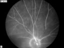
CRAO416 viewsCRAO in OD Late Phase of FA
|
|

Cilioretinal Artery Occlusion - Nasal (not macular)416 views77 year old female There is a shadow in the top half of her temporal vision in the left eye that came on suddenly yesterday morning. It has not changed since then and the right eye is OK. She has had sinus headaches. She had no focal neurological changes. Â
VA OD: Dcc20/32
VA OS: Dcc20/16-1
IOP: TP: OD:18 OS:19Â
|
|
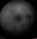
Branch retinal vein occlusion with macular edema416 views71 year old man Starting about 2 weeks ago the left eye was blurred. The right eye is seeing well.
VA OD: sc20/25+2 NccJ1+
VA OS: sc20/63+2 PH20/50-2 Ncc20/400
IOP: TP: OD:19 OS:16
|
|
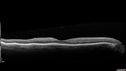
Chronic BRAO with Hemorrhages - Sclerosed Vessel415 views95 year old female with blurred vision in both eyes in for a checkup. History of perpapillary CNVM in the left eye 2 years prior not treated. History of stroke with multiple emboli 1 year ago.
Vision 20/32 OD, 20/40 OS
|
|
415 views
|
|
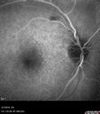
Branch Retinal Vein Occlusion with Edema - Failed avastin (bevacizumab) and did well with Eylea (Aflibercept)415 views67 year old female The vision in the left eye was good until about 4 months ago. Prior to that it was OK. The vision in the right eye is fine. She is not sure if she has had strokes from studies taken in December. She was, at that time, sent to the ER for high blood pressure. Â
VA OD: Dcc20/40 OS: 20/100
Four treatments with Avastin she was still 20/100 with edema. With two Eylea treatments she improved to 20/32 and has remained dry and with good vision
|
|
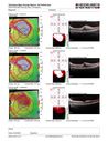
Branch Retinal Vein Occlusion with Edema - Failed avastin (bevacizumab) and did well with Eylea (Aflibercept)415 views67 year old female The vision in the left eye was good until about 4 months ago. Prior to that it was OK. The vision in the right eye is fine. She is not sure if she has had strokes from studies taken in December. She was, at that time, sent to the ER for high blood pressure. Â
VA OD: Dcc20/40 OS: 20/100
Four treatments with Avastin she was still 20/100 with edema. With two Eylea treatments she improved to 20/32 and has remained dry and with good vision
|
|

Branch Retinal Vein Occlusion - Both Eyes - Edema OD and NVE OS - Severe Non-perfusion Both Eyes415 views83 year old man with vision loss for 3 months not sure which eye. VA 20/100 OD and 20/50 OS. Tried avastin once without much improvement in the right eye and then lost to follow-up. FA in the left eye shows leaking NVE
|
|
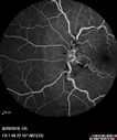
Central Retinal Vein Occlusion - No macular edema - Pre-foveal opercula414 views62 year old female with 20/20 vision. She had been seeing a spot out of the left eye for 4 years and then noticed splotches in her vision. She never devleoped edema or NV
|
|
| 1845 files on 19 page(s) |
 |
|
|
|
|
12 | |
|
|
|
|


