Most viewed - Retina Vascular Occlusions
|

Non-Ischemic Central Retinal Vein Occlusion in Young Man (30 years) - Many Cotton Wool Spots3075 views30-year-old man with a history of a central retinal vein occlusion for which he was first examined on 2 months ago and his vision then was 4/200. He responded nicely to two Avastin treatments. OD is 20/20, OS is 20/60
|
|

Asymptomatic Hollenhorst Plaque - Cholesterol Embolis2731 views71-year-old woman is not noticing any vision change. She does take pressure-lowering drops for glaucoma, both Timolol and Xalatan.
VISUAL ACUITY: Vision OD is 20/30, OS is 20/16
|
|
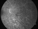
Non-Ischemic Central Retinal Vein Occlusion in Young Man (30 years) - Many Cotton Wool Spots2435 views30-year-old man with a history of a central retinal vein occlusion for which he was first examined on 2 months ago and his vision then was 4/200. He responded nicely to two Avastin treatments. OD is 20/20, OS is 20/60
|
|

Central Retinal Artery Occlusion - 2 months old by history1839 views77-year-old man was doing fine. His left eye was a little worse than the right eye, and then in the 2 months ago he had sudden severe vision loss in the right eye. Vision OD is 3/200; OS is 20/60. IOP: OD 30, OS 15.
OD: Vertical C/D ratio is 0.4. The macula is white from ischemia. The arteries are narrow.
Photos confirm clinical findings.
OCT SCAN: OCT scan of the right eye shows retinal thickening, consistent with macular edema associated with a central retinal artery occlusion. The left eye has severe inferior retinal thinning, consistent with an old hemicentral retinal artery occlusion in the left eye. The fovea in the left eye is a little bit thin but not too bad.
IMPRESSION: 1. CENTRAL RETINAL ARTERY OCCLUSION RIGHT EYE
2 OLD HEMICENTRAL RETINAL ARTERY OCCLUSION LEFT EYE
3 DIABETES WITHOUT RETINOPATHY
4 ELEVATED INTRAOCULAR PRESSURE RIGHT EYE STATUS POST INJECTION RIGHT EYE A WEEK AGO
DISCUSSION: I explained to the patient that the right eye unfortunately has had a central retinal artery and unfortunately we do not have anything to treat that. His vision will probably improve some over time. He needs to be watched though. It is possible he is developing rubeotic glaucoma, although it is probable the intraocular pressure is high from the injection last week. I asked him to restart the Alphagan, to stop the other two drops, and to return here in three weeks to make sure his pressure is more acceptable.
At this point, having lost almost all the retinal circulation in the right eye and half of the retinal circulation in the left eye, he only has half a retina in the left eye he is working off of. I told him it is very important he keep seeing you and anything that can be done to keep his blood pressure low and his cholesterol reasonable would be helpful to what remains of his retinal circulation.
|
|

Central Retinal Artery Occlusion1782 views60 year old male with LP vision due to extensive blood flow loss.
|
|

Retinal Artery Occlusion1713 viewsPatient comes in for eval on artery occlusions in both eyes. VA is 20/400, right eye and NLP, left eye. Fundus photos show paniretinal scars in the right eye with arterial narrowing and the left eye has arterial narrowing as well.
|
|

Inferotemporal and Inferonasal BRAO1660 viewsBranch Retinal Artery Occlusions - Multiple
|
|
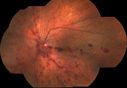
Hemi-retinal Vein Occlusion1615 views81 year old female seen in 2009 for a Hemi-retinal vein Occlusion OS with vision of 20/25.
|
|

Central Retinal Artery Occlusion - 2 months old by history1610 views77-year-old man was doing fine. His left eye was a little worse than the right eye, and then in the 2 months ago he had sudden severe vision loss in the right eye. Vision OD is 3/200; OS is 20/60. IOP: OD 30, OS 15.
OD: Vertical C/D ratio is 0.4. The macula is white from ischemia. The arteries are narrow.
Photos confirm clinical findings.
OCT SCAN: OCT scan of the right eye shows retinal thickening, consistent with macular edema associated with a central retinal artery occlusion. The left eye has severe inferior retinal thinning, consistent with an old hemicentral retinal artery occlusion in the left eye. The fovea in the left eye is a little bit thin but not too bad.
IMPRESSION: 1. CENTRAL RETINAL ARTERY OCCLUSION RIGHT EYE
2 OLD HEMICENTRAL RETINAL ARTERY OCCLUSION LEFT EYE
3 DIABETES WITHOUT RETINOPATHY
4 ELEVATED INTRAOCULAR PRESSURE RIGHT EYE STATUS POST INJECTION RIGHT EYE A WEEK AGO
DISCUSSION: I explained to the patient that the right eye unfortunately has had a central retinal artery and unfortunately we do not have anything to treat that. His vision will probably improve some over time. He needs to be watched though. It is possible he is developing rubeotic glaucoma, although it is probable the intraocular pressure is high from the injection last week. I asked him to restart the Alphagan, to stop the other two drops, and to return here in three weeks to make sure his pressure is more acceptable.
At this point, having lost almost all the retinal circulation in the right eye and half of the retinal circulation in the left eye, he only has half a retina in the left eye he is working off of. I told him it is very important he keep seeing you and anything that can be done to keep his blood pressure low and his cholesterol reasonable would be helpful to what remains of his retinal circulation.
|
|

Central Retinal Artery Occlusion with Cilioretinal Sparing - Acute - SD-OCT Line Scan Inner retinal edema1537 views 77-year-old man was doing fine until midnight of last night when he noticed sudden severe vision loss in the left eye. VISUAL ACUITY: OD 20/20, OS 2/200.
|
|

Macular Branch Retinal Vein Occlusions (2) Left Eye - No Macular Edema (yet) 1498 views77-year-old woman with two macular branch retinal vein occlusions in the left eye OD 20/25
|
|

Hemi Central Retinal Vein Occlusion 35 year old non-compliant man1476 views35-year-old man hazy vision in his left eye for about the last three days. OD 20/20, OS 20/30
|
|

Twig Branch Retinal Vein Occlusion - Recurrent Macular Edema with extended interval Lucentis Therapy after first 6 monthly Injections1424 views39-year-old man has a macular branch retinal vein occlusion with macular edema. He had an excellent response to Lucentis for his first 6 treatments. He was last here 6 weeks ago and his vision seems OK to him. OD 20/40.
|
|

Asymptomatic Hollenhorst Plaque - Cholesterol Embolis1375 views71-year-old woman is not noticing any vision change. She does take pressure-lowering drops for glaucoma, both Timolol and Xalatan.
VISUAL ACUITY: Vision OD is 20/30, OS is 20/16
|
|

Central Retinal Vein Occlusion1361 viewsPatient comes in with mild blurred vision in the right eye. Fundus exam shows CRVO with scattered retinal hemorrhages in the right eye.
|
|

Non-Ischemic Central Retinal Vein Occlusion in Young Man (30 years) - Many Cotton Wool Spots1354 views30-year-old man with a history of a central retinal vein occlusion for which he was first examined on 2 months ago and his vision then was 4/200. He responded nicely to two Avastin treatments. OD is 20/20, OS is 20/60
|
|

Asymptomatic Hollenhorst Plaque - Cholesterol Embolis1307 views71-year-old woman is not noticing any vision change. She does take pressure-lowering drops for glaucoma, both Timolol and Xalatan.
VISUAL ACUITY: Vision OD is 20/30, OS is 20/16
|
|

Non-Ischemic Central Retinal Vein Occlusion in Young Man (30 years) - Many Cotton Wool Spots1302 views30-year-old man with a history of a central retinal vein occlusion for which he was first examined on 2 months ago and his vision then was 4/200. He responded nicely to two Avastin treatments. OD is 20/20, OS is 20/60
|
|

Stasis Retinopathy, Central Retinal Vein Occlusion1281 viewsYoung male who complains of decreased vision in the left eye. VA is 20/20 in the left eye. Fundus photo shows mild central retinal vein occlusion in the left eye.
|
|

Cilioretinal artery occlusion and Ophthalmic Artery Occlusion with Relative Sparing of the Central Retinal Artery1277 views80-year-old woman who says she was looking in the mirror, covered her right eye and noticed she could not see well out of the left eye. That just happened yesterday. Her last eye check was a few years ago. She also had a carotid endarterectomy on the right side in 2006 shortly after coronary bypass surgery. OD is 20/40, OS is 20/200.
|
|

Hemicentral Retinal Vein Occlusion Vision 20/60 1268 views84-year-old man has a hemicentral retinal vein occlusion in the right eye. His vision has declined since then.
VISUAL ACUITY: Vision OD is 20/60.
|
|

Central Retinal Artery Occlusion with Cilioretinal Sparing - Acute1222 views 77-year-old man was doing fine until midnight of last night when he noticed sudden severe vision loss in the left eye. VISUAL ACUITY: OD 20/20, OS 2/200.
|
|

Disorganized Retinal Inner Layers (DRIL)1221 views72 year old man treated with Avastin for CRVO VA is still 20/200 with absent macular edema (Only 1 month post rx)
|
|

Hemi Central Retinal Vein Occlusion 35 year old non-compliant man1220 views35-year-old man hazy vision in his left eye for about the last three days. OD 20/20, OS 20/30
|
|
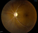
Retinal Artery Occlusion1205 viewsPatient comes in for eval on artery occlusions in both eyes. VA is 20/400, right eye and NLP, left eye. Fundus photos show paniretinal scars in the right eye with arterial narrowing and the left eye has arterial narrowing as well.
|
|

Hemicentral Retinal Vein Occlusion Vision 20/60 1195 views84-year-old man has a hemicentral retinal vein occlusion in the right eye. His vision has declined since then.
VISUAL ACUITY: Vision OD is 20/60.
|
|
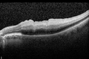
Central Retinal Artery Occlusion with Cilioretinal Sparing - Acute - SD-OCT Line Scan Inner retinal edema1185 views 77-year-old man was doing fine until midnight of last night when he noticed sudden severe vision loss in the left eye. VISUAL ACUITY: OD 20/20, OS 2/200.
|
|

BRVO with collaterals and cystoid macular edema1155 views66 Year Old Man with has had a branch retinal vein occlusion in the right eye with vision loss for the last few months. OD is 20/40, OS is 20/20. IOP: OD 18, OS 19. There is a superotemporal macular branch retinal vein occlusion with edema, hemorrhage, and exudate.
OCT SCAN: The right eye shows edema with a central foveal thickness of 354 microns FA: shows remodeling of the superotemporal vasculature. There are already venous-venous collaterals, suggesting the vein occlusion may be a little more than a few months old. There are multiple microaneurysms superotemporal to the fovea, and there is leakage in the late frames involving the fovea.
|
|

Branch Retinal Vein Occlusion1127 viewsPatient comes in for eval on retinal hemorrhage in the right eye. VA is 20/30 in the right eye. Fundus photography shows branch retinal vein occlusion inferior of the macula with retinal hemorrhage present. Will reevaluate in 2-months.
|
|

Central Retinal Artery Occlusion1108 viewsCentral Retinal Artery Occlusion with cilio artery perfusion.
|
|

Central Retinal Artery Occlusion less than 24 hours - 69 year old man VA light perception1107 views
|
|

branch retinal vein occlusion - fundus photo1095 views70 year old woman with 20/80 vision from a branch retinal vein occlusion and macular edema. She has had laser treatment and intravitreal steroids. Her vision with further laser improved to 20/50.
|
|

Old BRAO twig with plaque1082 views81-year-old woman Her left eye is amblyopic. Her right eye was doing fine until about last Sunday, which is five days ago. She had a change in vision where for five minutes she saw a shadow in her side vision out of the right eye. She describes it as being more up into the left. It lasted for just a brief time and then went away. She has had atrial fibrillation since January of 2008. She is on Coumadin for that. She is diabetic for eleven years. OD is 20/25, OS is 20/60. IOP: OD 15, OS 18.
|
|
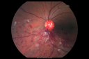
Hemi Central Retinal Vein Occlusion 35 year old non-compliant man1072 views35-year-old man hazy vision in his left eye for about the last three days. OD 20/20, OS 20/30
|
|

14.5 seconds1070 viewsCorresponding FA for Cilioretinal artery occlusion with early venous stasis retinopathy
|
|

Central Retinal Vein Occlusion - Severe Optic Nerve Edema1066 views15 yr old male presents with sudden onset of painless decreased vision in right eye x 6 days gradually worsening. No history of Trauma OD 20/30 OS 20/25, Diagnosis CRVO with ? Papillophlebitis.
|
|

Pattern Dystrophy - Adult Vitelliform Macular Degeneration1062 views72-year-old woman 2 months post ECCE with poor vision since surgery especially left eye.
VISUAL ACUITY: OD 20/50, OS 20/200
|
|

Central Retinal Artery Occlusion with Cilioretinal Sparing - Acute - FA Very Slow Filling CRA1052 views 77-year-old man was doing fine until midnight of last night when he noticed sudden severe vision loss in the left eye. VISUAL ACUITY: OD 20/20, OS 2/200.
|
|

Central Arterial Occlusion with Embolism Present1048 viewsPatient comes in with CRAO in the right eye. VA was hand motion. Fundus photo shows white veil over the retina with 2-emboli in a branch artery temporal in the right eye. She will be evaluated for emboli and followed up in a month
|
|
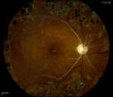
Retinal Artery Occlusion1041 viewsPatient comes in for eval on artery occlusions in both eyes. VA is 20/400, right eye and NLP, left eye. Fundus photos show paniretinal scars in the right eye with arterial narrowing and the left eye has arterial narrowing as well.
|
|

BRAO and PAMM1036 viewsLayers of retina are thickened showing ischemia to the middle retina (deep retinal capillary plexus)
|
|

Twig Branch Retinal Vein Occlusion - Recurrent Macular Edema with extended interval Lucentis Therapy after first 6 monthly Injections1026 views39-year-old man has a macular branch retinal vein occlusion with macular edema. He had an excellent response to Lucentis for his first 6 treatments. He was last here 6 weeks ago and his vision seems OK to him. OD 20/40.
|
|

Pattern Dystrophy - Adult Vitelliform Macular Degeneration1020 views72-year-old woman 2 months post ECCE with poor vision since surgery especially left eye.
VISUAL ACUITY: OD 20/50, OS 20/200
|
|

Central Retinal Vein Occlusion with Macular Edema1007 viewsPatient notices decreased vision in the left eye. VA is 20/60, left eye. Fundus and Fluorescence Angiogram shows CRVO in the left eye with scattered hemorrhages throughout the retina.
|
|
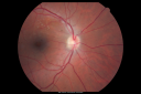
Twig Branch Retinal Vein Occlusion - Recurrent Macular Edema with extended interval Lucentis Therapy after first 6 monthly Injections997 views39-year-old man has a macular branch retinal vein occlusion with macular edema. He had an excellent response to Lucentis for his first 6 treatments. He was last here 6 weeks ago and his vision seems OK to him. OD 20/40.
|
|

Central Retinal Artery Occlusion with Cilioretinal Sparing - Acute - Line scan good eye997 views 77-year-old man was doing fine until midnight of last night when he noticed sudden severe vision loss in the left eye. VISUAL ACUITY: OD 20/20, OS 2/200.
|
|
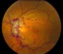
Central Retinal Vein Occlusion997 views
|
|

branch retinal vein occlusion - oct map987 views70 year old woman with 20/80 vision from a branch retinal vein occlusion and macular edema. She has had laser treatment and intravitreal steroids. Her vision with further laser improved to 20/50.
|
|

Branch Retinal Vein Occlusion with Macular Edema985 viewsCentral foveal thickness came to normal after laser and intravitreal Kenalog
|
|
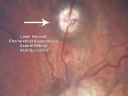
Central Retinal Vein Occlusion and Laser Induced Chorioretinal Anastomosis985 views84-year-old woman has age-related macular degeneration in both eyes with foveal atrophy. She also has a central retinal vein occlusion
in the left eye for which she had intravitreal Kenalog and finally chorioretinal anastomosis laser a year
ago. VISUAL ACUITY: Vision OD is 20/160, OS is 7/200
|
|
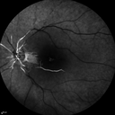
Central Retinal Artery Occlusion with Cilioretinal Sparing - Acute - FA Very Slow Filling CRA971 views 77-year-old man was doing fine until midnight of last night when he noticed sudden severe vision loss in the left eye. VISUAL ACUITY: OD 20/20, OS 2/200.
|
|

Sectoral Optic Atrophy from an old Branch Retinal Artery Occlusion 964 views74-year-old woman had cataract surgery 3 months ago. Her vision in the right eye has been abnormal since. She notices a shadow in the inferior vision in the right eye. OD is 20/60, OS is 20/50
|
|

branch retinal vein occlusion - fluorescein angiogram - arrow shows obstruction point with collateral vessels950 views70 year old woman with 20/80 vision from a branch retinal vein occlusion and macular edema. She has had laser treatment and intravitreal steroids. Her vision with further laser improved to 20/50.
|
|

BRVO with collaterals and cystoid macular edema950 views66 Year Old Man with has had a branch retinal vein occlusion in the right eye with vision loss for the last few months. OD is 20/40, OS is 20/20. IOP: OD 18, OS 19. There is a superotemporal macular branch retinal vein occlusion with edema, hemorrhage, and exudate.
OCT SCAN: The right eye shows edema with a central foveal thickness of 354 microns FA: shows remodeling of the superotemporal vasculature. There are already venous-venous collaterals, suggesting the vein occlusion may be a little more than a few months old. There are multiple microaneurysms superotemporal to the fovea, and there is leakage in the late frames involving the fovea.
|
|

BRVO with collaterals and cystoid macular edema949 views66 Year Old Man with has had a branch retinal vein occlusion in the right eye with vision loss for the last few months. OD is 20/40, OS is 20/20. IOP: OD 18, OS 19. There is a superotemporal macular branch retinal vein occlusion with edema, hemorrhage, and exudate.
OCT SCAN: The right eye shows edema with a central foveal thickness of 354 microns FA: shows remodeling of the superotemporal vasculature. There are already venous-venous collaterals, suggesting the vein occlusion may be a little more than a few months old. There are multiple microaneurysms superotemporal to the fovea, and there is leakage in the late frames involving the fovea.
|
|

Central Retinal Artery Occlusion less than 24 hours - 69 year old man VA light perception943 views
|
|

Pathology - Central Retinal Artery Occlusion - 6 weeks old - Inner Retinal Atrophy - Macular Pucker937 viewspatient died about 6 to 8 weeks after the artery occlusion, and he had no history of glaucoma. The inner retina atrophy, which looks very similar to that caused by COAG, is due to recent infarct.
|
|

Cilioretinal artery occlusion and Ophthalmic Artery Occlusion with Relative Sparing of the Central Retinal Artery933 views80-year-old woman who says she was looking in the mirror, covered her right eye and noticed she could not see well out of the left eye. That just happened yesterday. Her last eye check was a few years ago. She also had a carotid endarterectomy on the right side in 2006 shortly after coronary bypass surgery. OD is 20/40, OS is 20/200.
|
|
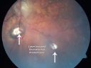
Central Retinal Vein Occlusion and Laser Induced Chorioretinal Anastomosis930 views84-year-old woman has age-related macular degeneration in both eyes with foveal atrophy. She also has a central retinal vein occlusion
in the left eye for which she had intravitreal Kenalog and finally chorioretinal anastomosis laser a year
ago. VISUAL ACUITY: Vision OD is 20/160, OS is 7/200
|
|

Central Retinal Artery Occlusion with Cilioretinal Sparing - Acute927 views 77-year-old man was doing fine until midnight of last night when he noticed sudden severe vision loss in the left eye. VISUAL ACUITY: OD 20/20, OS 2/200.
|
|

branch retinal vein occlusion - red free photo926 views70 year old woman with 20/80 vision from a branch retinal vein occlusion and macular edema. She has had laser treatment and intravitreal steroids. Her vision with further laser improved to 20/50.
|
|

Central Retinal Artery Occlusion with Cilioretinal Sparing - Acute - FA Very Slow Filling CRA925 views 77-year-old man was doing fine until midnight of last night when he noticed sudden severe vision loss in the left eye. VISUAL ACUITY: OD 20/20, OS 2/200.
|
|
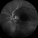
Central Retinal Artery Occlusion with Cilioretinal Sparing - Acute - FA Very Slow Filling CRA924 views 77-year-old man was doing fine until midnight of last night when he noticed sudden severe vision loss in the left eye. VISUAL ACUITY: OD 20/20, OS 2/200.
|
|
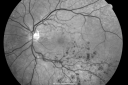
Macular Branch Retinal Vein Occlusions (2) Left Eye - No Macular Edema (yet) 918 views77-year-old woman with two macular branch retinal vein occlusions in the left eye OD 20/25
|
|
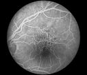
telangiectasia915 views20/20 pt with blurry area in va
|
|

Pathology - Central Retinal Artery Occlusion - 6 weeks old - Inner Retinal Atrophy - Macular Pucker914 viewspatient died about 6 to 8 weeks after the artery occlusion, and he had no history of glaucoma. The inner retina atrophy, which looks very similar to that caused by COAG, is due to recent infarct.
|
|

Branch Retinal Arterial Occlusion 20/20 Vision - Symptoms for 1 Day901 views
|
|
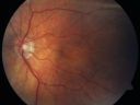
Cilioretinal artery occlusion and Ophthalmic Artery Occlusion with Relative Sparing of the Central Retinal Artery899 views80-year-old woman who says she was looking in the mirror, covered her right eye and noticed she could not see well out of the left eye. That just happened yesterday. Her last eye check was a few years ago. She also had a carotid endarterectomy on the right side in 2006 shortly after coronary bypass surgery. OD is 20/40, OS is 20/200.
|
|

Sectoral Optic Atrophy from an old Branch Retinal Artery Occlusion 896 views74-year-old woman had cataract surgery 3 months ago. Her vision in the right eye has been abnormal since. She notices a shadow in the inferior vision in the right eye. OD is 20/60, OS is 20/50
|
|
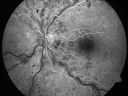
Non-Ischemic Central Retinal Vein Occlusion in Young Man (30 years) - Many Cotton Wool Spots894 views30-year-old man with a history of a central retinal vein occlusion for which he was first examined on 2 months ago and his vision then was 4/200. He responded nicely to two Avastin treatments. OD is 20/20, OS is 20/60
|
|

Classic BRVO894 viewsClassic branch retinal vein occlusion
|
|
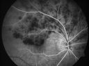
Hemicentral Retinal Vein Occlusion Vision 20/60 891 views84-year-old man has a hemicentral retinal vein occlusion in the right eye. His vision has declined since then.
VISUAL ACUITY: Vision OD is 20/60.
|
|

BRVO arrow showing obstruction point with collateral vessels - red free photo888 views70 year old woman with 20/80 vision from a branch retinal vein occlusion and macular edema. She has had laser treatment and intravitreal steroids. Her vision with further laser improved to 20/50.
|
|
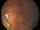
Cilioretinal artery occlusion and Ophthalmic Artery Occlusion with Relative Sparing of the Central Retinal Artery884 views80-year-old woman who says she was looking in the mirror, covered her right eye and noticed she could not see well out of the left eye. That just happened yesterday. Her last eye check was a few years ago. She also had a carotid endarterectomy on the right side in 2006 shortly after coronary bypass surgery. OD is 20/40, OS is 20/200.
|
|
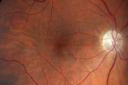
Sectoral Optic Atrophy from an old Branch Retinal Artery Occlusion 881 views74-year-old woman had cataract surgery 3 months ago. Her vision in the right eye has been abnormal since. She notices a shadow in the inferior vision in the right eye. OD is 20/60, OS is 20/50
|
|

Central Retinal Artery Occlusion - 2 months old by history878 views77-year-old man was doing fine. His left eye was a little worse than the right eye, and then in the 2 months ago he had sudden severe vision loss in the right eye. Vision OD is 3/200; OS is 20/60. IOP: OD 30, OS 15.
OD: Vertical C/D ratio is 0.4. The macula is white from ischemia. The arteries are narrow.
Photos confirm clinical findings.
OCT SCAN: OCT scan of the right eye shows retinal thickening, consistent with macular edema associated with a central retinal artery occlusion. The left eye has severe inferior retinal thinning, consistent with an old hemicentral retinal artery occlusion in the left eye. The fovea in the left eye is a little bit thin but not too bad.
IMPRESSION: 1. CENTRAL RETINAL ARTERY OCCLUSION RIGHT EYE
2 OLD HEMICENTRAL RETINAL ARTERY OCCLUSION LEFT EYE
3 DIABETES WITHOUT RETINOPATHY
4 ELEVATED INTRAOCULAR PRESSURE RIGHT EYE STATUS POST INJECTION RIGHT EYE A WEEK AGO
DISCUSSION: I explained to the patient that the right eye unfortunately has had a central retinal artery and unfortunately we do not have anything to treat that. His vision will probably improve some over time. He needs to be watched though. It is possible he is developing rubeotic glaucoma, although it is probable the intraocular pressure is high from the injection last week. I asked him to restart the Alphagan, to stop the other two drops, and to return here in three weeks to make sure his pressure is more acceptable.
At this point, having lost almost all the retinal circulation in the right eye and half of the retinal circulation in the left eye, he only has half a retina in the left eye he is working off of. I told him it is very important he keep seeing you and anything that can be done to keep his blood pressure low and his cholesterol reasonable would be helpful to what remains of his retinal circulation.
|
|

Ghost vessels with BRVO877 views
|
|
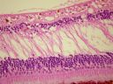
Pathology - Central Retinal Artery Occlusion - 6 weeks old - Inner Retinal Atrophy876 viewspatient died about 6 to 8 weeks after the artery occlusion, and he had no history of glaucoma. The inner retina atrophy, which looks very similar to that caused by COAG, is due to recent infarct.
|
|

Cilioretinal artery occlusion and Ophthalmic Artery Occlusion with Relative Sparing of the Central Retinal Artery872 views80-year-old woman who says she was looking in the mirror, covered her right eye and noticed she could not see well out of the left eye. That just happened yesterday. Her last eye check was a few years ago. She also had a carotid endarterectomy on the right side in 2006 shortly after coronary bypass surgery. OD is 20/40, OS is 20/200.
|
|

branch retinal vein occlusion - oct line scan865 views70 year old woman with 20/80 vision from a branch retinal vein occlusion and macular edema. She has had laser treatment and intravitreal steroids. Her vision with further laser improved to 20/50.
|
|

branch retinal vein occlusion - fluorescein angiogram861 views70 year old woman with 20/80 vision from a branch retinal vein occlusion and macular edema. She has had laser treatment and intravitreal steroids. Her vision with further laser improved to 20/50.
|
|
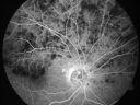
Hemicentral Retinal Vein Occlusion Vision 20/60 855 views84-year-old man has a hemicentral retinal vein occlusion in the right eye. His vision has declined since then.
VISUAL ACUITY: Vision OD is 20/60.
|
|

Embolic CRAO color852 views
|
|

849 views
|
|

DRIL - Disorganization Retinal Inner Layers848 viewsFollowing treatment for BRVO vision stayed at 3/200 despite resolution of edema
|
|

Central Retinal Artery Occlusion less than 24 hours - 69 year old man VA light perception847 views
|
|

Old Branch Retinal Vein Occlusion- Severe CME - Pucker - Diabetic Fundus Photo844 views61-year old man has diabetic retinopathy in the right eye. He also had a macular pucker and macular edema. I did a vitrectomy on September 5th. His vision was initially improving. His vision in the right eye seems to be a little more foggy.
VISUAL ACUITY: OD: 20/200
|
|

Branch Retinal Vein Occlusion with Macular Edema 3 Months after Intravitreal Kenalog and Grid Laser844 viewsCentral foveal thickness came to normal after laser and intravitreal Kenalog
|
|

BRVO with collaterals and cystoid macular edema843 views66 Year Old Man with has had a branch retinal vein occlusion in the right eye with vision loss for the last few months. OD is 20/40, OS is 20/20. IOP: OD 18, OS 19. There is a superotemporal macular branch retinal vein occlusion with edema, hemorrhage, and exudate.
OCT SCAN: The right eye shows edema with a central foveal thickness of 354 microns FA: shows remodeling of the superotemporal vasculature. There are already venous-venous collaterals, suggesting the vein occlusion may be a little more than a few months old. There are multiple microaneurysms superotemporal to the fovea, and there is leakage in the late frames involving the fovea.
|
|

Central Retinal Artery Occlusion with Cilioretinal Sparing - Acute - FA Very Slow Filling CRA842 views 77-year-old man was doing fine until midnight of last night when he noticed sudden severe vision loss in the left eye. VISUAL ACUITY: OD 20/20, OS 2/200.
|
|

FA: CRAO842 viewsCRAO with cilio artery perfusion
|
|

BRVO840 views42 year old female VA 20/25
Branch Retinal Vein Occlusion - Hot Spot on Vein at Occlusion Site
|
|

Twig Branch Retinal Vein Occlusion - Recurrent Macular Edema with extended interval Lucentis Therapy after first 6 monthly Injections838 views39-year-old man has a macular branch retinal vein occlusion with macular edema. He had an excellent response to Lucentis for his first 6 treatments. He was last here 6 weeks ago and his vision seems OK to him. OD 20/40.
|
|

Old Branch Retinal Vein Occlusion- Severe CME - Pucker - Diabetic Map OCT835 views61-year old man has diabetic retinopathy in the right eye. He also had a macular pucker and macular edema. I did a vitrectomy on September 5th. His vision was initially improving. His vision in the right eye seems to be a little more foggy.
VISUAL ACUITY: OD: 20/200
|
|
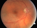
BRAO and PAMM835 views
|
|

Central Retinal Vein Occlusion and Cystoid Macular Edema - Acute - Vision 20/50 834 views68-year-old man six weeks of vision loss right eye from CRVO and CME, vision right eye 20/50
|
|

Branch Retinal Vein Occlusion with NVE (Neovascularization Elsewhere)833 views
|
|

BRVO with collaterals and cystoid macular edema828 views66 Year Old Man with has had a branch retinal vein occlusion in the right eye with vision loss for the last few months. OD is 20/40, OS is 20/20. IOP: OD 18, OS 19. There is a superotemporal macular branch retinal vein occlusion with edema, hemorrhage, and exudate.
OCT SCAN: The right eye shows edema with a central foveal thickness of 354 microns FA: shows remodeling of the superotemporal vasculature. There are already venous-venous collaterals, suggesting the vein occlusion may be a little more than a few months old. There are multiple microaneurysms superotemporal to the fovea, and there is leakage in the late frames involving the fovea.
|
|
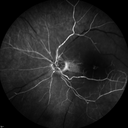
Central Retinal Artery Occlusion with Cilioretinal Sparing - Acute - FA Very Slow Filling CRA827 views 77-year-old man was doing fine until midnight of last night when he noticed sudden severe vision loss in the left eye. VISUAL ACUITY: OD 20/20, OS 2/200.
|
|

BRVO with collaterals and cystoid macular edema826 views66 Year Old Man with has had a branch retinal vein occlusion in the right eye with vision loss for the last few months. OD is 20/40, OS is 20/20. IOP: OD 18, OS 19. There is a superotemporal macular branch retinal vein occlusion with edema, hemorrhage, and exudate.
OCT SCAN: The right eye shows edema with a central foveal thickness of 354 microns FA: shows remodeling of the superotemporal vasculature. There are already venous-venous collaterals, suggesting the vein occlusion may be a little more than a few months old. There are multiple microaneurysms superotemporal to the fovea, and there is leakage in the late frames involving the fovea.
|
|
| 1845 files on 19 page(s) |
1 |
 |
|
|
|
|
|
|
|
|
|


