am meisten angesehen - Retina Gallery User Gallery
|
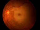
Cilio-Retinal Artery Occlusion with "Box Carring" and Cherry Red Spot1874 x angesehenCilioretinal Artery Occlusion with Cherry Red Spot
|
|

Nonproliferative Diabetic Retinopathy with Circinate Hard Exudates1529 x angesehen
|
|

Eales Disease - Fluorescein Angiogram1475 x angesehen
|
|

Optic Nerve Pit1437 x angesehenOptic Nerve Pit
|
|

Iris Neovascularization secondary to Central Retinal Artery Occlusion1410 x angesehen
|
|

Acute Multifocal Placoid Pigment Epitheliopathy (AMPPE) related scarring1335 x angesehen
|
|
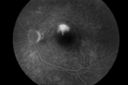
Central Serous Choroidopathy with Smoke Stack and Mushroom Cloud - Fluorescein Angiogram1295 x angesehen
|
|

Epiretinal Membrane with Flecks of Hemorrhage1255 x angesehen
|
|

Epiretinal Membrane with Pseudohole1203 x angesehen
|
|

Choroidal Rupture Secondary to Ocular Trauma1186 x angesehen
|
|

Macular Hole with Subretinal Hemorrhage Secondary to Ocular Trauma1174 x angesehen
|
|

Central Retinal Vein Occlusion with Disc Collaterals1165 x angesehen
|
|

Choriodal Rupture with Hemorrhage1142 x angesehen
|
|

Pseudophakic Cystoid Macular Edema FA late frame1112 x angesehen
|
|

Retinal Tear1108 x angesehen
|
|

Cavernous Hemangioma1099 x angesehen
|
|

Molteno Drainage Tube1089 x angesehen
|
|

Proliferative Diabetic Retinopathy with Tractional Retinal Detachment1067 x angesehenProliferative Diabetic Retinopathy with Tractional Retinal Detachment
|
|
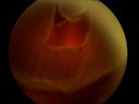
Retinal Detachment Secondary to Irregular Retinal Tear1057 x angesehenRetinal Detachment with Irregular Retinal Tear
|
|

Stargardt's Dystrophy - Silent Choroid - Fluorscein Angiogram1055 x angesehen
|
|

Nonprolific Diabetic Retinopathy with Circinate Ring1033 x angesehenNonprolific Diabetic Retinopathy with Circinate Ring
|
|

Retinal Tear Surrounded By Laser Border995 x angesehen
|
|
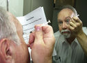
Intraocular Self injection985 x angesehenThe treatment of wet macular degeneration was revolutionized in late 2013 when Geneneron introduced its innovative and inexpensive take-home kit. Now, millions of patients around the world treat themselves on a PRN basis, using instructions written in 10 different languages.
April Fool!
|
|
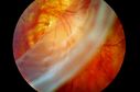
Giant Retinal Tear983 x angesehen
|
|

Choroidal Rupture Secondary to Ocular Trauma957 x angesehen
|
|

Central Retinal Venous Occlusion with Papilledema, Intraretinal Hemorrhage and Macular Edema953 x angesehen
|
|

Retinoschisis937 x angesehen
|
|

Ocular Trauma with Retinal Detachment924 x angesehenPatient suffered blunt force trauma to the eye resulting total retinal detachment. The retina is visible in the pupillary space.
|
|

Acute Multifocal Placoid Pigment Epitheliopathy917 x angesehen
|
|

ERM with Pseudohole883 x angesehen
|
|

Hollenhorst Plaque882 x angesehenMultile plaque spots in a middle aged patient.
|
|
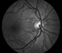
Red Free of Hollenhorst Plaque879 x angesehenRed free fundus photo of patient with multiple hollenhorst plaque
|
|
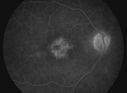
Pseudophakic Cystoid Macular Edema with Disc Hyperfluorescence875 x angesehen
|
|

Central Retinal Venous Occlusion with Papilledema, Irtraretinal Hemorrhage and Vascular Tortuosity820 x angesehen
|
|

Epiretinal Membrane811 x angesehen
|
|
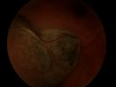
Melanoma803 x angesehen
|
|

Melanoma788 x angesehen
|
|
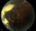
Traumatic Chorioretinal Scar764 x angesehen
|
|
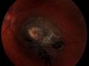
Elevated Nevus728 x angesehen
|
|

Asteroid Hyalosis - Benign Calcified Deposits in the Vitreous702 x angesehen
|
|

ARMD: Macular Degeneration with Subretinal Neovascular Membrane680 x angesehenARMD: Macular Degeneration with Subretinal Neovascular Membrane.
Upon presentation, this patient was found to have blots of subretinal hemorrhage extending into her FAZ, secondary to a subretinal neovascular membrane.
|
|
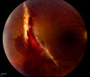
Choroidal Rupture with Subretinal Hemorrhage645 x angesehenThis patient was punched in the eye with a fist, resulting in a choroidal rupture.
|
|

ERM641 x angesehenERM: Epiretinal Membrane
Five years prior to presentation this patient was struck in the eye with a basketball, suffering a vitreal hemorrhage. He underwent vitrectomy and cataract surgery. He later developed a faint ERM. On recent presentation he complained of increased blurring and a floater in his O.D. Examination determined that the ERM had become more prominent.
|
|

Branch Retinal Artery Occlusion with Hollenhorst Plaque622 x angesehen
|
|

Branch Retinal Artery Occlusion with Hollenhorst Plaques598 x angesehenBranch Retinal Artery Occlusion with Hollenhorst Plaques
|
|

CRAO WITH SPARING OF CILIORETINAL ARTERY595 x angesehenThis patient was found to have retinal edema from a Central Retinal Artery Occlusion with sparing of the cilioretinal artery. There were no discernible Hollenhorst plaques
|
|

Diabetic Retinopathy with Hard Exudates592 x angesehen
|
|

August;Acute Ischemic Optic Neuropathy with Hemorrhage592 x angesehen
|
|

Idiopathic Intracranial Hypertension O.S.566 x angesehen
|
|
Choroidal Rupture Secondary to Ocular Trauma554 x angesehenChoroidal Rupture Secondary to Ocular Trauma
|
|

Epiretinal Membrane Stereo Pair549 x angesehen
|
|
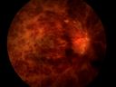
Central Retinal Vein Occlusion541 x angesehenCentral Retinal Vein Occlusion
|
|

Actively Bleeding NVE537 x angesehenThis male patient was referred for a diabetic examination. Upon dilated exam, he started to hemorrhage from a frond of NVE in the superior arcade. These six photographs follow the flow of the hemorrhage through the vitreous to the inferior arcade.
|
|
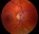
Idiopathic Intracranial Hypertension O.D.529 x angesehen
|
|

Ahmed Valve518 x angesehenAHMED VALVE: This 57 year old diabetic has end stage glaucoma. An Ahmed valve was inserted into her right anterior chamber to help control her IOP.
|
|

Branch Retinal Artery Occlusion with Hollenhorst Plaque514 x angesehenThis patient was found to have retinal edema along the superotemporal arcade secondary to Branch Retinal Artery Occlusion. A Hollenhorst plaque is visible at the first bifurcation of central artery.
|
|

Corneal Neovascularization495 x angesehen
|
|

Choroiditis482 x angesehenCHOROIDITIS: This 46 year old female patient was found to have a peripheral sector of old chorioretinal inflammation accompanied by low grade oozing, intraretinal telangietatic vessels and hard exudates. There was no active bleeding or overlying vitritis.
|
|

Calcification of the Choroid480 x angesehenCALCIFICATION OF THE CHOROID
Most patients with calcification of the choroid have it as an incidental finding. However, as a few have parathyroid abnormalities, this patient was screened for a serum calcium, that returned as normal.
|
|
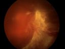
Diabetic Tractional Detachment477 x angesehen
|
|

CNVM470 x angesehenPossible Chorioretinitis
|
|

Papilledema468 x angesehen
|
|

CHOROIDAL MELANOMA Stereo Pair455 x angesehenThis patient was seen approximately five years ago with a suspicious choroidal nodule. The patient did not return for scheduled follow up appointments. When referred back to the practice, the nodule had erupted through Bruchs’ membrane to assume a brawny mushroom cloud configuration which is visible when the stereo images are merged.
|
|

Papilledema446 x angesehen
|
|

Macular Hole Photograph with superimposed OCT433 x angesehen
|
|
Superior Retinal Detachment415 x angesehen
|
|

414 x angesehen
|
|

Retinal Detachment394 x angesehen
|
|

hydroxychloroquine (Plaquenil) toxicity379 x angesehen
|
|
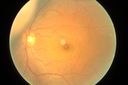
choroidal melanoma376 x angesehenAsian man aged 37 choroidal melanoma in left eye
|
|
362 x angesehen
|
|

357 x angesehen
|
|

Pucker344 x angesehen64female pucker
|
|

342 x angesehen
|
|

330 x angesehen
|
|
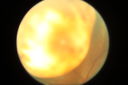
choroidal melanoma330 x angesehenAn Asian male, 37 years old, choroidal melanoma.
|
|

CNV outcomes325 x angesehenFibrotic scar in choroidal neovascularization after anti-VEGF treatment
|
|

325 x angesehen
|
|
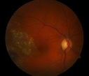
321 x angesehen
|
|

318 x angesehen
|
|

CNV outcomes317 x angesehenFibrotic scar in choroidal neovascularization after anti-VEGF treatmnet
|
|

309 x angesehen
|
|

CNV outcomes303 x angesehenFibrotic scar in choroidal neovascularization after anti-VEGF treatment
|
|

CNV outcomes303 x angesehenFibrotic scar in choroidal neovascularization after anti-VEGF treatment
|
|

CNV outcomes303 x angesehenFibrotic scar in choroidal neovascularization after anti-VEGF treatment
|
|

287 x angesehen
|
|

287 x angesehen
|
|

281 x angesehen
|
|

281 x angesehen
|
|

278 x angesehen
|
|

276 x angesehen
|
|

275 x angesehen
|
|
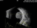
choroidal melanoma274 x angesehenAn Asian male, 37 years old, choroidal melanoma.
|
|

273 x angesehen
|
|
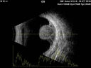
choroidal melanoma.270 x angesehenAn Asian male, 37 years old, choroidal melanoma.
|
|

267 x angesehen
|
|

Pucker266 x angesehen64female pucker
|
|

265 x angesehen
|
|

264 x angesehen
|
|
262 x angesehen
|
|
| 193 Dateien auf 2 Seite(n) |
1 |
|


