Retina Gallery ~ Full Sized Retina Images

Library of Free, Non-Copyrighted Retina Images and Videos

Choose your language:

Most viewed - Acute Macular Neuroretinopathy - 40 year old female

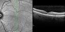
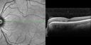
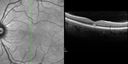
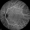
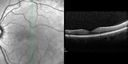
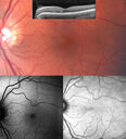
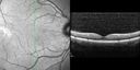
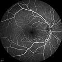
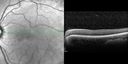
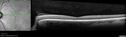
Sudden vision loss in the left eye in a 40 year old female. No prodromal symptoms. Vision was 20/50 left eye. She noticed that at first the vision was good but there was a moving spot in the vision in the left eye. She cleaned her glasses and took them off. She thought maybe the vision was off because of the glasses. Then the vision change persisted. When she closed the right eye there was a true blind spot that she could not see throught that was up and to the left (superotemporal) at first. Then eventually it moved so it is closer to the central vision and the spot has not gone away. She went to the ER on Saturday with the non-clearing blind spot and they did not do a brain scan. The patient also has Lupus for which she takes Plaquenil for about 3 years. She had a full workup yesterday with her ophthalmologist. There was concern about something neurological. Her Optometrist thougth she should come in for a checkup. PMHx: Systemic Lupus Erythematosus. Migraine. Hypertension, Systemic. SYSTEMIC MEDS: plaquenil. topomax. lisinopril 40mg. hydroxyzine HCL 25MG PRN. ZORVOLEX 35 MG PRN. ZOFREN 8 MG. NAPROXEN PRN 550MG. TRAMADOL HCL PRN 50 MG. GABAPENTIN PRN 600MG. TIZANIDINE 4 MG AT BEDTIME. Vit D2 1.25 MG. INDOMETHACIN 25 MG PRN. PRILOSEC ONCE A DAY 20 MG. RELPAX 40 MG. Propranolol Hydrochloride, 40 mg oral tablet. topiramate 100mg. hydroxychloroquine sulfate 200 MG (hydroxychloroquine 155 MG) Oral Tablet. Cymbalta 30 MG Enteric Coated Capsule.

amn_chto1_080514_11.jpg
Acute Macular Neuroretinopathy - (AMN) - Spot in Vision OS - 40 Year old Female1034 viewsSD OCT shows disruption of the outer retina nasal to the fovea
amn_chto1_080514_10.jpg
Acute Macular Neuroretinopathy - (AMN) - Spot in Vision OS - 40 Year old Female992 viewsSD OCT shows disruption of the outer retina nasal to the fovea
amn_chto1_080514_08.jpg
Acute Macular Neuroretinopathy - (AMN) - Spot in Vision OS - 40 Year old Female906 viewsSD OCT shows disruption of the outer retina nasal to the fovea
amn_chto1_080514_07.jpg
Acute Macular Neuroretinopathy - (AMN) - Spot in Vision OS - 40 Year old Female848 viewsSD OCT shows disruption of the outer retina nasal to the fovea
amn_chto1_080514_09.jpg
Acute Macular Neuroretinopathy - (AMN) - Spot in Vision OS - 40 Year old Female823 viewsSD OCT shows disruption of the outer retina nasal to the fovea
amn_chto1_080514_12.jpg
Acute Macular Neuroretinopathy - (AMN) - Spot in Vision OS - 40 Year old Female794 viewsSD OCT shows disruption of the outer retina nasal to the fovea
amn_chto1_080514_01.png
Acute Macular Neuroretinopathy - (AMN) - Spot in Vision OS - 40 Year old Female766 views
amn_chto1_080514_29.jpg
Acute Macular Neuroretinopathy - (AMN) - Spot in Vision OS - 40 Year old Female761 viewsICG angiogram is normal
amn_chto1_080514_32.jpg
Acute Macular Neuroretinopathy - (AMN) - Spot in Vision OS - 40 Year old Female743 viewsICG angiogram is normal
amn_chto2_091614_37.jpg
Acute Macular Neuroretinopathy - (AMN) - Spot in Vision OS - 40 Year old Female742 views4 months later spot is nearly gone and vision is back to normal (almost) - 20/20
amn_chto1_080514_06.jpg
Acute Macular Neuroretinopathy - (AMN) - Spot in Vision OS - 40 Year old Female740 viewsSD OCT shows disruption of the outer retina nasal to the fovea
amn_chto1_080514_01_copy.png
Acute Macular Neuroretinopathy - (AMN) - Spot in Vision OS - 40 Year old Female723 views
amn_chto1_080514_28.jpg
Acute Macular Neuroretinopathy - (AMN) - Spot in Vision OS - 40 Year old Female721 viewsICG angiogram is normal
amn_chto1_080514_33.jpg
Acute Macular Neuroretinopathy - (AMN) - Spot in Vision OS - 40 Year old Female720 viewsICG angiogram is normal
amn.jpg
Acute Macular Neuroretinopathy - 40 year old female718 views
amn_chto2_091614_35.jpg
Acute Macular Neuroretinopathy - (AMN) - Spot in Vision OS - 40 Year old Female717 views
amn_chto1_080514_02.png
Acute Macular Neuroretinopathy - (AMN) - Spot in Vision OS - 40 Year old Female709 viewsPhotos shows gray area nasal to the fovea
amn_chto1_080514_03.jpg
Acute Macular Neuroretinopathy - (AMN) - Spot in Vision OS - 40 Year old Female704 views
amn_chto1_080514_05.jpg
Acute Macular Neuroretinopathy - (AMN) - Spot in Vision OS - 40 Year old Female701 views
amn_chto1_080514_30.jpg
Acute Macular Neuroretinopathy - (AMN) - Spot in Vision OS - 40 Year old Female699 viewsICG angiogram is normal
amn_chto1_080514_19.jpg
Acute Macular Neuroretinopathy - (AMN) - Spot in Vision OS - 40 Year old Female697 viewsFluorescein Angiogram is normal
amn_chto1_080514_34.jpg
Acute Macular Neuroretinopathy - (AMN) - Spot in Vision OS - 40 Year old Female692 views
amn_chto1_080514_27.jpg
Acute Macular Neuroretinopathy - (AMN) - Spot in Vision OS - 40 Year old Female687 viewsICG angiogram is normal
amn_chto1_080514_31.jpg
Acute Macular Neuroretinopathy - (AMN) - Spot in Vision OS - 40 Year old Female684 viewsICG angiogram is normal
amn_chto1_080514_04.jpg
Acute Macular Neuroretinopathy - (AMN) - Spot in Vision OS - 40 Year old Female660 views
amn_chto1_080514_20.jpg
Acute Macular Neuroretinopathy - (AMN) - Spot in Vision OS - 40 Year old Female657 viewsFluorescein Angiogram is normal
amn_chto1_080514_15.jpg
Acute Macular Neuroretinopathy - (AMN) - Spot in Vision OS - 40 Year old Female654 viewsFAF is normal
amn_chto1_080514_18.jpg
Acute Macular Neuroretinopathy - (AMN) - Spot in Vision OS - 40 Year old Female630 viewsFluorescein Angiogram is normal
amn_chto1_080514_21.jpg
Acute Macular Neuroretinopathy - (AMN) - Spot in Vision OS - 40 Year old Female628 viewsFluorescein Angiogram is normal
amn_chto1_080514_13.jpg
Acute Macular Neuroretinopathy - (AMN) - Spot in Vision OS - 40 Year old Female620 views
amn_chto1_080514_16.jpg
Acute Macular Neuroretinopathy - (AMN) - Spot in Vision OS - 40 Year old Female619 viewsFAF is normal
amn_chto1_080514_14.jpg
Acute Macular Neuroretinopathy - (AMN) - Spot in Vision OS - 40 Year old Female616 viewsIR shows gray spot nasal to the fovea
amn_3yearlaterOS-ir.jpg
AMN 3 years later614 viewsShe still sees the blind spot one she looks for it.
amn_chto1_080514_17.jpg
Acute Macular Neuroretinopathy - (AMN) - Spot in Vision OS - 40 Year old Female611 viewsFluorescein Angiogram is normal
amn_chto1_080514_22.jpg
Acute Macular Neuroretinopathy - (AMN) - Spot in Vision OS - 40 Year old Female609 viewsFluorescein Angiogram is normal
amn_chto2_091614_36.jpg
Acute Macular Neuroretinopathy - (AMN) - Spot in Vision OS - 40 Year old Female601 viewsVision improved 1 month later
amn_chto1_080514_26.jpg
Acute Macular Neuroretinopathy - (AMN) - Spot in Vision OS - 40 Year old Female597 views
amn_3yearlaterOS.jpg
AMN 3 years later583 viewsShe still sees the blind spot one she looks for it.
amn_chto1_080514_25.jpg
Acute Macular Neuroretinopathy - (AMN) - Spot in Vision OS - 40 Year old Female577 viewsFluorescein Angiogram is normal
amn_chto1_080514_23.jpg
Acute Macular Neuroretinopathy - (AMN) - Spot in Vision OS - 40 Year old Female574 viewsFluorescein Angiogram is normal
amn_chto1_080514_24.jpg
Acute Macular Neuroretinopathy - (AMN) - Spot in Vision OS - 40 Year old Female546 viewsFluorescein Angiogram is normal
     
41 files on 1 page(s)

Sudden vision loss in the left eye in a 40 year old female. No prodromal symptoms. Vision was 20/50 left eye. She noticed that at first the vision was good but there was a moving spot in the vision in the left eye. She cleaned her glasses and took them off. She thought maybe the vision was off because of the glasses. Then the vision change persisted. When she closed the right eye there was a true blind spot that she could not see throught that was up and to the left (superotemporal) at first. Then eventually it moved so it is closer to the central vision and the spot has not gone away. She went to the ER on Saturday with the non-clearing blind spot and they did not do a brain scan. The patient also has Lupus for which she takes Plaquenil for about 3 years. She had a full workup yesterday with her ophthalmologist. There was concern about something neurological. Her Optometrist thougth she should come in for a checkup. PMHx: Systemic Lupus Erythematosus. Migraine. Hypertension, Systemic. SYSTEMIC MEDS: plaquenil. topomax. lisinopril 40mg. hydroxyzine HCL 25MG PRN. ZORVOLEX 35 MG PRN. ZOFREN 8 MG. NAPROXEN PRN 550MG. TRAMADOL HCL PRN 50 MG. GABAPENTIN PRN 600MG. TIZANIDINE 4 MG AT BEDTIME. Vit D2 1.25 MG. INDOMETHACIN 25 MG PRN. PRILOSEC ONCE A DAY 20 MG. RELPAX 40 MG. Propranolol Hydrochloride, 40 mg oral tablet. topiramate 100mg. hydroxychloroquine sulfate 200 MG (hydroxychloroquine 155 MG) Oral Tablet. Cymbalta 30 MG Enteric Coated Capsule.