Most viewed - Branch Retinal Vein Occlusion with retinal hemorrhages, Left eye
|
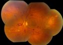
BRVO Inferiorly with Retinal Hemorrhages in the Left Eye593 viewsPatient comes in with complaint of "spot in vision", sort of a cloudy haze in the left eye. Patient's VA was 20/100-1 with no improvement with pinhole in the left eye. Fundus photos show multiple retinal hemorrhages scattered with a branch retinal vein occlusion inferiorily. Patient is also diabetic.
|
|
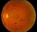
BRVO Inferiorly with Retinal Hemorrhages in the Left Eye542 viewsPatient comes in with complaint of "spot in vision", sort of a cloudy haze in the left eye. Patient's VA was 20/100-1 with no improvement with pinhole in the left eye. Fundus photos show multiple retinal hemorrhages scattered with a branch retinal vein occlusion inferiorily. Patient is also diabetic.
|
|
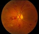
BRVO Inferiorly with Retinal Hemorrhages in the Left Eye476 viewsPatient comes in with complaint of "spot in vision", sort of a cloudy haze in the left eye. Patient's VA was 20/100-1 with no improvement with pinhole in the left eye. Fundus photos show multiple retinal hemorrhages scattered with a branch retinal vein occlusion inferiorily. Patient is also diabetic.
|
|
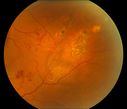
BRVO Inferiorly with Retinal Hemorrhages in the Left Eye476 viewsPatient comes in with complaint of "spot in vision", sort of a cloudy haze in the left eye. Patient's VA was 20/100-1 with no improvement with pinhole in the left eye. Fundus photos show multiple retinal hemorrhages scattered with a branch retinal vein occlusion inferiorily. Patient is also diabetic.
|
|
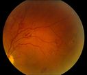
BRVO Inferiorly with Retinal Hemorrhages in the Left Eye441 viewsPatient comes in with complaint of "spot in vision", sort of a cloudy haze in the left eye. Patient's VA was 20/100-1 with no improvement with pinhole in the left eye. Fundus photos show multiple retinal hemorrhages scattered with a branch retinal vein occlusion inferiorily. Patient is also diabetic.
|
|
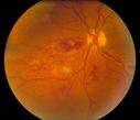
BRVO Inferiorly with Retinal Hemorrhages in the Left Eye410 viewsPatient comes in with complaint of "spot in vision", sort of a cloudy haze in the left eye. Patient's VA was 20/100-1 with no improvement with pinhole in the left eye. Fundus photos show multiple retinal hemorrhages scattered with a branch retinal vein occlusion inferiorily. Patient is also diabetic.
|
|
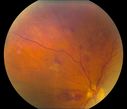
BRVO Inferiorly with Retinal Hemorrhages in the Left Eye401 viewsPatient comes in with complaint of "spot in vision", sort of a cloudy haze in the left eye. Patient's VA was 20/100-1 with no improvement with pinhole in the left eye. Fundus photos show multiple retinal hemorrhages scattered with a branch retinal vein occlusion inferiorily. Patient is also diabetic.
|
|
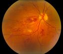
BRVO Inferiorly with Retinal Hemorrhages in the Left Eye386 viewsPatient comes in with complaint of "spot in vision", sort of a cloudy haze in the left eye. Patient's VA was 20/100-1 with no improvement with pinhole in the left eye. Fundus photos show multiple retinal hemorrhages scattered with a branch retinal vein occlusion inferiorily. Patient is also diabetic.
|
|
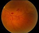
BRVO Inferiorly with Retinal Hemorrhages in the Left Eye366 viewsPatient comes in with complaint of "spot in vision", sort of a cloudy haze in the left eye. Patient's VA was 20/100-1 with no improvement with pinhole in the left eye. Fundus photos show multiple retinal hemorrhages scattered with a branch retinal vein occlusion inferiorily. Patient is also diabetic.
|
|
|
|
|
|
|


