Most viewed - Proliferative Diabetic Retinopathy - Lacy Neovascularization of the Disc - Photo of Fresh Panretinal (Scatter) Photocoagulation 64-year-old woman was seen 6 months ago, still with vision loss right eye. She has been diabetic for eleven years. Her hemoglobin A1Cs are running high lately. She had laser done in Texas. She relocated back to Florida now permanently. The right eye does have a central scotoma dating back to the laser treatment done about a year ago.
VISUAL ACUITY: OD 20/63, OS 20/40. IOP: OD 17, OS 13. There is 3+ nuclear sclerosis in both eyes.
EXTENDED OPHTHALMOSCOPY:
OD: Vertical C/D ratio is 0.0. There is no posterior vitreous separation and rare retinal hemorrhages.
OS: Vertical C/D ratio is 0.2. There is no posterior vitreous separation and rare retinal hemorrhages. There is new NVD.
SPECTRALIS-SD-OCT SCAN: The OCT scan of the right eye does show an epiretinal membrane where there is a central area of photoreceptor defect neatly under the center of the macula. The left eye shows an epiretinal membrane with vitreoretinal traction and a partial evolving posterior vitreous separation. The photos in the right eye show patchy retinal hemorrhages. In the left eye there is light laser and there is an epiretinal membrane.
FLUORESCEIN ANGIOGRAPHY: Fluorescein angiography shows multiple microaneurysms in each eye with light laser. The left eye does have a diffuse leakage from the macula, which looks more like tractional leakage than diabetic leakage. I see very few microaneurysms near the fovea. The left has leakage from NVD and nonperfusion inferonasally.
IMPRESSION:
1. BACKGROUND DIABETIC RETINOPATHY – OD; PDR OS.
2. VITREOMACULAR TRACTION – LEFT EYE
3. EPIRETINAL MEMBRANE – LEFT EYE
4. MACULAR EDEMA – LEFT EYE
5. CATARACT
6. CENTRAL SCOTOMA – RIGHT EYE
|

Proliferative Diabetic Retinopathy - Fresh and Old Scatter (Panretinal) Photocoagulation (Laser) - Neovascularization is regressing856 views1 month after initial laser
|
|

FA showing NVE, NVD, non- perfusion left eye inferonasally with neovascularization at the margin of the non-perfusion778 views
|
|

bdr762 views
|
|

Lacy Neovascularization of the Disk - Color Photo737 views
|
|
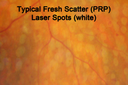
Panretinal (Scatter) Photocoagulation (Fresh)730 views
|
|

NVD - proliferative diabetic retinopathy Heidelberg False Color712 views
|
|

FA showing NVE, NVD, non- perfusion left eye inferonasally with neovascularization at the margin of the non-perfusion (heavy leakage from NV)711 views
|
|

Proliferative Diabetic Retinopathy - Fresh and Old Scatter (Panretinal) Photocoagulation (Laser) - Neovascularization is regressing706 views1 month after initial laser
|
|

FA showing NVE, NVD, non- perfusion left eye inferonasally with neovascularization at the margin of the non-perfusion700 views
|
|
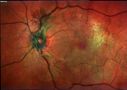
NVD - proliferative diabetic retinopathy Heidelberg False Color694 views
|
|
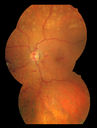
Proliferative Diabetic Retinopathy - Fresh and Old Scatter (Panretinal) Photocoagulation (Laser) - Neovascularization is regressing682 views1 month after initial laser
|
|
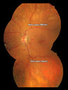
Proliferative Diabetic Retinopathy - Fresh and Old Scatter (Panretinal) Photocoagulation (Laser) - Neovascularization is regressing674 views1 month after initial laser
|
|
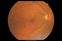
BDR657 views
|
|

NVD - proliferative diabetic retinopathy650 views
|
|
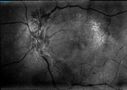
NVD - proliferative diabetic retinopathy Heidelberg False Color648 views
|
|
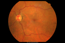
PDR638 views
|
|
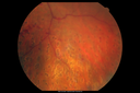
Proliferative Diabetic Retinopathy - Fresh and Old Scatter (Panretinal) Photocoagulation (Laser) - Neovascularization is regressing620 views1 month after initial laser
|
|
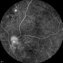
FA showing NVE, NVD, non- perfusion left eye inferonasally with neovascularization at the margin of the non-perfusion619 views
|
|
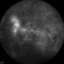
FA showing NVE, NVD, non- perfusion left eye inferonasally with neovascularization at the margin of the non-perfusion618 views
|
|
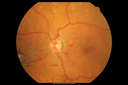
Proliferative Diabetic Retinopathy - Fresh and Old Scatter (Panretinal) Photocoagulation (Laser) - Neovascularization is regressing615 views1 month after initial laser
|
|

bdr613 views
|
|
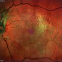
Heidelberg False Color607 views
|
|
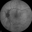
FAF598 views
|
|
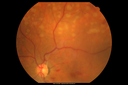
Proliferative Diabetic Retinopathy - Fresh and Old Scatter (Panretinal) Photocoagulation (Laser) - Neovascularization is regressing594 views1 month after initial laser
|
|
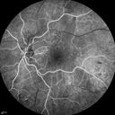
FA showing NVE, NVD, non- perfusion left eye inferonasally with neovascularization at the margin of the non-perfusion588 views
|
|
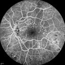
FA showing NVE, NVD, non- perfusion left eye inferonasally with neovascularization at the margin of the non-perfusion588 views
|
|
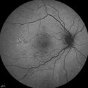
FAF583 views
|
|
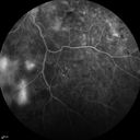
FA showing NVE, NVD, non- perfusion left eye inferonasally with neovascularization at the margin of the non-perfusion581 views
|
|
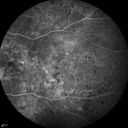
FA showing NVE, NVD, non- perfusion left eye inferonasally with neovascularization at the margin of the non-perfusion579 views
|
|
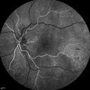
FA showing NVE, NVD, non- perfusion left eye inferonasally with neovascularization at the margin of the non-perfusion577 views
|
|
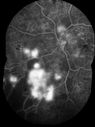
FA showing NVE, NVD, non- perfusion left eye inferonasally with neovascularization at the margin of the non-perfusion574 views
|
|
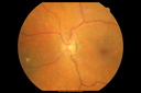
PDR572 views
|
|
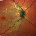
BDR571 views
|
|
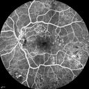
FA showing NVE, NVD, non- perfusion left eye inferonasally with neovascularization at the margin of the non-perfusion567 views
|
|
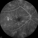
BDR right eye - previous laser564 views
|
|
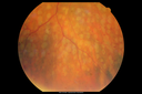
Panretinal (Scatter) Photocoagulation (Fresh)548 views
|
|
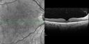
No significant macular edema546 views
|
|
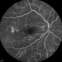
BDR right eye - previous laser541 views
|
|
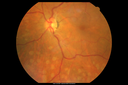
Panretinal (Scatter) Photocoagulation (Fresh)540 views
|
|
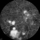
FA showing NVE, NVD, non- perfusion left eye inferonasally with neovascularization at the margin of the non-perfusion537 views
|
|

Neovascularization of the Disc - Heidelberg false color 530 views
|
|
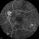
FA showing NVE, NVD, non- perfusion left eye inferonasally with neovascularization at the margin of the non-perfusion528 views
|
|
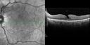
No significant macular edema510 views
|
|
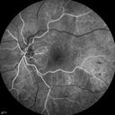
FA showing NVE, NVD, non- perfusion left eye inferonasally with neovascularization at the margin of the non-perfusion510 views
|
|
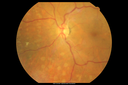
Panretinal (Scatter) Photocoagulation (Fresh)503 views
|
|
|
|
|
|
|
64-year-old woman was seen 6 months ago, still with vision loss right eye. She has been diabetic for eleven years. Her hemoglobin A1Cs are running high lately. She had laser done in Texas. She relocated back to Florida now permanently. The right eye does have a central scotoma dating back to the laser treatment done about a year ago.
VISUAL ACUITY: OD 20/63, OS 20/40. IOP: OD 17, OS 13. There is 3+ nuclear sclerosis in both eyes.
EXTENDED OPHTHALMOSCOPY:
OD: Vertical C/D ratio is 0.0. There is no posterior vitreous separation and rare retinal hemorrhages.
OS: Vertical C/D ratio is 0.2. There is no posterior vitreous separation and rare retinal hemorrhages. There is new NVD.
SPECTRALIS-SD-OCT SCAN: The OCT scan of the right eye does show an epiretinal membrane where there is a central area of photoreceptor defect neatly under the center of the macula. The left eye shows an epiretinal membrane with vitreoretinal traction and a partial evolving posterior vitreous separation. The photos in the right eye show patchy retinal hemorrhages. In the left eye there is light laser and there is an epiretinal membrane.
FLUORESCEIN ANGIOGRAPHY: Fluorescein angiography shows multiple microaneurysms in each eye with light laser. The left eye does have a diffuse leakage from the macula, which looks more like tractional leakage than diabetic leakage. I see very few microaneurysms near the fovea. The left has leakage from NVD and nonperfusion inferonasally.
IMPRESSION:
1. BACKGROUND DIABETIC RETINOPATHY – OD; PDR OS.
2. VITREOMACULAR TRACTION – LEFT EYE
3. EPIRETINAL MEMBRANE – LEFT EYE
4. MACULAR EDEMA – LEFT EYE
5. CATARACT
6. CENTRAL SCOTOMA – RIGHT EYE


