Más vistos - Branch Retinal Artery Occlusion BRAO 69 year old woman - She was doing well until Sunday when she was watching TV and boom she noticed that she could not see well out of the right eye. There was not pain. She noticed that she could not see on the top out of the right eye. The eye also felt a little swollen. There is a dark black thing in her vision OD. When she looks at letters she loses from the center of her vision and up. She had no focal neurological problems at the time of the vision loss. She has no history of stroke. VA 20/100 OD 20/25 OS
|

Branch Retinal Artery Occlusionvista 548 vecesAcute Inferotemporal Occlusion Right Eye - FA shows delayed circulation, OCT shows edema of superficial layers of retina
|
|
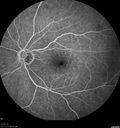
Branch Retinal Artery Occlusionvista 526 vecesAcute Inferotemporal Occlusion Right Eye - FA shows delayed circulation, OCT shows edema of superficial layers of retina
|
|
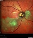
Branch Retinal Artery Occlusionvista 510 vecesAcute Inferotemporal Occlusion Right Eye - FA shows delayed circulation, OCT shows edema of superficial layers of retina
|
|
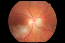
Branch Retinal Artery Occlusionvista 502 vecesAcute Inferotemporal Occlusion Right Eye - FA shows delayed circulation, OCT shows edema of superficial layers of retina
|
|
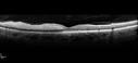
Branch Retinal Artery Occlusionvista 499 vecesAcute Inferotemporal Occlusion Right Eye - FA shows delayed circulation, OCT shows edema of superficial layers of retina
|
|

Branch Retinal Artery Occlusionvista 496 vecesAcute Inferotemporal Occlusion Right Eye - FA shows delayed circulation, OCT shows edema of superficial layers of retina
|
|
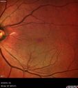
Branch Retinal Artery Occlusionvista 492 vecesAcute Inferotemporal Occlusion Right Eye - FA shows delayed circulation, OCT shows edema of superficial layers of retina
|
|

Branch Retinal Artery Occlusionvista 477 vecesAcute Inferotemporal Occlusion Right Eye - FA shows delayed circulation, OCT shows edema of superficial layers of retina
|
|

Branch Retinal Artery Occlusionvista 476 vecesAcute Inferotemporal Occlusion Right Eye - FA shows delayed circulation, OCT shows edema of superficial layers of retina
|
|
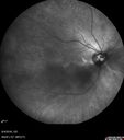
Branch Retinal Artery Occlusionvista 475 vecesAcute Inferotemporal Occlusion Right Eye - FA shows delayed circulation, OCT shows edema of superficial layers of retina
|
|
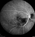
Branch Retinal Artery Occlusionvista 472 vecesAcute Inferotemporal Occlusion Right Eye - FA shows delayed circulation, OCT shows edema of superficial layers of retina
|
|
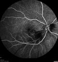
Branch Retinal Artery Occlusionvista 466 vecesAcute Inferotemporal Occlusion Right Eye - FA shows delayed circulation, OCT shows edema of superficial layers of retina
|
|

Branch Retinal Artery Occlusionvista 466 vecesAcute Inferotemporal Occlusion Right Eye - FA shows delayed circulation, OCT shows edema of superficial layers of retina
|
|

Branch Retinal Artery Occlusionvista 463 vecesAcute Inferotemporal Occlusion Right Eye - FA shows delayed circulation, OCT shows edema of superficial layers of retina
|
|

Branch Retinal Artery Occlusionvista 461 vecesAcute Inferotemporal Occlusion Right Eye - FA shows delayed circulation, OCT shows edema of superficial layers of retina
|
|
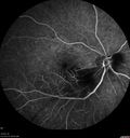
Branch Retinal Artery Occlusionvista 461 vecesAcute Inferotemporal Occlusion Right Eye - FA shows delayed circulation, OCT shows edema of superficial layers of retina
|
|
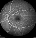
Branch Retinal Artery Occlusionvista 461 vecesAcute Inferotemporal Occlusion Right Eye - FA shows delayed circulation, OCT shows edema of superficial layers of retina
|
|
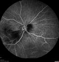
Branch Retinal Artery Occlusionvista 450 vecesAcute Inferotemporal Occlusion Right Eye - FA shows delayed circulation, OCT shows edema of superficial layers of retina
|
|
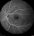
Branch Retinal Artery Occlusionvista 448 vecesAcute Inferotemporal Occlusion Right Eye - FA shows delayed circulation, OCT shows edema of superficial layers of retina
|
|
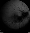
Branch Retinal Artery Occlusionvista 426 vecesAcute Inferotemporal Occlusion Right Eye - FA shows delayed circulation, OCT shows edema of superficial layers of retina
|
|
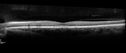
Branch Retinal Artery Occlusionvista 412 vecesAcute Inferotemporal Occlusion Right Eye - FA shows delayed circulation, OCT shows edema of superficial layers of retina
|
|

Branch Retinal Artery Occlusionvista 406 vecesAcute Inferotemporal Occlusion Right Eye - FA shows delayed circulation, OCT shows edema of superficial layers of retina
|
|
|
|
| 22 archivos en 1 página(s) |
|
69 year old woman - She was doing well until Sunday when she was watching TV and boom she noticed that she could not see well out of the right eye. There was not pain. She noticed that she could not see on the top out of the right eye. The eye also felt a little swollen. There is a dark black thing in her vision OD. When she looks at letters she loses from the center of her vision and up. She had no focal neurological problems at the time of the vision loss. She has no history of stroke. VA 20/100 OD 20/25 OS


