Last additions - Tumors, Masses, Pigmented Lesions
|
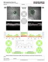
Advanced age-related macular degeneration188 views87 year old female. Severe macular bleed 15 years ago OD. Now with disciform scar. Left eye has progressing geographic atrophy.
VA OD: HM, OS: 5/200. IOP normal OUMar 17, 2024
|
|

Advanced age-related macular degeneration153 views87 year old female. Severe macular bleed 15 years ago OD. Now with disciform scar. Left eye has progressing geographic atrophy.
VA OD: HM, OS: 5/200. IOP normal OUMar 17, 2024
|
|
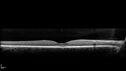
Advanced age-related macular degeneration185 views87 year old female. Severe macular bleed 15 years ago OD. Now with disciform scar. Left eye has progressing geographic atrophy.
VA OD: HM, OS: 5/200. IOP normal OUMar 17, 2024
|
|

Advanced age-related macular degeneration172 views87 year old female. Severe macular bleed 15 years ago OD. Now with disciform scar. Left eye has progressing geographic atrophy.
VA OD: HM, OS: 5/200. IOP normal OUMar 17, 2024
|
|
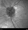
Advanced age-related macular degeneration151 views87 year old female. Severe macular bleed 15 years ago OD. Now with disciform scar. Left eye has progressing geographic atrophy.
VA OD: HM, OS: 5/200. IOP normal OUMar 17, 2024
|
|
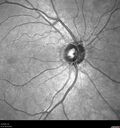
Advanced age-related macular degeneration174 views87 year old female. Severe macular bleed 15 years ago OD. Now with disciform scar. Left eye has progressing geographic atrophy.
VA OD: HM, OS: 5/200. IOP normal OUMar 17, 2024
|
|
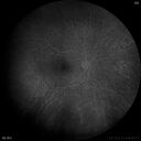
Retinal Angioma168 views46 year old female - Her original reason for an eye examination was that she had been in a car accident. Then she saw that the vision was blurry in just the left eye. Prior to that she had no known vision changes. Since the accident she notices that her vision is better and her headaches are getting less bothersome.
PMhx benign, Meds: none
VA OD: Dcc20/20
VA OS: Dcc20/25Mar 11, 2024
|
|
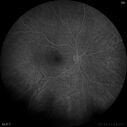
Retinal Angioma163 views46 year old female - Her original reason for an eye examination was that she had been in a car accident. Then she saw that the vision was blurry in just the left eye. Prior to that she had no known vision changes. Since the accident she notices that her vision is better and her headaches are getting less bothersome.
PMhx benign, Meds: none
VA OD: Dcc20/20
VA OS: Dcc20/25Mar 11, 2024
|
|

Retinal Angioma182 views46 year old female - Her original reason for an eye examination was that she had been in a car accident. Then she saw that the vision was blurry in just the left eye. Prior to that she had no known vision changes. Since the accident she notices that her vision is better and her headaches are getting less bothersome.
PMhx benign, Meds: none
VA OD: Dcc20/20
VA OS: Dcc20/25Mar 11, 2024
|
|
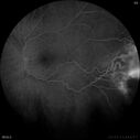
Retinal Angioma162 views46 year old female - Her original reason for an eye examination was that she had been in a car accident. Then she saw that the vision was blurry in just the left eye. Prior to that she had no known vision changes. Since the accident she notices that her vision is better and her headaches are getting less bothersome.
PMhx benign, Meds: none
VA OD: Dcc20/20
VA OS: Dcc20/25Mar 11, 2024
|
|

Retinal Angioma180 views46 year old female - Her original reason for an eye examination was that she had been in a car accident. Then she saw that the vision was blurry in just the left eye. Prior to that she had no known vision changes. Since the accident she notices that her vision is better and her headaches are getting less bothersome.
PMhx benign, Meds: none
VA OD: Dcc20/20
VA OS: Dcc20/25Mar 11, 2024
|
|

Retinal Angioma169 views46 year old female - Her original reason for an eye examination was that she had been in a car accident. Then she saw that the vision was blurry in just the left eye. Prior to that she had no known vision changes. Since the accident she notices that her vision is better and her headaches are getting less bothersome.
PMhx benign, Meds: none
VA OD: Dcc20/20
VA OS: Dcc20/25Mar 11, 2024
|
|

Retinal Angioma153 views46 year old female - Her original reason for an eye examination was that she had been in a car accident. Then she saw that the vision was blurry in just the left eye. Prior to that she had no known vision changes. Since the accident she notices that her vision is better and her headaches are getting less bothersome.
PMhx benign, Meds: none
VA OD: Dcc20/20
VA OS: Dcc20/25Mar 11, 2024
|
|

Retinal Angioma169 views46 year old female - Her original reason for an eye examination was that she had been in a car accident. Then she saw that the vision was blurry in just the left eye. Prior to that she had no known vision changes. Since the accident she notices that her vision is better and her headaches are getting less bothersome.
PMhx benign, Meds: none
VA OD: Dcc20/20
VA OS: Dcc20/25Mar 11, 2024
|
|
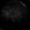
Retinal Angioma156 views46 year old female - Her original reason for an eye examination was that she had been in a car accident. Then she saw that the vision was blurry in just the left eye. Prior to that she had no known vision changes. Since the accident she notices that her vision is better and her headaches are getting less bothersome.
PMhx benign, Meds: none
VA OD: Dcc20/20
VA OS: Dcc20/25Mar 11, 2024
|
|

Retinal Angioma142 views46 year old female - Her original reason for an eye examination was that she had been in a car accident. Then she saw that the vision was blurry in just the left eye. Prior to that she had no known vision changes. Since the accident she notices that her vision is better and her headaches are getting less bothersome.
PMhx benign, Meds: none
VA OD: Dcc20/20
VA OS: Dcc20/25Mar 11, 2024
|
|

Retinal Angioma154 views46 year old female - Her original reason for an eye examination was that she had been in a car accident. Then she saw that the vision was blurry in just the left eye. Prior to that she had no known vision changes. Since the accident she notices that her vision is better and her headaches are getting less bothersome.
PMhx benign, Meds: none
VA OD: Dcc20/20
VA OS: Dcc20/25Mar 11, 2024
|
|

Retinal Angioma151 views46 year old female - Her original reason for an eye examination was that she had been in a car accident. Then she saw that the vision was blurry in just the left eye. Prior to that she had no known vision changes. Since the accident she notices that her vision is better and her headaches are getting less bothersome.
PMhx benign, Meds: none
VA OD: Dcc20/20
VA OS: Dcc20/25Mar 11, 2024
|
|

Retinal Angioma162 views46 year old female - Her original reason for an eye examination was that she had been in a car accident. Then she saw that the vision was blurry in just the left eye. Prior to that she had no known vision changes. Since the accident she notices that her vision is better and her headaches are getting less bothersome.
PMhx benign, Meds: none
VA OD: Dcc20/20
VA OS: Dcc20/25Mar 11, 2024
|
|
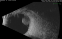
Class 2 choroidal melanoma224 views48 year old man with no visual complaints referred by optometrist.
Brachytherapy was done and biopsy showed a class 2 melanoma. 2 years later the patient developed liver metastases.Feb 12, 2024
|
|
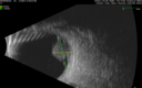
Class 2 choroidal melanoma200 views48 year old man with no visual complaints referred by optometrist.
Brachytherapy was done and biopsy showed a class 2 melanoma. 2 years later the patient developed liver metastases.Feb 12, 2024
|
|
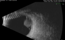
Class 2 choroidal melanoma211 views48 year old man with no visual complaints referred by optometrist.
Brachytherapy was done and biopsy showed a class 2 melanoma. 2 years later the patient developed liver metastases.Feb 12, 2024
|
|

Class 2 choroidal melanoma172 views48 year old man with no visual complaints referred by optometrist.
Brachytherapy was done and biopsy showed a class 2 melanoma. 2 years later the patient developed liver metastases.Feb 12, 2024
|
|
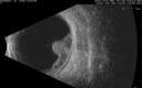
Class 2 choroidal melanoma193 views48 year old man with no visual complaints referred by optometrist.
Brachytherapy was done and biopsy showed a class 2 melanoma. 2 years later the patient developed liver metastases.Feb 12, 2024
|
|

Class 2 choroidal melanoma206 views48 year old man with no visual complaints referred by optometrist.
Brachytherapy was done and biopsy showed a class 2 melanoma. 2 years later the patient developed liver metastases.Feb 12, 2024
|
|

Class 2 choroidal melanoma253 views48 year old man with no visual complaints referred by optometrist.
Brachytherapy was done and biopsy showed a class 2 melanoma. 2 years later the patient developed liver metastases.Feb 12, 2024
|
|
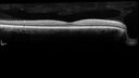
Class 2 choroidal melanoma197 views48 year old man with no visual complaints referred by optometrist.
Brachytherapy was done and biopsy showed a class 2 melanoma. 2 years later the patient developed liver metastases.Feb 12, 2024
|
|

Class 2 choroidal melanoma207 views48 year old man with no visual complaints referred by optometrist.
Brachytherapy was done and biopsy showed a class 2 melanoma. 2 years later the patient developed liver metastases.Feb 12, 2024
|
|

Class 2 choroidal melanoma231 views48 year old man with no visual complaints referred by optometrist.
Brachytherapy was done and biopsy showed a class 2 melanoma. 2 years later the patient developed liver metastases.Feb 12, 2024
|
|
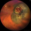
Class 2 choroidal melanoma217 views48 year old man with no visual complaints referred by optometrist.
Brachytherapy was done and biopsy showed a class 2 melanoma. 2 years later the patient developed liver metastases.Feb 12, 2024
|
|
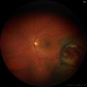
Class 2 choroidal melanoma192 views48 year old man with no visual complaints referred by optometrist.
Brachytherapy was done and biopsy showed a class 2 melanoma. 2 years later the patient developed liver metastases.Feb 12, 2024
|
|

Class 2 choroidal melanoma205 views48 year old man with no visual complaints referred by optometrist.
Brachytherapy was done and biopsy showed a class 2 melanoma. 2 years later the patient developed liver metastases.Feb 12, 2024
|
|

Radiation Retinopathy Both Eyes - 2023 - Two years after radiation for ocular lymphoma 254 viewsAug 21, 2023
|
|

Radiation Retinopathy Both Eyes - 2023 - Two years after radiation for ocular lymphoma (24)285 viewsAug 21, 2023
|
|

Radiation Retinopathy Both Eyes - 2023 - Two years after radiation for ocular lymphoma (23)255 viewsAug 21, 2023
|
|

Radiation Retinopathy Both Eyes - 2023 - Two years after radiation for ocular lymphoma (22)254 viewsAug 21, 2023
|
|

Radiation Retinopathy Both Eyes - 2023 - Two years after radiation for ocular lymphoma (21)307 viewsAug 21, 2023
|
|

Radiation Retinopathy Both Eyes - 2023 - Two years after radiation for ocular lymphoma (20)300 viewsAug 21, 2023
|
|

Radiation Retinopathy Both Eyes - 2023 - Two years after radiation for ocular lymphoma (19)244 viewsAug 21, 2023
|
|

Radiation Retinopathy Both Eyes - 2023 - Two years after radiation for ocular lymphoma (18)255 viewsAug 21, 2023
|
|

Radiation Retinopathy Both Eyes - 2023 - Two years after radiation for ocular lymphoma (1)263 viewsAug 21, 2023
|
|

Radiation Retinopathy Both Eyes - 2023 - Two years after radiation for ocular lymphoma (17)265 viewsAug 21, 2023
|
|

Radiation Retinopathy Both Eyes - 2023 - Two years after radiation for ocular lymphoma (16)251 viewsAug 21, 2023
|
|

Radiation Retinopathy Both Eyes - 2023 - Two years after radiation for ocular lymphoma (15)245 viewsAug 21, 2023
|
|

Radiation Retinopathy Both Eyes - 2023 - Two years after radiation for ocular lymphoma (14)278 viewsAug 21, 2023
|
|

Radiation Retinopathy Both Eyes - 2023 - Two years after radiation for ocular lymphoma (13)274 viewsAug 21, 2023
|
|

Radiation Retinopathy Both Eyes - 2023 - Two years after radiation for ocular lymphoma (12)259 viewsAug 21, 2023
|
|

Radiation Retinopathy Both Eyes - 2023 - Two years after radiation for ocular lymphoma (11)299 viewsAug 21, 2023
|
|

Radiation Retinopathy Both Eyes - 2023 - Two years after radiation for ocular lymphoma (10)258 viewsAug 21, 2023
|
|

Radiation Retinopathy Both Eyes - 2023 - Two years after radiation for ocular lymphoma (9)275 viewsAug 21, 2023
|
|

Radiation Retinopathy Both Eyes - 2023 - Two years after radiation for ocular lymphoma (8)280 viewsAug 21, 2023
|
|

Radiation Retinopathy Both Eyes - 2023 - Two years after radiation for ocular lymphoma (6)236 viewsAug 21, 2023
|
|

Radiation Retinopathy Both Eyes - 2023 - Two years after radiation for ocular lymphoma (7)251 viewsAug 21, 2023
|
|

Radiation Retinopathy Both Eyes - 2023 - Two years after radiation for ocular lymphoma (5)266 viewsAug 21, 2023
|
|

Radiation Retinopathy Both Eyes - 2023 - Two years after radiation for ocular lymphoma (4)253 viewsAug 21, 2023
|
|

Radiation Retinopathy Both Eyes - 2023 - Two years after radiation for ocular lymphoma (3)253 viewsAug 21, 2023
|
|

Radiation Retinopathy Both Eyes - 2023 - Two years after radiation for ocular lymphoma (2)276 viewsAug 21, 2023
|
|
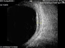
Large chronic choroidal nevus284 viewsAutofluorescence image shows guttering at the bottom of the lesion suggesting chronicity. This was documented 23 years prior to these photosJul 02, 2023
|
|
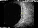
Large chronic choroidal nevus284 viewsAutofluorescence image shows guttering at the bottom of the lesion suggesting chronicity. This was documented 23 years prior to these photosJul 02, 2023
|
|
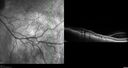
Large chronic choroidal nevus278 viewsAutofluorescence image shows guttering at the bottom of the lesion suggesting chronicity. This was documented 23 years prior to these photosJul 02, 2023
|
|
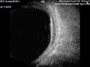
Large chronic choroidal nevus314 viewsAutofluorescence image shows guttering at the bottom of the lesion suggesting chronicity. This was documented 23 years prior to these photosJul 02, 2023
|
|
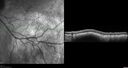
Large chronic choroidal nevus289 viewsAutofluorescence image shows guttering at the bottom of the lesion suggesting chronicity. This was documented 23 years prior to these photosJul 02, 2023
|
|
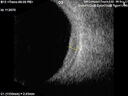
Large chronic choroidal nevus281 viewsAutofluorescence image shows guttering at the bottom of the lesion suggesting chronicity. This was documented 23 years prior to these photosJul 02, 2023
|
|

Large chronic choroidal nevus250 viewsAutofluorescence image shows guttering at the bottom of the lesion suggesting chronicity. This was documented 23 years prior to these photosJul 02, 2023
|
|

Large chronic choroidal nevus256 viewsAutofluorescence image shows guttering at the bottom of the lesion suggesting chronicity. This was documented 23 years prior to these photosJul 02, 2023
|
|
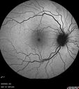
Large chronic choroidal nevus284 viewsAutofluorescence image shows guttering at the bottom of the lesion suggesting chronicity. This was documented 23 years prior to these photosJul 02, 2023
|
|

Large chronic choroidal nevus273 viewsAutofluorescence image shows guttering at the bottom of the lesion suggesting chronicity. This was documented 23 years prior to these photosJul 02, 2023
|
|
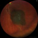
Large chronic choroidal nevus298 viewsAutofluorescence image shows guttering at the bottom of the lesion suggesting chronicity. This was documented 23 years prior to these photosJul 02, 2023
|
|
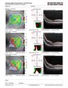
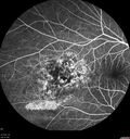
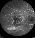
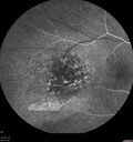
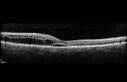
Choroidal Hemangioma Requiring PDT repeatedly304 views64 year old man. Diagnosed with hemangioma in 2015. PDT in 2016. Images are from 2020 when fluid started to reaccumulate. Needed PDT repeated two more times.
PDT treatments: [1] 2/19/2015 -Â Pavan (IOPÂ problem after PDT)Â AND [2] 11/2/21 PDT (7 mm temporal to fovea) - SMCÂ AND [3] 7/22/22 PDT 7.5 mm Temporal to the foveaJan 04, 2023
|
|

Choroidal Hemangioma Requiring PDT repeatedly370 views64 year old man. Diagnosed with hemangioma in 2015. PDT in 2016. Images are from 2020 when fluid started to reaccumulate. Needed PDT repeated two more times.
PDT treatments: [1] 2/19/2015 -Â Pavan (IOPÂ problem after PDT)Â AND [2] 11/2/21 PDT (7 mm temporal to fovea) - SMCÂ AND [3] 7/22/22 PDT 7.5 mm Temporal to the foveaJan 04, 2023
|
|
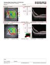
Choroidal Hemangioma Requiring PDT repeatedly287 views64 year old man. Diagnosed with hemangioma in 2015. PDT in 2016. Images are from 2020 when fluid started to reaccumulate. Needed PDT repeated two more times.
PDT treatments: [1] 2/19/2015 -Â Pavan (IOPÂ problem after PDT)Â AND [2] 11/2/21 PDT (7 mm temporal to fovea) - SMCÂ AND [3] 7/22/22 PDT 7.5 mm Temporal to the foveaJan 04, 2023
|
|

Choroidal Hemangioma Requiring PDT repeatedly286 views64 year old man. Diagnosed with hemangioma in 2015. PDT in 2016. Images are from 2020 when fluid started to reaccumulate. Needed PDT repeated two more times.
PDT treatments: [1] 2/19/2015 -Â Pavan (IOPÂ problem after PDT)Â AND [2] 11/2/21 PDT (7 mm temporal to fovea) - SMCÂ AND [3] 7/22/22 PDT 7.5 mm Temporal to the foveaJan 04, 2023
|
|

Choroidal Hemangioma Requiring PDT repeatedly246 views64 year old man. Diagnosed with hemangioma in 2015. PDT in 2016. Images are from 2020 when fluid started to reaccumulate. Needed PDT repeated two more times.
PDT treatments: [1] 2/19/2015 -Â Pavan (IOPÂ problem after PDT)Â AND [2] 11/2/21 PDT (7 mm temporal to fovea) - SMCÂ AND [3] 7/22/22 PDT 7.5 mm Temporal to the foveaJan 04, 2023
|
|
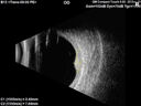
Choroidal Hemangioma Requiring PDT repeatedly286 views64 year old man. Diagnosed with hemangioma in 2015. PDT in 2016. Images are from 2020 when fluid started to reaccumulate. Needed PDT repeated two more times.
PDT treatments: [1] 2/19/2015 -Â Pavan (IOPÂ problem after PDT)Â AND [2] 11/2/21 PDT (7 mm temporal to fovea) - SMCÂ AND [3] 7/22/22 PDT 7.5 mm Temporal to the foveaJan 04, 2023
|
|
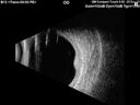
Choroidal Hemangioma Requiring PDT repeatedly260 views64 year old man. Diagnosed with hemangioma in 2015. PDT in 2016. Images are from 2020 when fluid started to reaccumulate. Needed PDT repeated two more times.
PDT treatments: [1] 2/19/2015 -Â Pavan (IOPÂ problem after PDT)Â AND [2] 11/2/21 PDT (7 mm temporal to fovea) - SMCÂ AND [3] 7/22/22 PDT 7.5 mm Temporal to the foveaJan 04, 2023
|
|

Choroidal Hemangioma Requiring PDT repeatedly239 views64 year old man. Diagnosed with hemangioma in 2015. PDT in 2016. Images are from 2020 when fluid started to reaccumulate. Needed PDT repeated two more times.
PDT treatments: [1] 2/19/2015 -Â Pavan (IOPÂ problem after PDT)Â AND [2] 11/2/21 PDT (7 mm temporal to fovea) - SMCÂ AND [3] 7/22/22 PDT 7.5 mm Temporal to the foveaJan 04, 2023
|
|
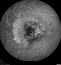
Choroidal Hemangioma Requiring PDT repeatedly254 views64 year old man. Diagnosed with hemangioma in 2015. PDT in 2016. Images are from 2020 when fluid started to reaccumulate. Needed PDT repeated two more times.
PDT treatments: [1] 2/19/2015 -Â Pavan (IOPÂ problem after PDT)Â AND [2] 11/2/21 PDT (7 mm temporal to fovea) - SMCÂ AND [3] 7/22/22 PDT 7.5 mm Temporal to the foveaJan 04, 2023
|
|
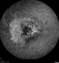
Choroidal Hemangioma Requiring PDT repeatedly275 views64 year old man. Diagnosed with hemangioma in 2015. PDT in 2016. Images are from 2020 when fluid started to reaccumulate. Needed PDT repeated two more times.
PDT treatments: [1] 2/19/2015 -Â Pavan (IOPÂ problem after PDT)Â AND [2] 11/2/21 PDT (7 mm temporal to fovea) - SMCÂ AND [3] 7/22/22 PDT 7.5 mm Temporal to the foveaJan 04, 2023
|
|
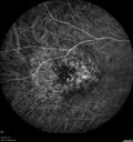
Choroidal Hemangioma Requiring PDT repeatedly245 views64 year old man. Diagnosed with hemangioma in 2015. PDT in 2016. Images are from 2020 when fluid started to reaccumulate. Needed PDT repeated two more times.
PDT treatments: [1] 2/19/2015 -Â Pavan (IOPÂ problem after PDT)Â AND [2] 11/2/21 PDT (7 mm temporal to fovea) - SMCÂ AND [3] 7/22/22 PDT 7.5 mm Temporal to the foveaJan 04, 2023
|
|
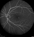
Choroidal Hemangioma Requiring PDT repeatedly241 views64 year old man. Diagnosed with hemangioma in 2015. PDT in 2016. Images are from 2020 when fluid started to reaccumulate. Needed PDT repeated two more times.
PDT treatments: [1] 2/19/2015 -Â Pavan (IOPÂ problem after PDT)Â AND [2] 11/2/21 PDT (7 mm temporal to fovea) - SMCÂ AND [3] 7/22/22 PDT 7.5 mm Temporal to the foveaJan 04, 2023
|
|
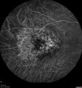
Choroidal Hemangioma Requiring PDT repeatedly276 views64 year old man. Diagnosed with hemangioma in 2015. PDT in 2016. Images are from 2020 when fluid started to reaccumulate. Needed PDT repeated two more times.
PDT treatments: [1] 2/19/2015 -Â Pavan (IOPÂ problem after PDT)Â AND [2] 11/2/21 PDT (7 mm temporal to fovea) - SMCÂ AND [3] 7/22/22 PDT 7.5 mm Temporal to the foveaJan 04, 2023
|
|
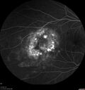
Choroidal Hemangioma Requiring PDT repeatedly250 views64 year old man. Diagnosed with hemangioma in 2015. PDT in 2016. Images are from 2020 when fluid started to reaccumulate. Needed PDT repeated two more times.
PDT treatments: [1] 2/19/2015 -Â Pavan (IOPÂ problem after PDT)Â AND [2] 11/2/21 PDT (7 mm temporal to fovea) - SMCÂ AND [3] 7/22/22 PDT 7.5 mm Temporal to the foveaJan 04, 2023
|
|
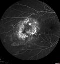
Choroidal Hemangioma Requiring PDT repeatedly276 views64 year old man. Diagnosed with hemangioma in 2015. PDT in 2016. Images are from 2020 when fluid started to reaccumulate. Needed PDT repeated two more times.
PDT treatments: [1] 2/19/2015 -Â Pavan (IOPÂ problem after PDT)Â AND [2] 11/2/21 PDT (7 mm temporal to fovea) - SMCÂ AND [3] 7/22/22 PDT 7.5 mm Temporal to the foveaJan 04, 2023
|
|
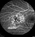
Choroidal Hemangioma Requiring PDT repeatedly285 views64 year old man. Diagnosed with hemangioma in 2015. PDT in 2016. Images are from 2020 when fluid started to reaccumulate. Needed PDT repeated two more times.
PDT treatments: [1] 2/19/2015 -Â Pavan (IOPÂ problem after PDT)Â AND [2] 11/2/21 PDT (7 mm temporal to fovea) - SMCÂ AND [3] 7/22/22 PDT 7.5 mm Temporal to the foveaJan 04, 2023
|
|
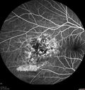
Choroidal Hemangioma Requiring PDT repeatedly256 views64 year old man. Diagnosed with hemangioma in 2015. PDT in 2016. Images are from 2020 when fluid started to reaccumulate. Needed PDT repeated two more times.
PDT treatments: [1] 2/19/2015 -Â Pavan (IOPÂ problem after PDT)Â AND [2] 11/2/21 PDT (7 mm temporal to fovea) - SMCÂ AND [3] 7/22/22 PDT 7.5 mm Temporal to the foveaJan 04, 2023
|
|
Choroidal Hemangioma Requiring PDT repeatedly249 views64 year old man. Diagnosed with hemangioma in 2015. PDT in 2016. Images are from 2020 when fluid started to reaccumulate. Needed PDT repeated two more times.
PDT treatments: [1] 2/19/2015 -Â Pavan (IOPÂ problem after PDT)Â AND [2] 11/2/21 PDT (7 mm temporal to fovea) - SMCÂ AND [3] 7/22/22 PDT 7.5 mm Temporal to the foveaJan 04, 2023
|
|
Choroidal Hemangioma Requiring PDT repeatedly224 views64 year old man. Diagnosed with hemangioma in 2015. PDT in 2016. Images are from 2020 when fluid started to reaccumulate. Needed PDT repeated two more times.
PDT treatments: [1] 2/19/2015 -Â Pavan (IOPÂ problem after PDT)Â AND [2] 11/2/21 PDT (7 mm temporal to fovea) - SMCÂ AND [3] 7/22/22 PDT 7.5 mm Temporal to the foveaJan 04, 2023
|
|
Choroidal Hemangioma Requiring PDT repeatedly260 views64 year old man. Diagnosed with hemangioma in 2015. PDT in 2016. Images are from 2020 when fluid started to reaccumulate. Needed PDT repeated two more times.
PDT treatments: [1] 2/19/2015 -Â Pavan (IOPÂ problem after PDT)Â AND [2] 11/2/21 PDT (7 mm temporal to fovea) - SMCÂ AND [3] 7/22/22 PDT 7.5 mm Temporal to the foveaJan 04, 2023
|
|

Choroidal Hemangioma Requiring PDT repeatedly240 views64 year old man. Diagnosed with hemangioma in 2015. PDT in 2016. Images are from 2020 when fluid started to reaccumulate. Needed PDT repeated two more times.
PDT treatments: [1] 2/19/2015 -Â Pavan (IOPÂ problem after PDT)Â AND [2] 11/2/21 PDT (7 mm temporal to fovea) - SMCÂ AND [3] 7/22/22 PDT 7.5 mm Temporal to the foveaJan 04, 2023
|
|

Choroidal Hemangioma Requiring PDT repeatedly241 views64 year old man. Diagnosed with hemangioma in 2015. PDT in 2016. Images are from 2020 when fluid started to reaccumulate. Needed PDT repeated two more times.
PDT treatments: [1] 2/19/2015 -Â Pavan (IOPÂ problem after PDT)Â AND [2] 11/2/21 PDT (7 mm temporal to fovea) - SMCÂ AND [3] 7/22/22 PDT 7.5 mm Temporal to the foveaJan 04, 2023
|
|
Choroidal Hemangioma Requiring PDT repeatedly253 views64 year old man. Diagnosed with hemangioma in 2015. PDT in 2016. Images are from 2020 when fluid started to reaccumulate. Needed PDT repeated two more times.
PDT treatments: [1] 2/19/2015 -Â Pavan (IOPÂ problem after PDT)Â AND [2] 11/2/21 PDT (7 mm temporal to fovea) - SMCÂ AND [3] 7/22/22 PDT 7.5 mm Temporal to the foveaJan 04, 2023
|
|
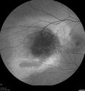
Choroidal Hemangioma Requiring PDT repeatedly258 views64 year old man. Diagnosed with hemangioma in 2015. PDT in 2016. Images are from 2020 when fluid started to reaccumulate. Needed PDT repeated two more times.
PDT treatments: [1] 2/19/2015 -Â Pavan (IOPÂ problem after PDT)Â AND [2] 11/2/21 PDT (7 mm temporal to fovea) - SMCÂ AND [3] 7/22/22 PDT 7.5 mm Temporal to the foveaJan 04, 2023
|
|
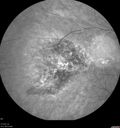
Choroidal Hemangioma Requiring PDT repeatedly239 views64 year old man. Diagnosed with hemangioma in 2015. PDT in 2016. Images are from 2020 when fluid started to reaccumulate. Needed PDT repeated two more times.
PDT treatments: [1] 2/19/2015 -Â Pavan (IOPÂ problem after PDT)Â AND [2] 11/2/21 PDT (7 mm temporal to fovea) - SMCÂ AND [3] 7/22/22 PDT 7.5 mm Temporal to the foveaJan 04, 2023
|
|
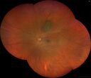
Choroidal nevus323 viewsStable for 8 years.Jan 02, 2023
|
|
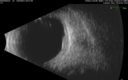
Choroidal nevus287 viewsStable for 8 years.Jan 02, 2023
|
|
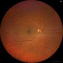
Choroidal nevus285 viewsStable for 8 years.Jan 02, 2023
|
|
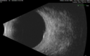
Choroidal nevus282 viewsStable for 8 years.Jan 02, 2023
|
|
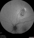
Choroidal nevus303 viewsStable for 8 years.Jan 02, 2023
|
|
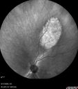
Choroidal nevus288 viewsStable for 8 years.Jan 02, 2023
|
|

Choroidal nevus314 viewsStable for 8 years.Jan 02, 2023
|
|
| 1098 files on 11 page(s) |
2 |  |
|
|
|
|
|
|
|
|


