Last additions - Iatrogenic Diseases
|
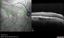
Phototoxicity following cataract surgery158 views70 year old female with nasal spot in vision since cataract surgery 3 years ago. VA 20/20 OU.Feb 14, 2024
|
|
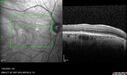
Phototoxicity following cataract surgery182 views70 year old female with nasal spot in vision since cataract surgery 3 years ago. VA 20/20 OU.Feb 14, 2024
|
|
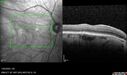
Phototoxicity following cataract surgery212 views70 year old female with nasal spot in vision since cataract surgery 3 years ago. VA 20/20 OU.Feb 14, 2024
|
|
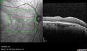
Phototoxicity following cataract surgery174 views70 year old female with nasal spot in vision since cataract surgery 3 years ago. VA 20/20 OU.Feb 14, 2024
|
|
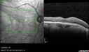
Phototoxicity following cataract surgery201 views70 year old female with nasal spot in vision since cataract surgery 3 years ago. VA 20/20 OU.Feb 14, 2024
|
|
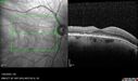
Phototoxicity following cataract surgery160 views70 year old female with nasal spot in vision since cataract surgery 3 years ago. VA 20/20 OU.Feb 14, 2024
|
|
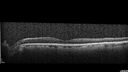
Phototoxicity following cataract surgery181 views70 year old female with nasal spot in vision since cataract surgery 3 years ago. VA 20/20 OU.Feb 14, 2024
|
|
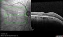
Phototoxicity following cataract surgery191 views70 year old female with nasal spot in vision since cataract surgery 3 years ago. VA 20/20 OU.Feb 14, 2024
|
|

Phototoxicity following cataract surgery164 views70 year old female with nasal spot in vision since cataract surgery 3 years ago. VA 20/20 OU.Feb 14, 2024
|
|
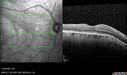
Phototoxicity following cataract surgery148 views70 year old female with nasal spot in vision since cataract surgery 3 years ago. VA 20/20 OU.Feb 14, 2024
|
|
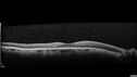
Phototoxicity following cataract surgery182 views70 year old female with nasal spot in vision since cataract surgery 3 years ago. VA 20/20 OU.Feb 14, 2024
|
|
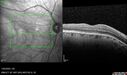
Phototoxicity following cataract surgery191 views70 year old female with nasal spot in vision since cataract surgery 3 years ago. VA 20/20 OU.Feb 14, 2024
|
|
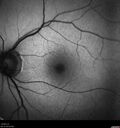
Phototoxicity following cataract surgery153 views70 year old female with nasal spot in vision since cataract surgery 3 years ago. VA 20/20 OU.Feb 14, 2024
|
|
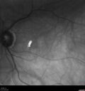
Phototoxicity following cataract surgery206 views70 year old female with nasal spot in vision since cataract surgery 3 years ago. VA 20/20 OU.Feb 14, 2024
|
|
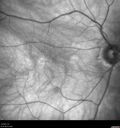
Phototoxicity following cataract surgery178 views70 year old female with nasal spot in vision since cataract surgery 3 years ago. VA 20/20 OU.Feb 14, 2024
|
|
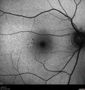
Phototoxicity following cataract surgery185 views70 year old female with nasal spot in vision since cataract surgery 3 years ago. VA 20/20 OU.Feb 14, 2024
|
|
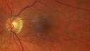
Phototoxicity following cataract surgery174 views70 year old female with nasal spot in vision since cataract surgery 3 years ago. VA 20/20 OU.Feb 14, 2024
|
|
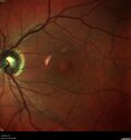
Phototoxicity following cataract surgery174 views70 year old female with nasal spot in vision since cataract surgery 3 years ago. VA 20/20 OU.Feb 14, 2024
|
|

Phototoxicity following cataract surgery175 views70 year old female with nasal spot in vision since cataract surgery 3 years ago. VA 20/20 OU.Feb 14, 2024
|
|
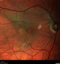
Phototoxicity following cataract surgery203 views70 year old female with nasal spot in vision since cataract surgery 3 years ago. VA 20/20 OU.Feb 14, 2024
|
|

Uveitis Glaucoma Hyphema (UGH) syndrome321 views65 year old female with intraocular lens half in the sulcus, iris atrophy, and recurrent uveitis since cataract surgery. She has a mild case and has been managed with topical steroids as needed.Jan 02, 2023
|
|

Uveitis Glaucoma Hyphema (UGH) syndrome316 views65 year old female with intraocular lens half in the sulcus, iris atrophy, and recurrent uveitis since cataract surgery. She has a mild case and has been managed with topical steroids as needed.Jan 02, 2023
|
|

Uveitis Glaucoma Hyphema (UGH) syndrome301 views65 year old female with intraocular lens half in the sulcus, iris atrophy, and recurrent uveitis since cataract surgery. She has a mild case and has been managed with topical steroids as needed.Jan 02, 2023
|
|

Choroidal Effusions following IOL exchange302 views67 year old man 10 years ago had retinal detachment with buckle, vitrectomy, laser, gas. Vision was 20/25 post-op. 2 months ago had sudden vision loss in the right eye from a dislocated PCIOL (in the bag). 3 weeks ago had a vitrectomy, remove IOL and scleral suture fixation of Akreos AO60 PCIOL with gortex sutures. IOP was 12 on post-op day 1 with a vitreous hemorrhage. Post-op day 7, vitreous hemorrhage cleared.
PMhx: HTN, High Cholesterol.
Med: Amlodipine, ASA, Atorvastatin, Metoprolol
Vision 20/200,Mar 09, 2022
|
|

Choroidal Effusions following IOL exchange308 views67 year old man 10 years ago had retinal detachment with buckle, vitrectomy, laser, gas. Vision was 20/25 post-op. 2 months ago had sudden vision loss in the right eye from a dislocated PCIOL (in the bag). 3 weeks ago had a vitrectomy, remove IOL and scleral suture fixation of Akreos AO60 PCIOL with gortex sutures. IOP was 12 on post-op day 1 with a vitreous hemorrhage. Post-op day 7, vitreous hemorrhage cleared.
PMhx: HTN, High Cholesterol.
Med: Amlodipine, ASA, Atorvastatin, Metoprolol
Vision 20/200,Mar 09, 2022
|
|

Choroidal Effusions following IOL exchange312 views67 year old man 10 years ago had retinal detachment with buckle, vitrectomy, laser, gas. Vision was 20/25 post-op. 2 months ago had sudden vision loss in the right eye from a dislocated PCIOL (in the bag). 3 weeks ago had a vitrectomy, remove IOL and scleral suture fixation of Akreos AO60 PCIOL with gortex sutures. IOP was 12 on post-op day 1 with a vitreous hemorrhage. Post-op day 7, vitreous hemorrhage cleared.
PMhx: HTN, High Cholesterol.
Med: Amlodipine, ASA, Atorvastatin, Metoprolol
Vision 20/200,Mar 09, 2022
|
|

Choroidal Effusions following IOL exchange282 views67 year old man 10 years ago had retinal detachment with buckle, vitrectomy, laser, gas. Vision was 20/25 post-op. 2 months ago had sudden vision loss in the right eye from a dislocated PCIOL (in the bag). 3 weeks ago had a vitrectomy, remove IOL and scleral suture fixation of Akreos AO60 PCIOL with gortex sutures. IOP was 12 on post-op day 1 with a vitreous hemorrhage. Post-op day 7, vitreous hemorrhage cleared.
PMhx: HTN, High Cholesterol.
Med: Amlodipine, ASA, Atorvastatin, Metoprolol
Vision 20/200,Mar 09, 2022
|
|

Choroidal Effusions following IOL exchange301 views67 year old man 10 years ago had retinal detachment with buckle, vitrectomy, laser, gas. Vision was 20/25 post-op. 2 months ago had sudden vision loss in the right eye from a dislocated PCIOL (in the bag). 3 weeks ago had a vitrectomy, remove IOL and scleral suture fixation of Akreos AO60 PCIOL with gortex sutures. IOP was 12 on post-op day 1 with a vitreous hemorrhage. Post-op day 7, vitreous hemorrhage cleared.
PMhx: HTN, High Cholesterol.
Med: Amlodipine, ASA, Atorvastatin, Metoprolol
Vision 20/200,Mar 09, 2022
|
|

Choroidal Effusions following IOL exchange322 views67 year old man 10 years ago had retinal detachment with buckle, vitrectomy, laser, gas. Vision was 20/25 post-op. 2 months ago had sudden vision loss in the right eye from a dislocated PCIOL (in the bag). 3 weeks ago had a vitrectomy, remove IOL and scleral suture fixation of Akreos AO60 PCIOL with gortex sutures. IOP was 12 on post-op day 1 with a vitreous hemorrhage. Post-op day 7, vitreous hemorrhage cleared.
PMhx: HTN, High Cholesterol.
Med: Amlodipine, ASA, Atorvastatin, Metoprolol
Vision 20/200,Mar 09, 2022
|
|

Choroidal Effusions following IOL exchange302 views67 year old man 10 years ago had retinal detachment with buckle, vitrectomy, laser, gas. Vision was 20/25 post-op. 2 months ago had sudden vision loss in the right eye from a dislocated PCIOL (in the bag). 3 weeks ago had a vitrectomy, remove IOL and scleral suture fixation of Akreos AO60 PCIOL with gortex sutures. IOP was 12 on post-op day 1 with a vitreous hemorrhage. Post-op day 7, vitreous hemorrhage cleared.
PMhx: HTN, High Cholesterol.
Med: Amlodipine, ASA, Atorvastatin, Metoprolol
Vision 20/200,Mar 09, 2022
|
|

395 viewsSep 30, 2020
|
|

444 viewsSep 30, 2020
|
|

397 viewsSep 30, 2020
|
|

416 viewsSep 30, 2020
|
|

391 viewsSep 30, 2020
|
|

425 viewsSep 30, 2020
|
|

Upside Down Lens Syndrome - Anterior Chamber IOL Upside Down700 views77 year old female who had vitrectomy for retained lens fragments and has had difficulty with recurrent uveitis since her cataract surgery. The anterior chamber intraocular lens is upside down. A lens exchange is planned.Mar 07, 2020
|
|
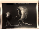
Choroidal Effusions following Glaucoma Surgery 9 months ago413 views67 year old female with glaucoma surgery 9 month ago. Her IOP is 12. She has peripheral choroidal effusions. VA 20/100 ph 20/32Feb 11, 2020
|
|
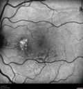
Choroidal Effusions following Glaucoma Surgery 9 months ago447 views67 year old female with glaucoma surgery 9 month ago. Her IOP is 12. She has peripheral choroidal effusions. VA 20/100 ph 20/32Feb 11, 2020
|
|
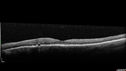
Choroidal Effusions following Glaucoma Surgery 9 months ago430 views67 year old female with glaucoma surgery 9 month ago. Her IOP is 12. She has peripheral choroidal effusions. VA 20/100 ph 20/32Feb 11, 2020
|
|
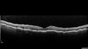
Choroidal Effusions following Glaucoma Surgery 9 months ago477 views67 year old female with glaucoma surgery 9 month ago. Her IOP is 12. She has peripheral choroidal effusions. VA 20/100 ph 20/32Feb 11, 2020
|
|

Choroidal Effusions following Glaucoma Surgery 9 months ago438 views67 year old female with glaucoma surgery 9 month ago. Her IOP is 12. She has peripheral choroidal effusions. VA 20/100 ph 20/32Feb 11, 2020
|
|
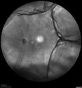
Choroidal Effusions following Glaucoma Surgery 9 months ago428 views67 year old female with glaucoma surgery 9 month ago. Her IOP is 12. She has peripheral choroidal effusions. VA 20/100 ph 20/32Feb 11, 2020
|
|

Choroidal Effusion One Day following macular pucker surgery411 views70 year old female one day following macular pucker surgery. Her VA was 20/100, IOP was 12. The air bubble was to pressurize the eye at the conclusion of the 25 gauge vitrectomy. The choroidal effusion is in the periphery.Feb 06, 2020
|
|

Perforating Needle injury retrobulbar anesthetic - excellent final vision432 views71 year old man who had a vitrectomy for macular pucker and then developed a retinal detachment. There was an accidental needle stick during a retrobulbar anesthetic. The posterior puncture site is just inferior to the macula. The patient had a vitrectomy the next day for the retinal detachment and then 3 months later a vitrectomy to remove the fibrous proliferation from the puncture site. The final visual acuity was 20/20.Aug 07, 2019
|
|

Perforating Needle injury retrobulbar anesthetic - excellent final vision466 views71 year old man who had a vitrectomy for macular pucker and then developed a retinal detachment. There was an accidental needle stick during a retrobulbar anesthetic. The posterior puncture site is just inferior to the macula. The patient had a vitrectomy the next day for the retinal detachment and then 3 months later a vitrectomy to remove the fibrous proliferation from the puncture site. The final visual acuity was 20/20.Aug 07, 2019
|
|

Perforating Needle injury retrobulbar anesthetic - excellent final vision602 views71 year old man who had a vitrectomy for macular pucker and then developed a retinal detachment. There was an accidental needle stick during a retrobulbar anesthetic. The posterior puncture site is just inferior to the macula. The patient had a vitrectomy the next day for the retinal detachment and then 3 months later a vitrectomy to remove the fibrous proliferation from the puncture site. The final visual acuity was 20/20.Aug 07, 2019
|
|

Perforating Needle injury retrobulbar anesthetic - excellent final vision596 views71 year old man who had a vitrectomy for macular pucker and then developed a retinal detachment. There was an accidental needle stick during a retrobulbar anesthetic. The posterior puncture site is just inferior to the macula. The patient had a vitrectomy the next day for the retinal detachment and then 3 months later a vitrectomy to remove the fibrous proliferation from the puncture site. The final visual acuity was 20/20.Aug 07, 2019
|
|
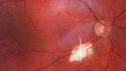
Perforating Needle injury retrobulbar anesthetic - excellent final vision545 views71 year old man who had a vitrectomy for macular pucker and then developed a retinal detachment. There was an accidental needle stick during a retrobulbar anesthetic. The posterior puncture site is just inferior to the macula. The patient had a vitrectomy the next day for the retinal detachment and then 3 months later a vitrectomy to remove the fibrous proliferation from the puncture site. The final visual acuity was 20/20.Aug 07, 2019
|
|

Outer retinal holes and cataract from YAG laser vitreolysis501 views44 year old man with 7 year history of multiple lasers for floaters. FAF and OCT show outer retinal holes and lens has focal defectsMar 26, 2019
|
|

Outer retinal holes and cataract from YAG laser vitreolysis482 views44 year old man with 7 year history of multiple lasers for floaters. FAF and OCT show outer retinal holes and lens has focal defectsMar 26, 2019
|
|

Outer retinal holes and cataract from YAG laser vitreolysis463 views44 year old man with 7 year history of multiple lasers for floaters. FAF and OCT show outer retinal holes and lens has focal defectsMar 26, 2019
|
|

Outer retinal holes and cataract from YAG laser vitreolysis542 views44 year old man with 7 year history of multiple lasers for floaters. FAF and OCT show outer retinal holes and lens has focal defectsMar 26, 2019
|
|

Outer retinal holes and cataract from YAG laser vitreolysis486 views44 year old man with 7 year history of multiple lasers for floaters. FAF and OCT show outer retinal holes and lens has focal defectsMar 26, 2019
|
|

Outer retinal holes and cataract from YAG laser vitreolysis448 views44 year old man with 7 year history of multiple lasers for floaters. FAF and OCT show outer retinal holes and lens has focal defectsMar 26, 2019
|
|

Outer retinal holes and cataract from YAG laser vitreolysis441 views44 year old man with 7 year history of multiple lasers for floaters. FAF and OCT show outer retinal holes and lens has focal defectsMar 26, 2019
|
|

Outer retinal holes and cataract from YAG laser vitreolysis492 views44 year old man with 7 year history of multiple lasers for floaters. FAF and OCT show outer retinal holes and lens has focal defectsMar 26, 2019
|
|

Outer retinal holes and cataract from YAG laser vitreolysis420 views44 year old man with 7 year history of multiple lasers for floaters. FAF and OCT show outer retinal holes and lens has focal defectsMar 26, 2019
|
|

Outer retinal holes and cataract from YAG laser vitreolysis420 views44 year old man with 7 year history of multiple lasers for floaters. FAF and OCT show outer retinal holes and lens has focal defectsMar 26, 2019
|
|

Outer retinal holes and cataract from YAG laser vitreolysis493 views44 year old man with 7 year history of multiple lasers for floaters. FAF and OCT show outer retinal holes and lens has focal defectsMar 26, 2019
|
|

Outer retinal holes and cataract from YAG laser vitreolysis467 views44 year old man with 7 year history of multiple lasers for floaters. FAF and OCT show outer retinal holes and lens has focal defectsMar 26, 2019
|
|

Outer retinal holes and cataract from YAG laser vitreolysis511 views44 year old man with 7 year history of multiple lasers for floaters. FAF and OCT show outer retinal holes and lens has focal defectsMar 26, 2019
|
|

Outer retinal holes and cataract from YAG laser vitreolysis482 views44 year old man with 7 year history of multiple lasers for floaters. FAF and OCT show outer retinal holes and lens has focal defectsMar 26, 2019
|
|

Outer retinal holes and cataract from YAG laser vitreolysis487 views44 year old man with 7 year history of multiple lasers for floaters. FAF and OCT show outer retinal holes and lens has focal defectsMar 26, 2019
|
|

Photic Maculopathy Following Cataract Surgery422 viewsInitial images are from 1 week and subsequent images from 1 month post surgery.Dec 18, 2018
|
|

Photic Maculopathy Following Cataract Surgery499 viewsInitial images are from 1 week and subsequent images from 1 month post surgery.Dec 18, 2018
|
|

Photic Maculopathy Following Cataract Surgery441 viewsInitial images are from 1 week and subsequent images from 1 month post surgery.Dec 18, 2018
|
|

Photic Maculopathy Following Cataract Surgery512 viewsInitial images are from 1 week and subsequent images from 1 month post surgery.Dec 18, 2018
|
|

Photic Maculopathy Following Cataract Surgery496 viewsInitial images are from 1 week and subsequent images from 1 month post surgery.Dec 18, 2018
|
|

Photic Maculopathy Following Cataract Surgery450 viewsInitial images are from 1 week and subsequent images from 1 month post surgery.Dec 18, 2018
|
|

Photic Maculopathy Following Cataract Surgery581 viewsInitial images are from 1 week and subsequent images from 1 month post surgery.Dec 18, 2018
|
|

Photic Maculopathy Following Cataract Surgery434 viewsInitial images are from 1 week and subsequent images from 1 month post surgery.Dec 18, 2018
|
|

Photic Maculopathy Following Cataract Surgery542 viewsInitial images are from 1 week and subsequent images from 1 month post surgery.Dec 18, 2018
|
|

Photic Maculopathy Following Cataract Surgery593 viewsInitial images are from 1 week and subsequent images from 1 month post surgery.Dec 18, 2018
|
|

Photic Maculopathy Following Cataract Surgery497 viewsInitial images are from 1 week and subsequent images from 1 month post surgery.Dec 18, 2018
|
|

Photic Maculopathy Following Cataract Surgery483 viewsInitial images are from 1 week and subsequent images from 1 month post surgery.Dec 18, 2018
|
|

Photic Maculopathy Following Cataract Surgery535 viewsInitial images are from 1 week and subsequent images from 1 month post surgery.Dec 18, 2018
|
|
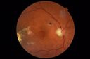
Macular Damage post Macular Hole Surgery517 viewsThere is a huge hole in the middle of the macula and several small holes in the posterior pole. The patient had 2 surgeries for an idiopathic stage II hole and then was offered a third procedure by the same surgeon and sought a second opinion (and was told to not have anything more done)Jul 03, 2018
|
|

Macular Damage post Macular Hole Surgery456 viewsThere is a huge hole in the middle of the macula and several small holes in the posterior pole. The patient had 2 surgeries for an idiopathic stage II hole and then was offered a third procedure by the same surgeon and sought a second opinion (and was told to not have anything more done)Jul 03, 2018
|
|
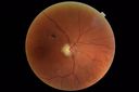
Macular Damage post Macular Hole Surgery502 viewsThere is a huge hole in the middle of the macula and several small holes in the posterior pole. The patient had 2 surgeries for an idiopathic stage II hole and then was offered a third procedure by the same surgeon and sought a second opinion (and was told to not have anything more done)Jul 03, 2018
|
|
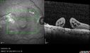
Macular Damage post Macular Hole Surgery485 viewsThere is a huge hole in the middle of the macula and several small holes in the posterior pole. The patient had 2 surgeries for an idiopathic stage II hole and then was offered a third procedure by the same surgeon and sought a second opinion (and was told to not have anything more done)Jul 03, 2018
|
|
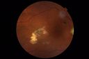
Macular Damage post Macular Hole Surgery528 viewsThere is a huge hole in the middle of the macula and several small holes in the posterior pole. The patient had 2 surgeries for an idiopathic stage II hole and then was offered a third procedure by the same surgeon and sought a second opinion (and was told to not have anything more done)Jul 03, 2018
|
|
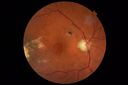
Macular Damage post Macular Hole Surgery481 viewsThere is a huge hole in the middle of the macula and several small holes in the posterior pole. The patient had 2 surgeries for an idiopathic stage II hole and then was offered a third procedure by the same surgeon and sought a second opinion (and was told to not have anything more done)Jul 03, 2018
|
|

Macular Damage post Macular Hole Surgery522 viewsThere is a huge hole in the middle of the macula and several small holes in the posterior pole. The patient had 2 surgeries for an idiopathic stage II hole and then was offered a third procedure by the same surgeon and sought a second opinion (and was told to not have anything more done)Jul 03, 2018
|
|

Macular Laser Scars899 views86 year old man who had Laser done from 2010 - 2018 for diabetic retinopathy. The right eye lost vision immediately after laser was done a few years ago. VA 1/200 OD; 20/25 OSMar 12, 2018
|
|

Macular Laser Scars579 views86 year old man who had Laser done from 2010 - 2018 for diabetic retinopathy. The right eye lost vision immediately after laser was done a few years ago. VA 1/200 OD; 20/25 OSMar 12, 2018
|
|

Macular Laser Scars631 views86 year old man who had Laser done from 2010 - 2018 for diabetic retinopathy. The right eye lost vision immediately after laser was done a few years ago. VA 1/200 OD; 20/25 OSMar 12, 2018
|
|
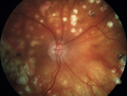
Macular Laser Scars579 views86 year old man who had Laser done from 2010 - 2018 for diabetic retinopathy. The right eye lost vision immediately after laser was done a few years ago. VA 1/200 OD; 20/25 OSMar 12, 2018
|
|

Absent Macula582 viewsRetina is balled up and dragged superiorly under silicone oil. This patient started with macular pucker surgery for a mild chronic pucker. She developed 2 retinal detachments. The OCT scan is through the center of the macula under oil. There is no retina present on the scanOct 02, 2017
|
|

Macular Laser Scars Left Eye - 5/200 Vision518 views83 year old woman who had extensive macular laser in the left eye about 15 years ago for DME. Vision is now 20/50 right eye and 3/200 left eyeMay 04, 2017
|
|

Macular Laser Scars Left Eye - 5/200 Vision497 views83 year old woman who had extensive macular laser in the left eye about 15 years ago for DME. Vision is now 20/50 right eye and 3/200 left eyeMay 04, 2017
|
|

Macular Laser Scars Left Eye - 5/200 Vision553 views83 year old woman who had extensive macular laser in the left eye about 15 years ago for DME. Vision is now 20/50 right eye and 3/200 left eyeMay 04, 2017
|
|

Macular Laser Scars Left Eye - 5/200 Vision561 views83 year old woman who had extensive macular laser in the left eye about 15 years ago for DME. Vision is now 20/50 right eye and 3/200 left eyeMay 04, 2017
|
|

Macular Laser Scars Left Eye - 5/200 Vision504 views83 year old woman who had extensive macular laser in the left eye about 15 years ago for DME. Vision is now 20/50 right eye and 3/200 left eyeMay 04, 2017
|
|

Macular Laser Scars Left Eye - 5/200 Vision548 views83 year old woman who had extensive macular laser in the left eye about 15 years ago for DME. Vision is now 20/50 right eye and 3/200 left eyeMay 04, 2017
|
|

Macular Laser Scars Left Eye - 5/200 Vision546 views83 year old woman who had extensive macular laser in the left eye about 15 years ago for DME. Vision is now 20/50 right eye and 3/200 left eyeMay 04, 2017
|
|

Macular Laser Scars Left Eye - 5/200 Vision566 views83 year old woman who had extensive macular laser in the left eye about 15 years ago for DME. Vision is now 20/50 right eye and 3/200 left eyeMay 04, 2017
|
|

Macular Atrophy - Phototoxicity - Macular Hole Surgery - Late ICG image677 views25 years ago patient suffered immediate severe vision loss following macular hole surgery. You can almost see light pipe tracks in the atrophy in the macula. Vision is 5/200Nov 07, 2016
|
|
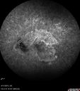
Macular Atrophy - Phototoxicity - Macular Hole Surgery628 views25 years ago patient suffered immediate severe vision loss following macular hole surgery. You can almost see light pipe tracks in the atrophy in the macula. Vision is 5/200Nov 07, 2016
|
|

Macular Atrophy - Phototoxicity - Macular Hole Surgery686 views25 years ago patient suffered immediate severe vision loss following macular hole surgery. You can almost see light pipe tracks in the atrophy in the macula. Vision is 5/200Nov 07, 2016
|
|
|
|


