Last additions - Peripheral Retinal Degeneration
|

Peripheral Exudative Hemorrhagic Chorioretinopathy456 views93 year old female with shadow in side vision in the left eye. Referred for retinal detachment - Exudative lesion subsided in about 6 months without treatment.
Vision 20/63 OD and 20/100 OS (from dry AMD)Feb 11, 2020
|
|

Peripheral Exudative Hemorrhagic Chorioretinopathy519 views93 year old female with shadow in side vision in the left eye. Referred for retinal detachment - Exudative lesion subsided in about 6 months without treatment.
Vision 20/63 OD and 20/100 OS (from dry AMD)Feb 11, 2020
|
|

Peripheral Exudative Hemorrhagic Chorioretinopathy432 views93 year old female with shadow in side vision in the left eye. Referred for retinal detachment - Exudative lesion subsided in about 6 months without treatment.
Vision 20/63 OD and 20/100 OS (from dry AMD)Feb 11, 2020
|
|

Peripheral Exudative Hemorrhagic Chorioretinopathy456 views93 year old female with shadow in side vision in the left eye. Referred for retinal detachment - Exudative lesion subsided in about 6 months without treatment.
Vision 20/63 OD and 20/100 OS (from dry AMD)Feb 11, 2020
|
|

Peripheral Exudative Hemorrhagic Chorioretinopathy477 views93 year old female with shadow in side vision in the left eye. Referred for retinal detachment - Exudative lesion subsided in about 6 months without treatment.
Vision 20/63 OD and 20/100 OS (from dry AMD)Feb 11, 2020
|
|

Peripheral Exudative Hemorrhagic Chorioretinopathy480 views93 year old female with shadow in side vision in the left eye. Referred for retinal detachment - Exudative lesion subsided in about 6 months without treatment.
Vision 20/63 OD and 20/100 OS (from dry AMD)Feb 11, 2020
|
|

Peripheral Exudative Hemorrhagic Chorioretinopathy711 views93 year old female with shadow in side vision in the left eye. Referred for retinal detachment - Exudative lesion subsided in about 6 months without treatment.
Vision 20/63 OD and 20/100 OS (from dry AMD)Feb 11, 2020
|
|

Peripheral Exudative Hemorrhagic Chorioretinopathy508 views93 year old female with shadow in side vision in the left eye. Referred for retinal detachment - Exudative lesion subsided in about 6 months without treatment.
Vision 20/63 OD and 20/100 OS (from dry AMD)Feb 11, 2020
|
|

Peripheral Exudative Hemorrhagic Chorioretinopathy581 views93 year old female with shadow in side vision in the left eye. Referred for retinal detachment - Exudative lesion subsided in about 6 months without treatment.
Vision 20/63 OD and 20/100 OS (from dry AMD)Feb 11, 2020
|
|

Peripheral Exudative Hemorrhagic Chorioretinopathy507 views93 year old female with shadow in side vision in the left eye. Referred for retinal detachment - Exudative lesion subsided in about 6 months without treatment.
Vision 20/63 OD and 20/100 OS (from dry AMD)Feb 11, 2020
|
|

Peripheral Exudative Hemorrhagic Chorioretinopathy510 views93 year old female with shadow in side vision in the left eye. Referred for retinal detachment - Exudative lesion subsided in about 6 months without treatment.
Vision 20/63 OD and 20/100 OS (from dry AMD)Feb 11, 2020
|
|

Peripheral Exudative Hemorrhagic Chorioretinopathy455 views93 year old female with shadow in side vision in the left eye. Referred for retinal detachment - Exudative lesion subsided in about 6 months without treatment.
Vision 20/63 OD and 20/100 OS (from dry AMD)Feb 11, 2020
|
|

Peripheral Exudative Hemorrhagic Chorioretinopathy410 views93 year old female with shadow in side vision in the left eye. Referred for retinal detachment - Exudative lesion subsided in about 6 months without treatment.
Vision 20/63 OD and 20/100 OS (from dry AMD)Feb 11, 2020
|
|

Peripheral Exudative Hemorrhagic Chorioretinopathy443 views93 year old female with shadow in side vision in the left eye. Referred for retinal detachment - Exudative lesion subsided in about 6 months without treatment.
Vision 20/63 OD and 20/100 OS (from dry AMD)Feb 11, 2020
|
|

Peripheral Exudative Hemorrhagic Chorioretinopathy437 views93 year old female with shadow in side vision in the left eye. Referred for retinal detachment - Exudative lesion subsided in about 6 months without treatment.
Vision 20/63 OD and 20/100 OS (from dry AMD)Feb 11, 2020
|
|

Peripheral Exudative Hemorrhagic Chorioretinopathy414 views93 year old female with shadow in side vision in the left eye. Referred for retinal detachment - Exudative lesion subsided in about 6 months without treatment.
Vision 20/63 OD and 20/100 OS (from dry AMD)Feb 11, 2020
|
|

Peripheral Exudative Hemorrhagic Chorioretinopathy462 views93 year old female with shadow in side vision in the left eye. Referred for retinal detachment - Exudative lesion subsided in about 6 months without treatment.
Vision 20/63 OD and 20/100 OS (from dry AMD)Feb 11, 2020
|
|

Peripheral Exudative Hemorrhagic Chorioretinopathy562 views93 year old female with shadow in side vision in the left eye. Referred for retinal detachment - Exudative lesion subsided in about 6 months without treatment.
Vision 20/63 OD and 20/100 OS (from dry AMD)Feb 11, 2020
|
|

Peripheral Exudative Hemorrhagic Chorioretinopathy642 views93 year old female with shadow in side vision in the left eye. Referred for retinal detachment - Exudative lesion subsided in about 6 months without treatment.
Vision 20/63 OD and 20/100 OS (from dry AMD)Feb 11, 2020
|
|

Peripheral Exudative Hemorrhagic Chorioretinopathy432 views93 year old female with shadow in side vision in the left eye. Referred for retinal detachment - Exudative lesion subsided in about 6 months without treatment.
Vision 20/63 OD and 20/100 OS (from dry AMD)Feb 11, 2020
|
|

Peripheral Exudative Hemorrhagic Chorioretinopathy445 views93 year old female with shadow in side vision in the left eye. Referred for retinal detachment - Exudative lesion subsided in about 6 months without treatment.
Vision 20/63 OD and 20/100 OS (from dry AMD)Feb 11, 2020
|
|

Reticular peripheral pigment degeneration1711 viewsJun 11, 2019
|
|

Reticular pigment degeneration1613 views78 year old man with wet AMD- Peripheral retina show reticular pigment degenerationJun 11, 2019
|
|

Lattice Degeneration649 viewsAsymptomaticJun 10, 2019
|
|

Pigmented Lattice Degeneration1074 viewsPigmented Lattice - Incidental FindingJun 10, 2019
|
|

Pigmented Lattice Degeneration625 viewsPigmented Lattice - Incidental FindingJun 10, 2019
|
|

Equatorial Drusen576 views61 year old female with normal visoinMar 27, 2018
|
|

Equatorial Drusen465 views61 year old female with normal visoinMar 27, 2018
|
|

Equatorial Drusen500 views61 year old female with normal visoinMar 27, 2018
|
|

Equatorial Drusen468 views61 year old female with normal visoinMar 27, 2018
|
|

Equatorial Drusen540 views61 year old female with normal visoinMar 27, 2018
|
|

Equatorial Drusen524 views61 year old female with normal visoinMar 27, 2018
|
|

Equatorial Drusen577 views61 year old female with normal visoinMar 27, 2018
|
|

Equatorial Drusen511 views61 year old female with normal visoinMar 27, 2018
|
|

Equatorial Drusen811 views61 year old female with normal visoinMar 27, 2018
|
|

Equatorial Drusen572 views61 year old female with normal visoinMar 27, 2018
|
|

Equatorial Drusen495 views61 year old female with normal visoinMar 27, 2018
|
|

Equatorial Drusen589 views61 year old female with normal visoinMar 27, 2018
|
|

Equatorial Drusen533 views61 year old female with normal visoinMar 27, 2018
|
|

Equatorial Drusen688 views61 year old female with normal visoinMar 27, 2018
|
|

Equatorial Drusen855 views61 year old female with normal visoinMar 27, 2018
|
|

Bullous Retinoschisis - Confirmed with SD OCT675 viewsAtypical location for retinoschisis - SD-OCT is used to confirm that this is retinoschisis and not a retinal detachmentJul 07, 2016
|
|

Bullous Retinoschisis - Confirmed with SD OCT602 viewsAtypical location for retinoschisis - SD-OCT is used to confirm that this is retinoschisis and not a retinal detachmentJul 07, 2016
|
|

Bullous Retinoschisis - Confirmed with SD OCT666 viewsAtypical location for retinoschisis - SD-OCT is used to confirm that this is retinoschisis and not a retinal detachmentJul 07, 2016
|
|

Bullous Retinoschisis - Confirmed with SD OCT532 viewsAtypical location for retinoschisis - SD-OCT is used to confirm that this is retinoschisis and not a retinal detachmentJul 07, 2016
|
|

Bullous Retinoschisis - Confirmed with SD OCT639 viewsAtypical location for retinoschisis - SD-OCT is used to confirm that this is retinoschisis and not a retinal detachmentJul 07, 2016
|
|

Bullous Retinoschisis - Confirmed with SD OCT560 viewsAtypical location for retinoschisis - SD-OCT is used to confirm that this is retinoschisis and not a retinal detachmentJul 07, 2016
|
|

Bullous Retinoschisis - Confirmed with SD OCT676 viewsAtypical location for retinoschisis - SD-OCT is used to confirm that this is retinoschisis and not a retinal detachmentJul 07, 2016
|
|

Bullous Retinoschisis - Confirmed with SD OCT670 viewsAtypical location for retinoschisis - SD-OCT is used to confirm that this is retinoschisis and not a retinal detachmentJul 07, 2016
|
|

Pseudoxanthoma Elasticum with Angioid Streaks568 viewsYoung female patient who comes in for routine eye exam, VA 20/20 in both eyes. Fundus photos shows a Peu'd orange appearance in the periphery of the retina and angioid streaks around the optic nerve. Apr 28, 2014
|
|

Pseudoxanthoma Elasticum with Angioid Streaks609 viewsYoung female patient who comes in for routine eye exam, VA 20/20 in both eyes. Fundus photos shows a Peu'd orange appearance in the periphery of the retina and angioid streaks around the optic nerve. Apr 28, 2014
|
|

Pseudoxanthoma Elasticum with Angioid Streaks566 viewsYoung female patient who comes in for routine eye exam, VA 20/20 in both eyes. Fundus photos shows a Peu'd orange appearance in the periphery of the retina and angioid streaks around the optic nerve. Apr 28, 2014
|
|

Pseudoxanthoma Elasticum with Angioid Streaks666 viewsYoung female patient who comes in for routine eye exam, VA 20/20 in both eyes. Fundus photos shows a Peu'd orange appearance in the periphery of the retina and angioid streaks around the optic nerve. Apr 28, 2014
|
|
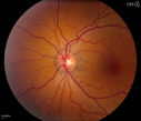
Pseudoxanthoma Elasticum with Angioid Streaks598 viewsYoung female patient who comes in for routine eye exam, VA 20/20 in both eyes. Fundus photos shows a Peu'd orange appearance in the periphery of the retina and angioid streaks around the optic nerve. Apr 28, 2014
|
|

Pseudoxanthoma Elasticum with Angioid Streaks1199 viewsYoung female patient who comes in for routine eye exam, VA 20/20 in both eyes. Fundus photos shows a Peu'd orange appearance in the periphery of the retina and angioid streaks around the optic nerve. Apr 28, 2014
|
|
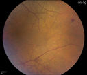
Peripheral Retinal Degeneration499 viewsPatient comes in with double vision. VA was 20/20 in both eyes. Fundus exam shows Retinal Degenerative changes in both eyes. Offer to correct double vision with temporary Fresnel prismMar 28, 2013
|
|
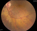
Peripheral Retinal Degeneration429 viewsPatient comes in with double vision. VA was 20/20 in both eyes. Fundus exam shows Retinal Degenerative changes in both eyes. Offer to correct double vision with temporary Fresnel prismMar 28, 2013
|
|
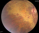
Peripheral Retinal Degeneration419 viewsPatient comes in with double vision. VA was 20/20 in both eyes. Fundus exam shows Retinal Degenerative changes in both eyes. Offer to correct double vision with temporary Fresnel prismMar 28, 2013
|
|

Peripheral Retinal Degeneration490 viewsPatient comes in with double vision. VA was 20/20 in both eyes. Fundus exam shows Retinal Degenerative changes in both eyes. Offer to correct double vision with temporary Fresnel prismMar 28, 2013
|
|
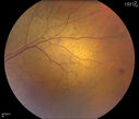
Peripheral Retinal Degeneration455 viewsPatient comes in with double vision. VA was 20/20 in both eyes. Fundus exam shows Retinal Degenerative changes in both eyes. Offer to correct double vision with temporary Fresnel prismMar 28, 2013
|
|
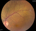
Peripheral Retinal Degeneration505 viewsPatient comes in with double vision. VA was 20/20 in both eyes. Fundus exam shows Retinal Degenerative changes in both eyes. Offer to correct double vision with temporary Fresnel prismMar 28, 2013
|
|
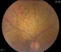
Peripheral Retinal Degeneration453 viewsPatient comes in with double vision. VA was 20/20 in both eyes. Fundus exam shows Retinal Degenerative changes in both eyes. Offer to correct double vision with temporary Fresnel prismMar 28, 2013
|
|
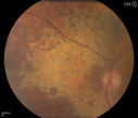
Peripheral Retinal Degeneration450 viewsPatient comes in with double vision. VA was 20/20 in both eyes. Fundus exam shows Retinal Degenerative changes in both eyes. Offer to correct double vision with temporary Fresnel prismMar 28, 2013
|
|
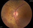
Peripheral Retinal Degeneration453 viewsPatient comes in with double vision. VA was 20/20 in both eyes. Fundus exam shows Retinal Degenerative changes in both eyes. Offer to correct double vision with temporary Fresnel prismMar 28, 2013
|
|
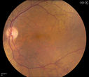
Peripheral Retinal Degeneration465 viewsPatient comes in with double vision. VA was 20/20 in both eyes. Fundus exam shows Retinal Degenerative changes in both eyes. Offer to correct double vision with temporary Fresnel prismMar 28, 2013
|
|
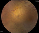
Peripheral Retinal Degeneration460 viewsPatient comes in with double vision. VA was 20/20 in both eyes. Fundus exam shows Retinal Degenerative changes in both eyes. Offer to correct double vision with temporary Fresnel prismMar 28, 2013
|
|
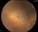
Peripheral Retinal Degeneration463 viewsPatient comes in with double vision. VA was 20/20 in both eyes. Fundus exam shows Retinal Degenerative changes in both eyes. Offer to correct double vision with temporary Fresnel prismMar 28, 2013
|
|
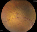
Peripheral Retinal Degeneration501 viewsPatient comes in with double vision. VA was 20/20 in both eyes. Fundus exam shows Retinal Degenerative changes in both eyes. Offer to correct double vision with temporary Fresnel prismMar 28, 2013
|
|
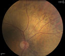
Peripheral Retinal Degeneration537 viewsPatient comes in with double vision. VA was 20/20 in both eyes. Fundus exam shows Retinal Degenerative changes in both eyes. Offer to correct double vision with temporary Fresnel prismMar 28, 2013
|
|
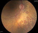
Peripheral Retinal Degeneration663 viewsPatient comes in with double vision. VA was 20/20 in both eyes. Fundus exam shows Retinal Degenerative changes in both eyes. Offer to correct double vision with temporary Fresnel prismMar 28, 2013
|
|
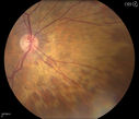
Peripheral Retinal Degeneration674 viewsPatient comes in with double vision. VA was 20/20 in both eyes. Fundus exam shows Retinal Degenerative changes in both eyes. Offer to correct double vision with temporary Fresnel prismMar 28, 2013
|
|
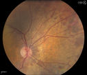
Peripheral Retinal Degeneration611 viewsPatient comes in with double vision. VA was 20/20 in both eyes. Fundus exam shows Retinal Degenerative changes in both eyes. Offer to correct double vision with temporary Fresnel prismMar 28, 2013
|
|
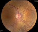
Peripheral Retinal Degeneration595 viewsPatient comes in with double vision. VA was 20/20 in both eyes. Fundus exam shows Retinal Degenerative changes in both eyes. Offer to correct double vision with temporary Fresnel prismMar 28, 2013
|
|
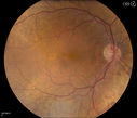
Peripheral Retinal Degeneration570 viewsPatient comes in with double vision. VA was 20/20 in both eyes. Fundus exam shows Retinal Degenerative changes in both eyes. Offer to correct double vision with temporary Fresnel prismMar 28, 2013
|
|
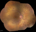
Peripheral Retinal Degeneration668 viewsPatient comes in with double vision. VA was 20/20 in both eyes. Fundus exam shows Retinal Degenerative changes in both eyes. Offer to correct double vision with temporary Fresnel prismMar 28, 2013
|
|

Peripheral Retinal Degeneration851 viewsPatient comes in with double vision. VA was 20/20 in both eyes. Fundus exam shows Retinal Degenerative changes in both eyes. Offer to correct double vision with temporary Fresnel prismMar 28, 2013
|
|

Peripheral Exudative Hemorrhagic Chorioretinopathy780 views70 year old man with recurrent peripheral choroidal hemorrhages - no treatment - b-scan shows hemorrhage shrinking over a monthNov 02, 2012
|
|

Peripheral Exudative Hemorrhagic Chorioretinopathy723 views70 year old man with recurrent peripheral choroidal hemorrhages - no treatment - b-scan shows hemorrhage shrinking over a monthNov 02, 2012
|
|

Peripheral Exudative Hemorrhagic Chorioretinopathy759 views70 year old man with recurrent peripheral choroidal hemorrhages - no treatment - b-scan shows hemorrhage shrinking over a monthNov 02, 2012
|
|

Peripheral Exudative Hemorrhagic Chorioretinopathy902 views70 year old man with recurrent peripheral choroidal hemorrhages - no treatment - b-scan shows hemorrhage shrinking over a monthNov 02, 2012
|
|

Peripheral Exudative Hemorrhagic Chorioretinopathy1063 views70 year old man with recurrent peripheral choroidal hemorrhages - no treatment - b-scan shows hemorrhage shrinking over a monthNov 02, 2012
|
|

Peripheral Exudative Hemorrhagic Chorioretinopathy1132 views70 year old man with recurrent peripheral choroidal hemorrhages - no treatment - b-scan shows hemorrhage shrinking over a monthNov 02, 2012
|
|

Peripheral Exudative Hemorrhagic Chorioretinopathy691 views70 year old man with recurrent peripheral choroidal hemorrhages - no treatment - b-scan shows hemorrhage shrinking over a monthNov 02, 2012
|
|

Peripheral Exudative Hemorrhagic Chorioretinopathy733 views70 year old man with recurrent peripheral choroidal hemorrhages - no treatment - b-scan shows hemorrhage shrinking over a monthNov 02, 2012
|
|
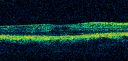
ECTOPIC DISCIFORM SCAR/PERIPHERAL CHOROIDAL NEOVASCULAR MEMBRANE – EXUDATE –INFLAMMATION IN REACTION TO THE PERIPHERAL VASCULAR LESION 733 views53-year-old woman has noticed some blurred vision in the right eye for a few years. In retrospect, she thinks it is possible since she was a teenager the right eye was always the weaker eye and that the left eye has always been the better eye. VISUAL ACUITY: Vision OD is 20/400Dec 20, 2011
|
|

ECTOPIC DISCIFORM SCAR/PERIPHERAL CHOROIDAL NEOVASCULAR MEMBRANE – EXUDATE –INFLAMMATION IN REACTION TO THE PERIPHERAL VASCULAR LESION 700 views53-year-old woman has noticed some blurred vision in the right eye for a few years. In retrospect, she thinks it is possible since she was a teenager the right eye was always the weaker eye and that the left eye has always been the better eye. VISUAL ACUITY: Vision OD is 20/400Dec 20, 2011
|
|

ECTOPIC DISCIFORM SCAR/PERIPHERAL CHOROIDAL NEOVASCULAR MEMBRANE – EXUDATE –INFLAMMATION IN REACTION TO THE PERIPHERAL VASCULAR LESION 686 views53-year-old woman has noticed some blurred vision in the right eye for a few years. In retrospect, she thinks it is possible since she was a teenager the right eye was always the weaker eye and that the left eye has always been the better eye. VISUAL ACUITY: Vision OD is 20/400Dec 20, 2011
|
|

ECTOPIC DISCIFORM SCAR/PERIPHERAL CHOROIDAL NEOVASCULAR MEMBRANE – EXUDATE –INFLAMMATION IN REACTION TO THE PERIPHERAL VASCULAR LESION 708 views53-year-old woman has noticed some blurred vision in the right eye for a few years. In retrospect, she thinks it is possible since she was a teenager the right eye was always the weaker eye and that the left eye has always been the better eye. VISUAL ACUITY: Vision OD is 20/400Dec 20, 2011
|
|

ECTOPIC DISCIFORM SCAR/PERIPHERAL CHOROIDAL NEOVASCULAR MEMBRANE – EXUDATE –INFLAMMATION IN REACTION TO THE PERIPHERAL VASCULAR LESION 642 views53-year-old woman has noticed some blurred vision in the right eye for a few years. In retrospect, she thinks it is possible since she was a teenager the right eye was always the weaker eye and that the left eye has always been the better eye. VISUAL ACUITY: Vision OD is 20/400Dec 20, 2011
|
|

ECTOPIC DISCIFORM SCAR/PERIPHERAL CHOROIDAL NEOVASCULAR MEMBRANE – EXUDATE –INFLAMMATION IN REACTION TO THE PERIPHERAL VASCULAR LESION 705 views53-year-old woman has noticed some blurred vision in the right eye for a few years. In retrospect, she thinks it is possible since she was a teenager the right eye was always the weaker eye and that the left eye has always been the better eye. VISUAL ACUITY: Vision OD is 20/400Dec 20, 2011
|
|
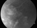
ECTOPIC DISCIFORM SCAR/PERIPHERAL CHOROIDAL NEOVASCULAR MEMBRANE – EXUDATE –INFLAMMATION IN REACTION TO THE PERIPHERAL VASCULAR LESION 591 views53-year-old woman has noticed some blurred vision in the right eye for a few years. In retrospect, she thinks it is possible since she was a teenager the right eye was always the weaker eye and that the left eye has always been the better eye. VISUAL ACUITY: Vision OD is 20/400Dec 20, 2011
|
|

ECTOPIC DISCIFORM SCAR/PERIPHERAL CHOROIDAL NEOVASCULAR MEMBRANE – EXUDATE –INFLAMMATION IN REACTION TO THE PERIPHERAL VASCULAR LESION 607 views53-year-old woman has noticed some blurred vision in the right eye for a few years. In retrospect, she thinks it is possible since she was a teenager the right eye was always the weaker eye and that the left eye has always been the better eye. VISUAL ACUITY: Vision OD is 20/400Dec 20, 2011
|
|

ECTOPIC DISCIFORM SCAR/PERIPHERAL CHOROIDAL NEOVASCULAR MEMBRANE – EXUDATE –INFLAMMATION IN REACTION TO THE PERIPHERAL VASCULAR LESION 634 views53-year-old woman has noticed some blurred vision in the right eye for a few years. In retrospect, she thinks it is possible since she was a teenager the right eye was always the weaker eye and that the left eye has always been the better eye. VISUAL ACUITY: Vision OD is 20/400Dec 20, 2011
|
|

ECTOPIC DISCIFORM SCAR/PERIPHERAL CHOROIDAL NEOVASCULAR MEMBRANE – EXUDATE –INFLAMMATION IN REACTION TO THE PERIPHERAL VASCULAR LESION 609 views53-year-old woman has noticed some blurred vision in the right eye for a few years. In retrospect, she thinks it is possible since she was a teenager the right eye was always the weaker eye and that the left eye has always been the better eye. VISUAL ACUITY: Vision OD is 20/400Dec 20, 2011
|
|

ECTOPIC DISCIFORM SCAR/PERIPHERAL CHOROIDAL NEOVASCULAR MEMBRANE – EXUDATE –INFLAMMATION IN REACTION TO THE PERIPHERAL VASCULAR LESION 660 views53-year-old woman has noticed some blurred vision in the right eye for a few years. In retrospect, she thinks it is possible since she was a teenager the right eye was always the weaker eye and that the left eye has always been the better eye. VISUAL ACUITY: Vision OD is 20/400Dec 20, 2011
|
|

ECTOPIC DISCIFORM SCAR/PERIPHERAL CHOROIDAL NEOVASCULAR MEMBRANE – EXUDATE –INFLAMMATION IN REACTION TO THE PERIPHERAL VASCULAR LESION 911 views53-year-old woman has noticed some blurred vision in the right eye for a few years. In retrospect, she thinks it is possible since she was a teenager the right eye was always the weaker eye and that the left eye has always been the better eye. VISUAL ACUITY: Vision OD is 20/400Dec 20, 2011
|
|

ECTOPIC DISCIFORM SCAR/PERIPHERAL CHOROIDAL NEOVASCULAR MEMBRANE – EXUDATE –INFLAMMATION IN REACTION TO THE PERIPHERAL VASCULAR LESION 860 views53-year-old woman has noticed some blurred vision in the right eye for a few years. In retrospect, she thinks it is possible since she was a teenager the right eye was always the weaker eye and that the left eye has always been the better eye. VISUAL ACUITY: Vision OD is 20/400Dec 20, 2011
|
|
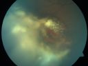
ECTOPIC DISCIFORM SCAR/PERIPHERAL CHOROIDAL NEOVASCULAR MEMBRANE – EXUDATE –INFLAMMATION IN REACTION TO THE PERIPHERAL VASCULAR LESION 888 views53-year-old woman has noticed some blurred vision in the right eye for a few years. In retrospect, she thinks it is possible since she was a teenager the right eye was always the weaker eye and that the left eye has always been the better eye. VISUAL ACUITY: Vision OD is 20/400Dec 20, 2011
|
|

ECTOPIC DISCIFORM SCAR/PERIPHERAL CHOROIDAL NEOVASCULAR MEMBRANE – EXUDATE –INFLAMMATION IN REACTION TO THE PERIPHERAL VASCULAR LESION 873 views53-year-old woman has noticed some blurred vision in the right eye for a few years. In retrospect, she thinks it is possible since she was a teenager the right eye was always the weaker eye and that the left eye has always been the better eye. VISUAL ACUITY: Vision OD is 20/400Dec 20, 2011
|
|

Eccentric Disciform Scar - Breakthrough Vitreous Hemorrhage - InferoTemporal Subretinal Fibrosis - Ectopic Choroidal Neovascular Membranes789 views88-year-old man has noticed for the last few months the vision in the right eye is foggy, like there is a film in the eye or a haze, His vision is 20/16 in each eye.
Right eye - inferotemporal periphery there is a hemorrhage at the equator at about 7:00 o’clock, which is about 4-disc diameters across and adjacent to that there is scarring.
Left eye - large disciform scar inferotemporallyDec 11, 2011
|
|
|
|


