Last additions - Central Retinal Artery Occlusion
|
412 viewsApr 23, 2020
|
|

445 viewsApr 23, 2020
|
|

475 viewsApr 23, 2020
|
|
442 viewsApr 23, 2020
|
|
413 viewsApr 23, 2020
|
|

497 viewsApr 23, 2020
|
|

436 viewsApr 23, 2020
|
|
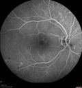
Acute Central Retinal Artery Occlusion439 views1 day of vision loss 79 year old man to HM. NVG developed 2 months later.Mar 26, 2019
|
|

Acute Central Retinal Artery Occlusion483 views1 day of vision loss 79 year old man to HM. NVG developed 2 months later.Mar 26, 2019
|
|
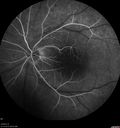
Acute Central Retinal Artery Occlusion445 views1 day of vision loss 79 year old man to HM. NVG developed 2 months later.Mar 26, 2019
|
|
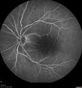
Acute Central Retinal Artery Occlusion471 views1 day of vision loss 79 year old man to HM. NVG developed 2 months later.Mar 26, 2019
|
|
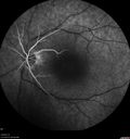
Acute Central Retinal Artery Occlusion454 views1 day of vision loss 79 year old man to HM. NVG developed 2 months later.Mar 26, 2019
|
|

Acute Central Retinal Artery Occlusion468 views1 day of vision loss 79 year old man to HM. NVG developed 2 months later.Mar 26, 2019
|
|

Acute Central Retinal Artery Occlusion444 views1 day of vision loss 79 year old man to HM. NVG developed 2 months later.Mar 26, 2019
|
|

Acute Central Retinal Artery Occlusion407 views1 day of vision loss 79 year old man to HM. NVG developed 2 months later.Mar 26, 2019
|
|

Acute Central Retinal Artery Occlusion467 views1 day of vision loss 79 year old man to HM. NVG developed 2 months later.Mar 26, 2019
|
|

Acute Central Retinal Artery Occlusion503 views1 day of vision loss 79 year old man to HM. NVG developed 2 months later.Mar 26, 2019
|
|

Acute Central Retinal Artery Occlusion450 views1 day of vision loss 79 year old man to HM. NVG developed 2 months later.Mar 26, 2019
|
|

Acute Central Retinal Artery Occlusion510 views1 day of vision loss 79 year old man to HM. NVG developed 2 months later.Mar 26, 2019
|
|
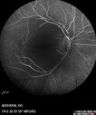
Central Retinal Artery Occlusion - Diabetic Retinopathy - NO cherry red spot452 views58 year old man with sudden vision loss in the right eye. The vision had been 20/40 and dropped to 20/200 the day prior to presentation.
VA 20/200 OD, 20/20 OS
FA shows CRAO. No cherry red spot.Feb 27, 2019
|
|
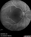
Central Retinal Artery Occlusion - Diabetic Retinopathy - NO cherry red spot540 views58 year old man with sudden vision loss in the right eye. The vision had been 20/40 and dropped to 20/200 the day prior to presentation.
VA 20/200 OD, 20/20 OS
FA shows CRAO. No cherry red spot.Feb 27, 2019
|
|
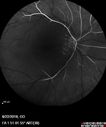
Central Retinal Artery Occlusion - Diabetic Retinopathy - NO cherry red spot480 views58 year old man with sudden vision loss in the right eye. The vision had been 20/40 and dropped to 20/200 the day prior to presentation.
VA 20/200 OD, 20/20 OS
FA shows CRAO. No cherry red spot.Feb 27, 2019
|
|
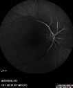
Central Retinal Artery Occlusion - Diabetic Retinopathy - NO cherry red spot532 views58 year old man with sudden vision loss in the right eye. The vision had been 20/40 and dropped to 20/200 the day prior to presentation.
VA 20/200 OD, 20/20 OS
FA shows CRAO. No cherry red spot.Feb 27, 2019
|
|
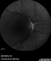
Central Retinal Artery Occlusion - Diabetic Retinopathy - NO cherry red spot498 views58 year old man with sudden vision loss in the right eye. The vision had been 20/40 and dropped to 20/200 the day prior to presentation.
VA 20/200 OD, 20/20 OS
FA shows CRAO. No cherry red spot.Feb 27, 2019
|
|
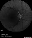
Central Retinal Artery Occlusion - Diabetic Retinopathy - NO cherry red spot552 views58 year old man with sudden vision loss in the right eye. The vision had been 20/40 and dropped to 20/200 the day prior to presentation.
VA 20/200 OD, 20/20 OS
FA shows CRAO. No cherry red spot.Feb 27, 2019
|
|

Central Retinal Artery Occlusion - Diabetic Retinopathy - NO cherry red spot487 views58 year old man with sudden vision loss in the right eye. The vision had been 20/40 and dropped to 20/200 the day prior to presentation.
VA 20/200 OD, 20/20 OS
FA shows CRAO. No cherry red spot.Feb 27, 2019
|
|
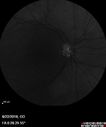
Central Retinal Artery Occlusion - Diabetic Retinopathy - NO cherry red spot529 views58 year old man with sudden vision loss in the right eye. The vision had been 20/40 and dropped to 20/200 the day prior to presentation.
VA 20/200 OD, 20/20 OS
FA shows CRAO. No cherry red spot.Feb 27, 2019
|
|

Central Retinal Artery Occlusion - Diabetic Retinopathy - NO cherry red spot446 views58 year old man with sudden vision loss in the right eye. The vision had been 20/40 and dropped to 20/200 the day prior to presentation.
VA 20/200 OD, 20/20 OS
FA shows CRAO. No cherry red spot.Feb 27, 2019
|
|

Central Retinal Artery Occlusion - Diabetic Retinopathy - NO cherry red spot565 views58 year old man with sudden vision loss in the right eye. The vision had been 20/40 and dropped to 20/200 the day prior to presentation.
VA 20/200 OD, 20/20 OS
FA shows CRAO. No cherry red spot.Feb 27, 2019
|
|

Central Retinal Artery Occlusion - Diabetic Retinopathy - NO cherry red spot575 views58 year old man with sudden vision loss in the right eye. The vision had been 20/40 and dropped to 20/200 the day prior to presentation.
VA 20/200 OD, 20/20 OS
FA shows CRAO. No cherry red spot.Feb 27, 2019
|
|

Central Retinal Artery Occlusion - Diabetic Retinopathy - NO cherry red spot623 views58 year old man with sudden vision loss in the right eye. The vision had been 20/40 and dropped to 20/200 the day prior to presentation.
VA 20/200 OD, 20/20 OS
FA shows CRAO. No cherry red spot.Feb 27, 2019
|
|
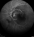
Central Retinal Artery Occlusion then Neovascular Glaucoma500 views81 year old male with sudden vision loss. 2 months later developed NVGJul 27, 2018
|
|
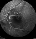
Central Retinal Artery Occlusion then Neovascular Glaucoma571 views81 year old male with sudden vision loss. 2 months later developed NVGJul 27, 2018
|
|
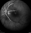
Central Retinal Artery Occlusion then Neovascular Glaucoma474 views81 year old male with sudden vision loss. 2 months later developed NVGJul 27, 2018
|
|
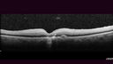
Central Retinal Artery Occlusion then Neovascular Glaucoma433 views81 year old male with sudden vision loss. 2 months later developed NVGJul 27, 2018
|
|

Central Retinal Artery Occlusion then Neovascular Glaucoma420 views81 year old male with sudden vision loss. 2 months later developed NVGJul 27, 2018
|
|

Central Retinal Artery Occlusion then Neovascular Glaucoma514 views81 year old male with sudden vision loss. 2 months later developed NVGJul 27, 2018
|
|

Central Retinal Artery Occlusion then Neovascular Glaucoma550 views81 year old male with sudden vision loss. 2 months later developed NVGJul 27, 2018
|
|

Central Retinal Artery Occlusion then Neovascular Glaucoma441 views81 year old male with sudden vision loss. 2 months later developed NVGJul 27, 2018
|
|

Central Retinal Artery Occlusion then Neovascular Glaucoma505 views81 year old male with sudden vision loss. 2 months later developed NVGJul 27, 2018
|
|

Central Retinal Artery Occlusion then Neovascular Glaucoma498 views81 year old male with sudden vision loss. 2 months later developed NVGJul 27, 2018
|
|
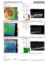
Central Retinal Artery Occlusion - MAP764 viewsProgression from acute to atrophy - decreasing retinal thickenssNov 28, 2016
|
|
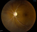
Retinal Artery Occlusion1198 viewsPatient comes in for eval on artery occlusions in both eyes. VA is 20/400, right eye and NLP, left eye. Fundus photos show paniretinal scars in the right eye with arterial narrowing and the left eye has arterial narrowing as well.Sep 23, 2014
|
|

Retinal Artery Occlusion1708 viewsPatient comes in for eval on artery occlusions in both eyes. VA is 20/400, right eye and NLP, left eye. Fundus photos show paniretinal scars in the right eye with arterial narrowing and the left eye has arterial narrowing as well.Sep 23, 2014
|
|
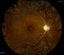
Retinal Artery Occlusion1033 viewsPatient comes in for eval on artery occlusions in both eyes. VA is 20/400, right eye and NLP, left eye. Fundus photos show paniretinal scars in the right eye with arterial narrowing and the left eye has arterial narrowing as well.Sep 23, 2014
|
|
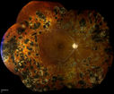
Retinal Artery Occlusion594 viewsPatient comes in for eval on artery occlusions in both eyes. VA is 20/400, right eye and NLP, left eye. Fundus photos show paniretinal scars in the right eye with arterial narrowing and the left eye has arterial narrowing as well.Sep 23, 2014
|
|

Central Retinal Artery Occlusion1105 viewsCentral Retinal Artery Occlusion with cilio artery perfusion. May 15, 2014
|
|

FA: CRAO838 viewsCRAO with cilio artery perfusionMay 15, 2014
|
|

Embolic CRAO Mid560 viewsOct 24, 2013
|
|

Embolic CRAO Late510 viewsOct 24, 2013
|
|

Embolic CRAO Early619 viewsOct 24, 2013
|
|

Embolic CRAO color846 viewsOct 24, 2013
|
|
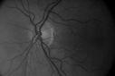
Embolic CRAO B&W529 viewsOct 24, 2013
|
|

Central Arterial Occlusion with Embolism Present1045 viewsPatient comes in with CRAO in the right eye. VA was hand motion. Fundus photo shows white veil over the retina with 2-emboli in a branch artery temporal in the right eye. She will be evaluated for emboli and followed up in a monthJul 16, 2013
|
|

Central Arterial Occlusion with Embolism Present486 viewsPatient comes in with CRAO in the right eye. VA was hand motion. Fundus photo shows white veil over the retina with 2-emboli in a branch artery temporal in the right eye. She will be evaluated for emboli and followed up in a monthJul 16, 2013
|
|
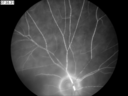
CRAO405 viewsCRAO in ODMay 30, 2013
|
|

CRAO493 viewsCRAO Color MontageJan 11, 2013
|
|
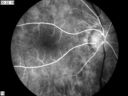
CRAO414 viewsCRAO in OD Tranisit Phase of FAJan 11, 2013
|
|

CRAO465 viewsCRAO in OD Tranisit Phase of FAJan 11, 2013
|
|
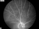
CRAO413 viewsCRAO in OD Late Phase of FAJan 11, 2013
|
|

CRAO475 viewsCRAO in OD of a 37 yr old Male, Transit PhaseJan 08, 2013
|
|

Central Retinal Artery Occlusion with Cilioretinal Sparing - Acute - Line scan good eye993 views 77-year-old man was doing fine until midnight of last night when he noticed sudden severe vision loss in the left eye. VISUAL ACUITY: OD 20/20, OS 2/200.Sep 05, 2012
|
|

Central Retinal Artery Occlusion with Cilioretinal Sparing - Acute - SD-OCT Line Scan Inner retinal edema1532 views 77-year-old man was doing fine until midnight of last night when he noticed sudden severe vision loss in the left eye. VISUAL ACUITY: OD 20/20, OS 2/200.Sep 05, 2012
|
|
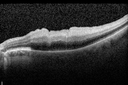
Central Retinal Artery Occlusion with Cilioretinal Sparing - Acute - SD-OCT Line Scan Inner retinal edema1181 views 77-year-old man was doing fine until midnight of last night when he noticed sudden severe vision loss in the left eye. VISUAL ACUITY: OD 20/20, OS 2/200.Sep 05, 2012
|
|
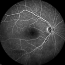
Central Retinal Artery Occlusion with Cilioretinal Sparing - Acute781 views 77-year-old man was doing fine until midnight of last night when he noticed sudden severe vision loss in the left eye. VISUAL ACUITY: OD 20/20, OS 2/200.Sep 05, 2012
|
|

Central Retinal Artery Occlusion with Cilioretinal Sparing - Acute696 views 77-year-old man was doing fine until midnight of last night when he noticed sudden severe vision loss in the left eye. VISUAL ACUITY: OD 20/20, OS 2/200.Sep 05, 2012
|
|
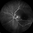
Central Retinal Artery Occlusion with Cilioretinal Sparing - Acute - FA Very Slow Filling CRA821 views 77-year-old man was doing fine until midnight of last night when he noticed sudden severe vision loss in the left eye. VISUAL ACUITY: OD 20/20, OS 2/200.Sep 05, 2012
|
|

Central Retinal Artery Occlusion with Cilioretinal Sparing - Acute - FA Very Slow Filling CRA840 views 77-year-old man was doing fine until midnight of last night when he noticed sudden severe vision loss in the left eye. VISUAL ACUITY: OD 20/20, OS 2/200.Sep 05, 2012
|
|

Central Retinal Artery Occlusion with Cilioretinal Sparing - Acute - FA Very Slow Filling CRA921 views 77-year-old man was doing fine until midnight of last night when he noticed sudden severe vision loss in the left eye. VISUAL ACUITY: OD 20/20, OS 2/200.Sep 05, 2012
|
|
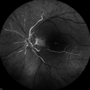
Central Retinal Artery Occlusion with Cilioretinal Sparing - Acute - FA Very Slow Filling CRA918 views 77-year-old man was doing fine until midnight of last night when he noticed sudden severe vision loss in the left eye. VISUAL ACUITY: OD 20/20, OS 2/200.Sep 05, 2012
|
|
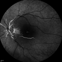
Central Retinal Artery Occlusion with Cilioretinal Sparing - Acute - FA Very Slow Filling CRA966 views 77-year-old man was doing fine until midnight of last night when he noticed sudden severe vision loss in the left eye. VISUAL ACUITY: OD 20/20, OS 2/200.Sep 05, 2012
|
|

Central Retinal Artery Occlusion with Cilioretinal Sparing - Acute - FA Very Slow Filling CRA1047 views 77-year-old man was doing fine until midnight of last night when he noticed sudden severe vision loss in the left eye. VISUAL ACUITY: OD 20/20, OS 2/200.Sep 05, 2012
|
|

Central Retinal Artery Occlusion with Cilioretinal Sparing - Acute924 views 77-year-old man was doing fine until midnight of last night when he noticed sudden severe vision loss in the left eye. VISUAL ACUITY: OD 20/20, OS 2/200.Sep 05, 2012
|
|

Central Retinal Artery Occlusion with Cilioretinal Sparing - Acute1218 views 77-year-old man was doing fine until midnight of last night when he noticed sudden severe vision loss in the left eye. VISUAL ACUITY: OD 20/20, OS 2/200.Sep 05, 2012
|
|

Montage of Hemi-Central Retinal Artery Occlusion728 viewsJul 06, 2012
|
|

Montage of Hemi-Central Retinal Artery Occlusion701 viewsJul 06, 2012
|
|

660 viewsMay 07, 2012
|
|

752 viewsMay 07, 2012
|
|

781 viewsMay 07, 2012
|
|

Central Retinal Artery Occlusion1775 views60 year old male with LP vision due to extensive blood flow loss.Apr 25, 2012
|
|
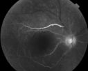
698 views60 year old male with LP vision due to extensive blood flow loss.Apr 25, 2012
|
|

Pathology - Central Retinal Artery Occlusion - 6 weeks old - Inner Retinal Atrophy778 viewspatient died about 6 to 8 weeks after the artery occlusion, and he had no history of glaucoma. The inner retina atrophy, which looks very similar to that caused by COAG, is due to recent infarct. Jul 06, 2011
|
|
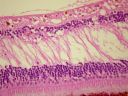
Pathology - Central Retinal Artery Occlusion - 6 weeks old - Inner Retinal Atrophy872 viewspatient died about 6 to 8 weeks after the artery occlusion, and he had no history of glaucoma. The inner retina atrophy, which looks very similar to that caused by COAG, is due to recent infarct. Jul 06, 2011
|
|

Pathology - Central Retinal Artery Occlusion - 6 weeks old - Inner Retinal Atrophy - Macular Pucker931 viewspatient died about 6 to 8 weeks after the artery occlusion, and he had no history of glaucoma. The inner retina atrophy, which looks very similar to that caused by COAG, is due to recent infarct. Jul 06, 2011
|
|

Pathology - Central Retinal Artery Occlusion - 6 weeks old - Inner Retinal Atrophy - Macular Pucker905 viewspatient died about 6 to 8 weeks after the artery occlusion, and he had no history of glaucoma. The inner retina atrophy, which looks very similar to that caused by COAG, is due to recent infarct. Jul 06, 2011
|
|

80 Year old man with 3 day history of vision loss right eye. Vision 4/200.561 viewsFeb 10, 2011
|
|
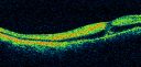
80 Year old man with 3 day history of vision loss right eye. Vision 4/200.537 viewsFeb 10, 2011
|
|
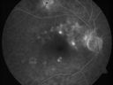
80 Year old man with 3 day history of vision loss right eye. Vision 4/200.494 viewsFeb 10, 2011
|
|

80 Year old man with 3 day history of vision loss right eye. Vision 4/200.592 viewsFeb 10, 2011
|
|
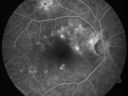
80 Year old man with 3 day history of vision loss right eye. Vision 4/200.499 viewsFeb 10, 2011
|
|
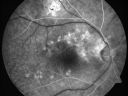
80 Year old man with 3 day history of vision loss right eye. Vision 4/200.523 viewsFeb 10, 2011
|
|
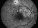
80 Year old man with 3 day history of vision loss right eye. Vision 4/200.488 viewsFeb 10, 2011
|
|
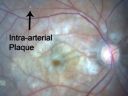
80 Year old man with 3 day history of vision loss right eye. Vision 4/200.589 viewsFeb 10, 2011
|
|

80 Year old man with 3 day history of vision loss right eye. Vision 4/200.582 viewsFeb 10, 2011
|
|

80 Year old man with 3 day history of vision loss right eye. Vision 4/200.594 viewsFeb 10, 2011
|
|

Central Retinal Artery Occlusion less than 24 hours - 69 year old man VA light perception614 viewsFeb 07, 2011
|
|

Central Retinal Artery Occlusion less than 24 hours - 69 year old man VA light perception843 viewsFeb 07, 2011
|
|

Central Retinal Artery Occlusion less than 24 hours - 69 year old man VA light perception577 viewsFeb 07, 2011
|
|

Central Retinal Artery Occlusion less than 24 hours - 69 year old man VA light perception494 viewsFeb 07, 2011
|
|
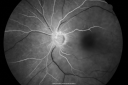
Central Retinal Artery Occlusion less than 24 hours - 69 year old man VA light perception554 viewsFeb 07, 2011
|
|
|
|


