Last additions - Macular Degeneration - Age Related, Myopic, Choroidal Neovascular Membranes
|

Asymptomatic wet AMD - Subfoveal CNM both eyes483 views82 year old woman - 20/25 OU. Images show occult subfoveal CNVM both eyes best seen on late ICG. The left eye also has probable polypoidal choroidal vasculopathy. No treatment was recommended on the first visit - follow-up at 1 month.May 05, 2017
|
|

Asymptomatic wet AMD - Subfoveal CNM both eyes433 views82 year old woman - 20/25 OU. Images show occult subfoveal CNVM both eyes best seen on late ICG. The left eye also has probable polypoidal choroidal vasculopathy. No treatment was recommended on the first visit - follow-up at 1 month.May 05, 2017
|
|
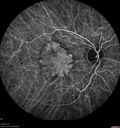
Asymptomatic wet AMD - Subfoveal CNM both eyes470 views82 year old woman - 20/25 OU. Images show occult subfoveal CNVM both eyes best seen on late ICG. The left eye also has probable polypoidal choroidal vasculopathy. No treatment was recommended on the first visit - follow-up at 1 month.May 05, 2017
|
|
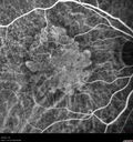
Asymptomatic wet AMD - Subfoveal CNM both eyes479 views82 year old woman - 20/25 OU. Images show occult subfoveal CNVM both eyes best seen on late ICG. The left eye also has probable polypoidal choroidal vasculopathy. No treatment was recommended on the first visit - follow-up at 1 month.May 05, 2017
|
|

Asymptomatic wet AMD - Subfoveal CNM both eyes407 views82 year old woman - 20/25 OU. Images show occult subfoveal CNVM both eyes best seen on late ICG. The left eye also has probable polypoidal choroidal vasculopathy. No treatment was recommended on the first visit - follow-up at 1 month.May 05, 2017
|
|

Asymptomatic wet AMD - Subfoveal CNM both eyes509 views82 year old woman - 20/25 OU. Images show occult subfoveal CNVM both eyes best seen on late ICG. The left eye also has probable polypoidal choroidal vasculopathy. No treatment was recommended on the first visit - follow-up at 1 month.May 05, 2017
|
|
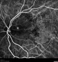
Asymptomatic wet AMD - Subfoveal CNM both eyes409 views82 year old woman - 20/25 OU. Images show occult subfoveal CNVM both eyes best seen on late ICG. The left eye also has probable polypoidal choroidal vasculopathy. No treatment was recommended on the first visit - follow-up at 1 month.May 05, 2017
|
|

Asymptomatic wet AMD - Subfoveal CNM both eyes498 views82 year old woman - 20/25 OU. Images show occult subfoveal CNVM both eyes best seen on late ICG. The left eye also has probable polypoidal choroidal vasculopathy. No treatment was recommended on the first visit - follow-up at 1 month.May 05, 2017
|
|

Asymptomatic wet AMD - Subfoveal CNM both eyes439 views82 year old woman - 20/25 OU. Images show occult subfoveal CNVM both eyes best seen on late ICG. The left eye also has probable polypoidal choroidal vasculopathy. No treatment was recommended on the first visit - follow-up at 1 month.May 05, 2017
|
|

Asymptomatic wet AMD - Subfoveal CNM both eyes459 views82 year old woman - 20/25 OU. Images show occult subfoveal CNVM both eyes best seen on late ICG. The left eye also has probable polypoidal choroidal vasculopathy. No treatment was recommended on the first visit - follow-up at 1 month.May 05, 2017
|
|
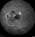
Asymptomatic wet AMD - Subfoveal CNM both eyes499 views82 year old woman - 20/25 OU. Images show occult subfoveal CNVM both eyes best seen on late ICG. The left eye also has probable polypoidal choroidal vasculopathy. No treatment was recommended on the first visit - follow-up at 1 month.May 05, 2017
|
|
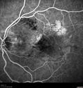
Asymptomatic wet AMD - Subfoveal CNM both eyes425 views82 year old woman - 20/25 OU. Images show occult subfoveal CNVM both eyes best seen on late ICG. The left eye also has probable polypoidal choroidal vasculopathy. No treatment was recommended on the first visit - follow-up at 1 month.May 05, 2017
|
|
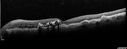
Asymptomatic wet AMD - Subfoveal CNM both eyes405 views82 year old woman - 20/25 OU. Images show occult subfoveal CNVM both eyes best seen on late ICG. The left eye also has probable polypoidal choroidal vasculopathy. No treatment was recommended on the first visit - follow-up at 1 month.May 05, 2017
|
|
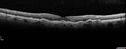
Asymptomatic wet AMD - Subfoveal CNM both eyes409 views82 year old woman - 20/25 OU. Images show occult subfoveal CNVM both eyes best seen on late ICG. The left eye also has probable polypoidal choroidal vasculopathy. No treatment was recommended on the first visit - follow-up at 1 month.May 05, 2017
|
|

Asymptomatic wet AMD - Subfoveal CNM both eyes399 views82 year old woman - 20/25 OU. Images show occult subfoveal CNVM both eyes best seen on late ICG. The left eye also has probable polypoidal choroidal vasculopathy. No treatment was recommended on the first visit - follow-up at 1 month.May 05, 2017
|
|
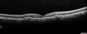
Asymptomatic wet AMD - Subfoveal CNM both eyes416 views82 year old woman - 20/25 OU. Images show occult subfoveal CNVM both eyes best seen on late ICG. The left eye also has probable polypoidal choroidal vasculopathy. No treatment was recommended on the first visit - follow-up at 1 month.May 05, 2017
|
|

Asymptomatic wet AMD - Subfoveal CNM both eyes401 views82 year old woman - 20/25 OU. Images show occult subfoveal CNVM both eyes best seen on late ICG. The left eye also has probable polypoidal choroidal vasculopathy. No treatment was recommended on the first visit - follow-up at 1 month.May 05, 2017
|
|

Asymptomatic wet AMD - Subfoveal CNM both eyes428 views82 year old woman - 20/25 OU. Images show occult subfoveal CNVM both eyes best seen on late ICG. The left eye also has probable polypoidal choroidal vasculopathy. No treatment was recommended on the first visit - follow-up at 1 month.May 05, 2017
|
|
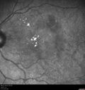
Asymptomatic wet AMD - Subfoveal CNM both eyes406 views82 year old woman - 20/25 OU. Images show occult subfoveal CNVM both eyes best seen on late ICG. The left eye also has probable polypoidal choroidal vasculopathy. No treatment was recommended on the first visit - follow-up at 1 month.May 05, 2017
|
|

Asymptomatic wet AMD - Subfoveal CNM both eyes472 views82 year old woman - 20/25 OU. Images show occult subfoveal CNVM both eyes best seen on late ICG. The left eye also has probable polypoidal choroidal vasculopathy. No treatment was recommended on the first visit - follow-up at 1 month.May 05, 2017
|
|
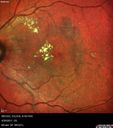
Asymptomatic wet AMD - Subfoveal CNM both eyes542 views82 year old woman - 20/25 OU. Images show occult subfoveal CNVM both eyes best seen on late ICG. The left eye also has probable polypoidal choroidal vasculopathy. No treatment was recommended on the first visit - follow-up at 1 month.May 05, 2017
|
|
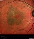
Asymptomatic wet AMD - Subfoveal CNM both eyes584 views82 year old woman - 20/25 OU. Images show occult subfoveal CNVM both eyes best seen on late ICG. The left eye also has probable polypoidal choroidal vasculopathy. No treatment was recommended on the first visit - follow-up at 1 month.May 05, 2017
|
|
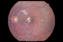
Asymptomatic wet AMD - Subfoveal CNM both eyes523 views82 year old woman - 20/25 OU. Images show occult subfoveal CNVM both eyes best seen on late ICG. The left eye also has probable polypoidal choroidal vasculopathy. No treatment was recommended on the first visit - follow-up at 1 month.May 05, 2017
|
|

Asymptomatic wet AMD - Subfoveal CNM both eyes476 views82 year old woman - 20/25 OU. Images show occult subfoveal CNVM both eyes best seen on late ICG. The left eye also has probable polypoidal choroidal vasculopathy. No treatment was recommended on the first visit - follow-up at 1 month.May 05, 2017
|
|

Confluent Soft Drusen with Subetinal Fluid - Right Eye663 viewsAsymptomatic 85 year old man. OCT shows pocket of subfoveal fluid in the center of the macula. This could be mechanical from the drusen preventing the retina from settling down. Testing did not reveal a CNVM and subsequent OCT 6 weeks later showed no change in structure.Feb 06, 2017
|
|
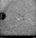
Confluent Soft Drusen with Subetinal Fluid - Right Eye465 viewsAsymptomatic 85 year old man. OCT shows pocket of subfoveal fluid in the center of the macula. This could be mechanical from the drusen preventing the retina from settling down. Testing did not reveal a CNVM and subsequent OCT 6 weeks later showed no change in structure.Feb 06, 2017
|
|

Confluent Soft Drusen with Subetinal Fluid - Right Eye463 viewsAsymptomatic 85 year old man. OCT shows pocket of subfoveal fluid in the center of the macula. This could be mechanical from the drusen preventing the retina from settling down. Testing did not reveal a CNVM and subsequent OCT 6 weeks later showed no change in structure.Feb 06, 2017
|
|
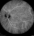
Confluent Soft Drusen with Subetinal Fluid - Right Eye574 viewsAsymptomatic 85 year old man. OCT shows pocket of subfoveal fluid in the center of the macula. This could be mechanical from the drusen preventing the retina from settling down. Testing did not reveal a CNVM and subsequent OCT 6 weeks later showed no change in structure.Feb 06, 2017
|
|
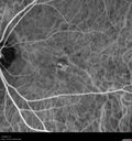
Confluent Soft Drusen with Subetinal Fluid - Right Eye494 viewsAsymptomatic 85 year old man. OCT shows pocket of subfoveal fluid in the center of the macula. This could be mechanical from the drusen preventing the retina from settling down. Testing did not reveal a CNVM and subsequent OCT 6 weeks later showed no change in structure.Feb 06, 2017
|
|

Confluent Soft Drusen with Subetinal Fluid - Right Eye571 viewsAsymptomatic 85 year old man. OCT shows pocket of subfoveal fluid in the center of the macula. This could be mechanical from the drusen preventing the retina from settling down. Testing did not reveal a CNVM and subsequent OCT 6 weeks later showed no change in structure.Feb 06, 2017
|
|

Confluent Soft Drusen with Subetinal Fluid - Right Eye607 viewsAsymptomatic 85 year old man. OCT shows pocket of subfoveal fluid in the center of the macula. This could be mechanical from the drusen preventing the retina from settling down. Testing did not reveal a CNVM and subsequent OCT 6 weeks later showed no change in structure.Feb 06, 2017
|
|

Confluent Soft Drusen with Subetinal Fluid - Right Eye524 viewsAsymptomatic 85 year old man. OCT shows pocket of subfoveal fluid in the center of the macula. This could be mechanical from the drusen preventing the retina from settling down. Testing did not reveal a CNVM and subsequent OCT 6 weeks later showed no change in structure.Feb 06, 2017
|
|

Confluent Soft Drusen with Subetinal Fluid - Right Eye534 viewsAsymptomatic 85 year old man. OCT shows pocket of subfoveal fluid in the center of the macula. This could be mechanical from the drusen preventing the retina from settling down. Testing did not reveal a CNVM and subsequent OCT 6 weeks later showed no change in structure.Feb 06, 2017
|
|
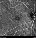
Confluent Soft Drusen with Subetinal Fluid - Right Eye469 viewsAsymptomatic 85 year old man. OCT shows pocket of subfoveal fluid in the center of the macula. This could be mechanical from the drusen preventing the retina from settling down. Testing did not reveal a CNVM and subsequent OCT 6 weeks later showed no change in structure.Feb 06, 2017
|
|
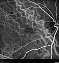
Confluent Soft Drusen with Subetinal Fluid - Right Eye460 viewsAsymptomatic 85 year old man. OCT shows pocket of subfoveal fluid in the center of the macula. This could be mechanical from the drusen preventing the retina from settling down. Testing did not reveal a CNVM and subsequent OCT 6 weeks later showed no change in structure.Feb 06, 2017
|
|

Confluent Soft Drusen with Subetinal Fluid - Right Eye452 viewsAsymptomatic 85 year old man. OCT shows pocket of subfoveal fluid in the center of the macula. This could be mechanical from the drusen preventing the retina from settling down. Testing did not reveal a CNVM and subsequent OCT 6 weeks later showed no change in structure.Feb 06, 2017
|
|

Confluent Soft Drusen with Subetinal Fluid - Right Eye524 viewsAsymptomatic 85 year old man. OCT shows pocket of subfoveal fluid in the center of the macula. This could be mechanical from the drusen preventing the retina from settling down. Testing did not reveal a CNVM and subsequent OCT 6 weeks later showed no change in structure.Feb 06, 2017
|
|
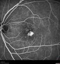
Confluent Soft Drusen with Subetinal Fluid - Right Eye485 viewsAsymptomatic 85 year old man. OCT shows pocket of subfoveal fluid in the center of the macula. This could be mechanical from the drusen preventing the retina from settling down. Testing did not reveal a CNVM and subsequent OCT 6 weeks later showed no change in structure.Feb 06, 2017
|
|

Confluent Soft Drusen with Subetinal Fluid - Right Eye438 viewsAsymptomatic 85 year old man. OCT shows pocket of subfoveal fluid in the center of the macula. This could be mechanical from the drusen preventing the retina from settling down. Testing did not reveal a CNVM and subsequent OCT 6 weeks later showed no change in structure.Feb 06, 2017
|
|
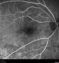
Confluent Soft Drusen with Subetinal Fluid - Right Eye488 viewsAsymptomatic 85 year old man. OCT shows pocket of subfoveal fluid in the center of the macula. This could be mechanical from the drusen preventing the retina from settling down. Testing did not reveal a CNVM and subsequent OCT 6 weeks later showed no change in structure.Feb 06, 2017
|
|

Confluent Soft Drusen with Subetinal Fluid - Right Eye457 viewsAsymptomatic 85 year old man. OCT shows pocket of subfoveal fluid in the center of the macula. This could be mechanical from the drusen preventing the retina from settling down. Testing did not reveal a CNVM and subsequent OCT 6 weeks later showed no change in structure.Feb 06, 2017
|
|
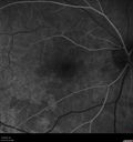
Confluent Soft Drusen with Subetinal Fluid - Right Eye476 viewsAsymptomatic 85 year old man. OCT shows pocket of subfoveal fluid in the center of the macula. This could be mechanical from the drusen preventing the retina from settling down. Testing did not reveal a CNVM and subsequent OCT 6 weeks later showed no change in structure.Feb 06, 2017
|
|
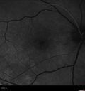
Confluent Soft Drusen with Subetinal Fluid - Right Eye491 viewsAsymptomatic 85 year old man. OCT shows pocket of subfoveal fluid in the center of the macula. This could be mechanical from the drusen preventing the retina from settling down. Testing did not reveal a CNVM and subsequent OCT 6 weeks later showed no change in structure.Feb 06, 2017
|
|
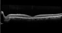
Confluent Soft Drusen with Subetinal Fluid - Right Eye485 viewsAsymptomatic 85 year old man. OCT shows pocket of subfoveal fluid in the center of the macula. This could be mechanical from the drusen preventing the retina from settling down. Testing did not reveal a CNVM and subsequent OCT 6 weeks later showed no change in structure.Feb 06, 2017
|
|
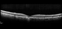
Confluent Soft Drusen with Subetinal Fluid - Right Eye474 viewsAsymptomatic 85 year old man. OCT shows pocket of subfoveal fluid in the center of the macula. This could be mechanical from the drusen preventing the retina from settling down. Testing did not reveal a CNVM and subsequent OCT 6 weeks later showed no change in structure.Feb 06, 2017
|
|

Confluent Soft Drusen with Subetinal Fluid - Right Eye759 viewsAsymptomatic 85 year old man. OCT shows pocket of subfoveal fluid in the center of the macula. This could be mechanical from the drusen preventing the retina from settling down. Testing did not reveal a CNVM and subsequent OCT 6 weeks later showed no change in structure.Feb 06, 2017
|
|
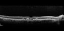
Confluent Soft Drusen with Subetinal Fluid - Right Eye595 viewsAsymptomatic 85 year old man. OCT shows pocket of subfoveal fluid in the center of the macula. This could be mechanical from the drusen preventing the retina from settling down. Testing did not reveal a CNVM and subsequent OCT 6 weeks later showed no change in structure.Feb 06, 2017
|
|

Confluent Soft Drusen with Subetinal Fluid - Right Eye525 viewsAsymptomatic 85 year old man. OCT shows pocket of subfoveal fluid in the center of the macula. This could be mechanical from the drusen preventing the retina from settling down. Testing did not reveal a CNVM and subsequent OCT 6 weeks later showed no change in structure.Feb 06, 2017
|
|
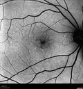
Confluent Soft Drusen with Subetinal Fluid - Right Eye501 viewsAsymptomatic 85 year old man. OCT shows pocket of subfoveal fluid in the center of the macula. This could be mechanical from the drusen preventing the retina from settling down. Testing did not reveal a CNVM and subsequent OCT 6 weeks later showed no change in structure.Feb 06, 2017
|
|
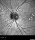
Confluent Soft Drusen with Subetinal Fluid - Right Eye481 viewsAsymptomatic 85 year old man. OCT shows pocket of subfoveal fluid in the center of the macula. This could be mechanical from the drusen preventing the retina from settling down. Testing did not reveal a CNVM and subsequent OCT 6 weeks later showed no change in structure.Feb 06, 2017
|
|
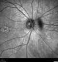
Confluent Soft Drusen with Subetinal Fluid - Right Eye511 viewsAsymptomatic 85 year old man. OCT shows pocket of subfoveal fluid in the center of the macula. This could be mechanical from the drusen preventing the retina from settling down. Testing did not reveal a CNVM and subsequent OCT 6 weeks later showed no change in structure.Feb 06, 2017
|
|
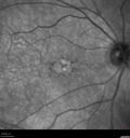
Confluent Soft Drusen with Subetinal Fluid - Right Eye513 viewsAsymptomatic 85 year old man. OCT shows pocket of subfoveal fluid in the center of the macula. This could be mechanical from the drusen preventing the retina from settling down. Testing did not reveal a CNVM and subsequent OCT 6 weeks later showed no change in structure.Feb 06, 2017
|
|

Confluent Soft Drusen with Subetinal Fluid - Right Eye559 viewsAsymptomatic 85 year old man. OCT shows pocket of subfoveal fluid in the center of the macula. This could be mechanical from the drusen preventing the retina from settling down. Testing did not reveal a CNVM and subsequent OCT 6 weeks later showed no change in structure.Feb 06, 2017
|
|

Confluent Soft Drusen with Subetinal Fluid - Right Eye548 viewsAsymptomatic 85 year old man. OCT shows pocket of subfoveal fluid in the center of the macula. This could be mechanical from the drusen preventing the retina from settling down. Testing did not reveal a CNVM and subsequent OCT 6 weeks later showed no change in structure.Feb 06, 2017
|
|

Confluent Soft Drusen with Subetinal Fluid - Right Eye573 viewsAsymptomatic 85 year old man. OCT shows pocket of subfoveal fluid in the center of the macula. This could be mechanical from the drusen preventing the retina from settling down. Testing did not reveal a CNVM and subsequent OCT 6 weeks later showed no change in structure.Feb 06, 2017
|
|

Confluent Soft Drusen with Subetinal Fluid - Right Eye798 viewsAsymptomatic 85 year old man. OCT shows pocket of subfoveal fluid in the center of the macula. This could be mechanical from the drusen preventing the retina from settling down. Testing did not reveal a CNVM and subsequent OCT 6 weeks later showed no change in structure.Feb 06, 2017
|
|

Multifocal Pattern Dystrophy453 views51 year old with normal vision - images look a lot like fundus flavimaculatusJul 07, 2016
|
|

Multifocal Pattern Dystrophy402 views51 year old with normal vision - images look a lot like fundus flavimaculatusJul 07, 2016
|
|

Multifocal Pattern Dystrophy479 views51 year old with normal vision - images look a lot like fundus flavimaculatusJul 07, 2016
|
|
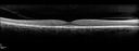
Multifocal Pattern Dystrophy487 views51 year old with normal vision - images look a lot like fundus flavimaculatusJul 07, 2016
|
|

Multifocal Pattern Dystrophy500 views51 year old with normal vision - images look a lot like fundus flavimaculatusJul 07, 2016
|
|

Multifocal Pattern Dystrophy634 views51 year old with normal vision - images look a lot like fundus flavimaculatusJul 07, 2016
|
|

Multifocal Pattern Dystrophy599 views51 year old with normal vision - images look a lot like fundus flavimaculatusJul 07, 2016
|
|
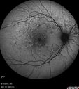
Multifocal Pattern Dystrophy508 views51 year old with normal vision - images look a lot like fundus flavimaculatusJul 07, 2016
|
|

Multifocal Pattern Dystrophy935 views51 year old with normal vision - images look a lot like fundus flavimaculatusJul 07, 2016
|
|

Multifocal Pattern Dystrophy469 views51 year old with normal vision - images look a lot like fundus flavimaculatusJul 07, 2016
|
|
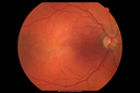
Multifocal Pattern Dystrophy667 views51 year old with normal vision - images look a lot like fundus flavimaculatusJul 07, 2016
|
|
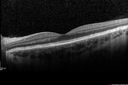
Cuticular Drusen - 26 year old female - Glomerulonephritis524 viewsVA 20/25 OU. Images are over 4 yearsFeb 16, 2016
|
|
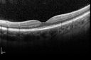
Cuticular Drusen - 26 year old female - Glomerulonephritis442 viewsVA 20/25 OU. Images are over 4 yearsFeb 16, 2016
|
|
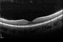
Cuticular Drusen - 26 year old female - Glomerulonephritis486 viewsVA 20/25 OU. Images are over 4 yearsFeb 16, 2016
|
|
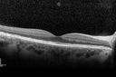
Cuticular Drusen - 26 year old female - Glomerulonephritis434 viewsVA 20/25 OU. Images are over 4 yearsFeb 16, 2016
|
|
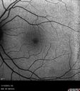
Cuticular Drusen - 26 year old female - Glomerulonephritis433 viewsVA 20/25 OU. Images are over 4 yearsFeb 16, 2016
|
|
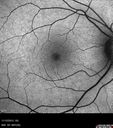
Cuticular Drusen - 26 year old female - Glomerulonephritis473 viewsVA 20/25 OU. Images are over 4 yearsFeb 16, 2016
|
|
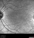
Cuticular Drusen - 26 year old female - Glomerulonephritis429 viewsVA 20/25 OU. Images are over 4 yearsFeb 16, 2016
|
|
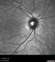
Cuticular Drusen - 26 year old female - Glomerulonephritis430 viewsVA 20/25 OU. Images are over 4 yearsFeb 16, 2016
|
|
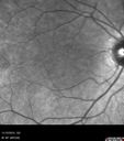
Cuticular Drusen - 26 year old female - Glomerulonephritis453 viewsVA 20/25 OU. Images are over 4 yearsFeb 16, 2016
|
|
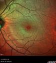
Cuticular Drusen - 26 year old female - Glomerulonephritis464 viewsVA 20/25 OU. Images are over 4 yearsFeb 16, 2016
|
|
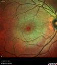
Cuticular Drusen - 26 year old female - Glomerulonephritis465 viewsVA 20/25 OU. Images are over 4 yearsFeb 16, 2016
|
|

Cuticular Drusen - 26 year old female - Glomerulonephritis456 viewsVA 20/25 OU. Images are over 4 yearsFeb 16, 2016
|
|

Cuticular Drusen - 26 year old female - Glomerulonephritis429 viewsVA 20/25 OU. Images are over 4 yearsFeb 16, 2016
|
|
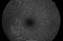
Cuticular Drusen - 26 year old female - Glomerulonephritis432 viewsVA 20/25 OU. Images are over 4 years - FA shows starry sky early images - This is because of loss of RPE over the drusen creating very small window defects.Feb 16, 2016
|
|
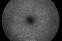
Cuticular Drusen - 26 year old female - Glomerulonephritis439 viewsVA 20/25 OU. Images are over 4 years. FA shows starry sky early images - This is because of loss of RPE over the drusen creating very small window defects.Feb 16, 2016
|
|
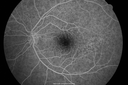
Cuticular Drusen - 26 year old female - Glomerulonephritis477 viewsVA 20/25 OU. Images are over 4 years. FA shows starry sky early images - This is because of loss of RPE over the drusen creating very small window defects.Feb 16, 2016
|
|
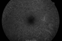
Cuticular Drusen - 26 year old female - Glomerulonephritis452 viewsVA 20/25 OU. Images are over 4 years. FA shows starry sky early images - This is because of loss of RPE over the drusen creating very small window defects.Feb 16, 2016
|
|

Cuticular Drusen - 26 year old female - Glomerulonephritis400 viewsVA 20/25 OU. Images are over 4 years FA shows starry sky early images - This is because of loss of RPE over the drusen creating very small window defects.Feb 16, 2016
|
|
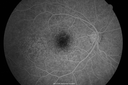
Cuticular Drusen - 26 year old female - Glomerulonephritis401 viewsVA 20/25 OU. Images are over 4 years FA shows starry sky early images - This is because of loss of RPE over the drusen creating very small window defects.Feb 16, 2016
|
|
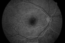
Cuticular Drusen - 26 year old female - Glomerulonephritis458 viewsVA 20/25 OU. Images are over 4 years FA shows starry sky early images - This is because of loss of RPE over the drusen creating very small window defects.Feb 16, 2016
|
|
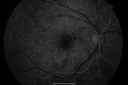
Cuticular Drusen - 26 year old female - Glomerulonephritis393 viewsVA 20/25 OU. Images are over 4 yearsFeb 16, 2016
|
|
Cuticular Drusen - 26 year old female - Glomerulonephritis466 viewsVA 20/25 OU. Images are over 4 yearsFeb 16, 2016
|
|
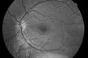
Cuticular Drusen - 26 year old female - Glomerulonephritis517 viewsVA 20/25 OU. Images are over 4 yearsFeb 16, 2016
|
|
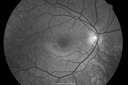
Cuticular Drusen - 26 year old female - Glomerulonephritis483 viewsVA 20/25 OU. Images are over 4 yearsFeb 16, 2016
|
|

Cuticular Drusen - 26 year old female - Glomerulonephritis603 viewsVA 20/25 OU. Images are over 4 yearsFeb 16, 2016
|
|

Cuticular Drusen - 26 year old female - Glomerulonephritis486 viewsVA 20/25 OU. Images are over 4 yearsFeb 16, 2016
|
|
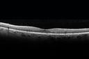
Adult Vitelliform Macular Dystrophy - Pattern Dystrophy422 viewsNov 17, 2015
|
|
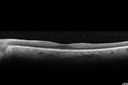
Adult Vitelliform Macular Dystrophy - Pattern Dystrophy422 viewsNov 17, 2015
|
|

Adult Vitelliform Macular Dystrophy - Pattern Dystrophy586 viewsSD OCT shows material to be subretinal not sub-RPE as you would see in typical AMDNov 17, 2015
|
|
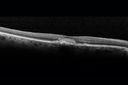
Adult Vitelliform Macular Dystrophy - Pattern Dystrophy496 viewsSD OCT shows material to be subretinal not sub-RPE as you would see in typical AMDNov 17, 2015
|
|

Adult Vitelliform Macular Dystrophy - Pattern Dystrophy395 viewsNov 17, 2015
|
|
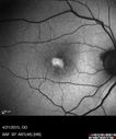
Adult Vitelliform Macular Dystrophy - Pattern Dystrophy447 viewsBright subfoveal FAF from lipofuscinNov 17, 2015
|
|
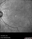
Adult Vitelliform Macular Dystrophy - Pattern Dystrophy392 viewsNov 17, 2015
|
|
| 2694 files on 27 page(s) |
 |
|
|
|
|
16 | |
|
|
|
|


