Viimased lisandused - Myopic Macular Degeneration - Wet
|

Myopic CNVM in the left eye. New Classic subfoveal lesionvaatamisi: 40764 year old female. She had retinal detachment repair in the right eye in 1996 (25 years ago). The left eye has new distortion for a week or two.
VA 20/40 OU.
Left eye has a very small subfoveal CNVMapr 02, 2020
|
|

Myopic CNVM in the left eye. New Classic subfoveal lesionvaatamisi: 47764 year old female. She had retinal detachment repair in the right eye in 1996 (25 years ago). The left eye has new distortion for a week or two.
VA 20/40 OU.
Left eye has a very small subfoveal CNVMapr 02, 2020
|
|

Myopic CNVM in the left eye. New Classic subfoveal lesionvaatamisi: 43364 year old female. She had retinal detachment repair in the right eye in 1996 (25 years ago). The left eye has new distortion for a week or two.
VA 20/40 OU.
Left eye has a very small subfoveal CNVMapr 02, 2020
|
|

Myopic CNVM in the left eye. New Classic subfoveal lesionvaatamisi: 48664 year old female. She had retinal detachment repair in the right eye in 1996 (25 years ago). The left eye has new distortion for a week or two.
VA 20/40 OU.
Left eye has a very small subfoveal CNVMapr 02, 2020
|
|

Myopic CNVM in the left eye. New Classic subfoveal lesionvaatamisi: 42164 year old female. She had retinal detachment repair in the right eye in 1996 (25 years ago). The left eye has new distortion for a week or two.
VA 20/40 OU.
Left eye has a very small subfoveal CNVMapr 02, 2020
|
|

Myopic CNVM in the left eye. New Classic subfoveal lesionvaatamisi: 44764 year old female. She had retinal detachment repair in the right eye in 1996 (25 years ago). The left eye has new distortion for a week or two.
VA 20/40 OU.
Left eye has a very small subfoveal CNVMapr 02, 2020
|
|

Myopic CNVM in the left eye. New Classic subfoveal lesionvaatamisi: 50164 year old female. She had retinal detachment repair in the right eye in 1996 (25 years ago). The left eye has new distortion for a week or two.
VA 20/40 OU.
Left eye has a very small subfoveal CNVMapr 02, 2020
|
|
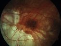
Myopic CNVM in the left eye. New Classic subfoveal lesionvaatamisi: 50964 year old female. She had retinal detachment repair in the right eye in 1996 (25 years ago). The left eye has new distortion for a week or two.
VA 20/40 OU.
Left eye has a very small subfoveal CNVMapr 02, 2020
|
|

Myopic CNVM in the left eye. New Classic subfoveal lesionvaatamisi: 50464 year old female. She had retinal detachment repair in the right eye in 1996 (25 years ago). The left eye has new distortion for a week or two.
VA 20/40 OU.
Left eye has a very small subfoveal CNVMapr 02, 2020
|
|
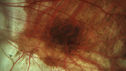
Myopic CNVM in the left eye. New Classic subfoveal lesionvaatamisi: 38664 year old female. She had retinal detachment repair in the right eye in 1996 (25 years ago). The left eye has new distortion for a week or two.
VA 20/40 OU.
Left eye has a very small subfoveal CNVMapr 02, 2020
|
|

Myopic CNVM in the left eye. New Classic subfoveal lesionvaatamisi: 49364 year old female. She had retinal detachment repair in the right eye in 1996 (25 years ago). The left eye has new distortion for a week or two.
VA 20/40 OU.
Left eye has a very small subfoveal CNVMapr 02, 2020
|
|
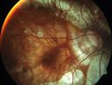
Myopic CNVM in the left eye. New Classic subfoveal lesionvaatamisi: 38364 year old female. She had retinal detachment repair in the right eye in 1996 (25 years ago). The left eye has new distortion for a week or two.
VA 20/40 OU.
Left eye has a very small subfoveal CNVMapr 02, 2020
|
|
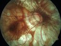
Myopic CNVM in the left eye. New Classic subfoveal lesionvaatamisi: 49464 year old female. She had retinal detachment repair in the right eye in 1996 (25 years ago). The left eye has new distortion for a week or two.
VA 20/40 OU.
Left eye has a very small subfoveal CNVMapr 02, 2020
|
|
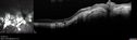
Rapid macular atrophy with anti-VEGF therapy for myopic CNVMvaatamisi: 37164 year old female with 20/32 vision and distortion in the left and better eye. She was treated with monthly lucentis and her vision declined to 20/100. Medicine was stopped and she improved a little to 20/80.nov 17, 2019
|
|

Rapid macular atrophy with anti-VEGF therapy for myopic CNVMvaatamisi: 40364 year old female with 20/32 vision and distortion in the left and better eye. She was treated with monthly lucentis and her vision declined to 20/100. Medicine was stopped and she improved a little to 20/80.nov 17, 2019
|
|

Rapid macular atrophy with anti-VEGF therapy for myopic CNVMvaatamisi: 40864 year old female with 20/32 vision and distortion in the left and better eye. She was treated with monthly lucentis and her vision declined to 20/100. Medicine was stopped and she improved a little to 20/80.nov 17, 2019
|
|

Rapid macular atrophy with anti-VEGF therapy for myopic CNVMvaatamisi: 39364 year old female with 20/32 vision and distortion in the left and better eye. She was treated with monthly lucentis and her vision declined to 20/100. Medicine was stopped and she improved a little to 20/80.nov 17, 2019
|
|
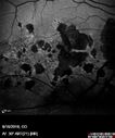
Rapid macular atrophy with anti-VEGF therapy for myopic CNVMvaatamisi: 42564 year old female with 20/32 vision and distortion in the left and better eye. She was treated with monthly lucentis and her vision declined to 20/100. Medicine was stopped and she improved a little to 20/80.nov 17, 2019
|
|

Rapid macular atrophy with anti-VEGF therapy for myopic CNVMvaatamisi: 39564 year old female with 20/32 vision and distortion in the left and better eye. She was treated with monthly lucentis and her vision declined to 20/100. Medicine was stopped and she improved a little to 20/80.nov 17, 2019
|
|
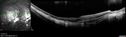
Rapid macular atrophy with anti-VEGF therapy for myopic CNVMvaatamisi: 41264 year old female with 20/32 vision and distortion in the left and better eye. She was treated with monthly lucentis and her vision declined to 20/100. Medicine was stopped and she improved a little to 20/80.nov 17, 2019
|
|
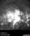
Rapid macular atrophy with anti-VEGF therapy for myopic CNVMvaatamisi: 39264 year old female with 20/32 vision and distortion in the left and better eye. She was treated with monthly lucentis and her vision declined to 20/100. Medicine was stopped and she improved a little to 20/80.nov 17, 2019
|
|
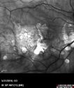
Rapid macular atrophy with anti-VEGF therapy for myopic CNVMvaatamisi: 39864 year old female with 20/32 vision and distortion in the left and better eye. She was treated with monthly lucentis and her vision declined to 20/100. Medicine was stopped and she improved a little to 20/80.nov 17, 2019
|
|
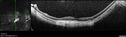
Rapid macular atrophy with anti-VEGF therapy for myopic CNVMvaatamisi: 41864 year old female with 20/32 vision and distortion in the left and better eye. She was treated with monthly lucentis and her vision declined to 20/100. Medicine was stopped and she improved a little to 20/80.nov 17, 2019
|
|

Rapid macular atrophy with anti-VEGF therapy for myopic CNVMvaatamisi: 39964 year old female with 20/32 vision and distortion in the left and better eye. She was treated with monthly lucentis and her vision declined to 20/100. Medicine was stopped and she improved a little to 20/80.nov 17, 2019
|
|

Rapid macular atrophy with anti-VEGF therapy for myopic CNVMvaatamisi: 40364 year old female with 20/32 vision and distortion in the left and better eye. She was treated with monthly lucentis and her vision declined to 20/100. Medicine was stopped and she improved a little to 20/80.nov 17, 2019
|
|
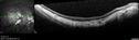
Rapid macular atrophy with anti-VEGF therapy for myopic CNVMvaatamisi: 40364 year old female with 20/32 vision and distortion in the left and better eye. She was treated with monthly lucentis and her vision declined to 20/100. Medicine was stopped and she improved a little to 20/80.nov 17, 2019
|
|
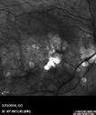
Rapid macular atrophy with anti-VEGF therapy for myopic CNVMvaatamisi: 41664 year old female with 20/32 vision and distortion in the left and better eye. She was treated with monthly lucentis and her vision declined to 20/100. Medicine was stopped and she improved a little to 20/80.nov 17, 2019
|
|
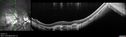
Rapid macular atrophy with anti-VEGF therapy for myopic CNVMvaatamisi: 39964 year old female with 20/32 vision and distortion in the left and better eye. She was treated with monthly lucentis and her vision declined to 20/100. Medicine was stopped and she improved a little to 20/80.nov 17, 2019
|
|

Rapid macular atrophy with anti-VEGF therapy for myopic CNVMvaatamisi: 39564 year old female with 20/32 vision and distortion in the left and better eye. She was treated with monthly lucentis and her vision declined to 20/100. Medicine was stopped and she improved a little to 20/80.nov 17, 2019
|
|
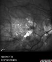
Rapid macular atrophy with anti-VEGF therapy for myopic CNVMvaatamisi: 44764 year old female with 20/32 vision and distortion in the left and better eye. She was treated with monthly lucentis and her vision declined to 20/100. Medicine was stopped and she improved a little to 20/80.nov 17, 2019
|
|
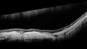
Rapid macular atrophy with anti-VEGF therapy for myopic CNVMvaatamisi: 40864 year old female with 20/32 vision and distortion in the left and better eye. She was treated with monthly lucentis and her vision declined to 20/100. Medicine was stopped and she improved a little to 20/80.nov 17, 2019
|
|
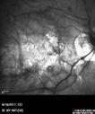
Rapid macular atrophy with anti-VEGF therapy for myopic CNVMvaatamisi: 37564 year old female with 20/32 vision and distortion in the left and better eye. She was treated with monthly lucentis and her vision declined to 20/100. Medicine was stopped and she improved a little to 20/80.nov 17, 2019
|
|

Rapid macular atrophy with anti-VEGF therapy for myopic CNVMvaatamisi: 46064 year old female with 20/32 vision and distortion in the left and better eye. She was treated with monthly lucentis and her vision declined to 20/100. Medicine was stopped and she improved a little to 20/80.nov 17, 2019
|
|
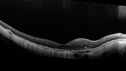
Rapid macular atrophy with anti-VEGF therapy for myopic CNVMvaatamisi: 38764 year old female with 20/32 vision and distortion in the left and better eye. She was treated with monthly lucentis and her vision declined to 20/100. Medicine was stopped and she improved a little to 20/80.nov 17, 2019
|
|
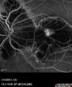
Rapid macular atrophy with anti-VEGF therapy for myopic CNVMvaatamisi: 41464 year old female with 20/32 vision and distortion in the left and better eye. She was treated with monthly lucentis and her vision declined to 20/100. Medicine was stopped and she improved a little to 20/80.nov 17, 2019
|
|

Rapid macular atrophy with anti-VEGF therapy for myopic CNVMvaatamisi: 40964 year old female with 20/32 vision and distortion in the left and better eye. She was treated with monthly lucentis and her vision declined to 20/100. Medicine was stopped and she improved a little to 20/80.nov 17, 2019
|
|
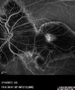
Rapid macular atrophy with anti-VEGF therapy for myopic CNVMvaatamisi: 42964 year old female with 20/32 vision and distortion in the left and better eye. She was treated with monthly lucentis and her vision declined to 20/100. Medicine was stopped and she improved a little to 20/80.nov 17, 2019
|
|
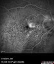
Rapid macular atrophy with anti-VEGF therapy for myopic CNVMvaatamisi: 41764 year old female with 20/32 vision and distortion in the left and better eye. She was treated with monthly lucentis and her vision declined to 20/100. Medicine was stopped and she improved a little to 20/80.nov 17, 2019
|
|
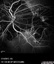
Rapid macular atrophy with anti-VEGF therapy for myopic CNVMvaatamisi: 42464 year old female with 20/32 vision and distortion in the left and better eye. She was treated with monthly lucentis and her vision declined to 20/100. Medicine was stopped and she improved a little to 20/80.nov 17, 2019
|
|
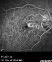
Rapid macular atrophy with anti-VEGF therapy for myopic CNVMvaatamisi: 41464 year old female with 20/32 vision and distortion in the left and better eye. She was treated with monthly lucentis and her vision declined to 20/100. Medicine was stopped and she improved a little to 20/80.nov 17, 2019
|
|
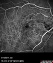
Rapid macular atrophy with anti-VEGF therapy for myopic CNVMvaatamisi: 40164 year old female with 20/32 vision and distortion in the left and better eye. She was treated with monthly lucentis and her vision declined to 20/100. Medicine was stopped and she improved a little to 20/80.nov 17, 2019
|
|
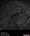
Rapid macular atrophy with anti-VEGF therapy for myopic CNVMvaatamisi: 39464 year old female with 20/32 vision and distortion in the left and better eye. She was treated with monthly lucentis and her vision declined to 20/100. Medicine was stopped and she improved a little to 20/80.nov 17, 2019
|
|

Rapid macular atrophy with anti-VEGF therapy for myopic CNVMvaatamisi: 39664 year old female with 20/32 vision and distortion in the left and better eye. She was treated with monthly lucentis and her vision declined to 20/100. Medicine was stopped and she improved a little to 20/80.nov 17, 2019
|
|

Rapid macular atrophy with anti-VEGF therapy for myopic CNVMvaatamisi: 39464 year old female with 20/32 vision and distortion in the left and better eye. She was treated with monthly lucentis and her vision declined to 20/100. Medicine was stopped and she improved a little to 20/80.nov 17, 2019
|
|

Rapid macular atrophy with anti-VEGF therapy for myopic CNVMvaatamisi: 41664 year old female with 20/32 vision and distortion in the left and better eye. She was treated with monthly lucentis and her vision declined to 20/100. Medicine was stopped and she improved a little to 20/80.nov 17, 2019
|
|

Rapid macular atrophy with anti-VEGF therapy for myopic CNVMvaatamisi: 36964 year old female with 20/32 vision and distortion in the left and better eye. She was treated with monthly lucentis and her vision declined to 20/100. Medicine was stopped and she improved a little to 20/80.nov 17, 2019
|
|
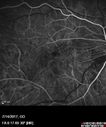
Rapid macular atrophy with anti-VEGF therapy for myopic CNVMvaatamisi: 41264 year old female with 20/32 vision and distortion in the left and better eye. She was treated with monthly lucentis and her vision declined to 20/100. Medicine was stopped and she improved a little to 20/80.nov 17, 2019
|
|
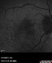
Rapid macular atrophy with anti-VEGF therapy for myopic CNVMvaatamisi: 39464 year old female with 20/32 vision and distortion in the left and better eye. She was treated with monthly lucentis and her vision declined to 20/100. Medicine was stopped and she improved a little to 20/80.nov 17, 2019
|
|
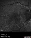
Rapid macular atrophy with anti-VEGF therapy for myopic CNVMvaatamisi: 38964 year old female with 20/32 vision and distortion in the left and better eye. She was treated with monthly lucentis and her vision declined to 20/100. Medicine was stopped and she improved a little to 20/80.nov 17, 2019
|
|
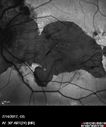
Rapid macular atrophy with anti-VEGF therapy for myopic CNVMvaatamisi: 42464 year old female with 20/32 vision and distortion in the left and better eye. She was treated with monthly lucentis and her vision declined to 20/100. Medicine was stopped and she improved a little to 20/80.nov 17, 2019
|
|

Rapid macular atrophy with anti-VEGF therapy for myopic CNVMvaatamisi: 40164 year old female with 20/32 vision and distortion in the left and better eye. She was treated with monthly lucentis and her vision declined to 20/100. Medicine was stopped and she improved a little to 20/80.nov 17, 2019
|
|

Rapid macular atrophy with anti-VEGF therapy for myopic CNVMvaatamisi: 43264 year old female with 20/32 vision and distortion in the left and better eye. She was treated with monthly lucentis and her vision declined to 20/100. Medicine was stopped and she improved a little to 20/80.nov 17, 2019
|
|
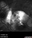
Rapid macular atrophy with anti-VEGF therapy for myopic CNVMvaatamisi: 40664 year old female with 20/32 vision and distortion in the left and better eye. She was treated with monthly lucentis and her vision declined to 20/100. Medicine was stopped and she improved a little to 20/80.nov 17, 2019
|
|

Myopic Macular Degeneration - Staphyloma - Recent Vision Loss Right Eye - Possible CNVMvaatamisi: 59083-year-old woman is a high myope with myopic macular degeneration. She had a retinal detachment repair in the right eye in 1968. I saw her in June of 2003. She had posterior colobomas and areas of pigment epithelial atrophy. She has noticed over the last six weeks substantial declining vision in the right eye. Prior to that, the vision in the two eyes was about the same. She uses Xalatan in the left eye and Alphagan in both eyes.
VISUAL ACUITY: Vision OD is 20/160, OS is 20/40juuni 28, 2011
|
|

Myopic Macular Degeneration - Staphyloma - Recent Vision Loss Right Eye - Possible CNVMvaatamisi: 57683-year-old woman is a high myope with myopic macular degeneration. She had a retinal detachment repair in the right eye in 1968. I saw her in June of 2003. She had posterior colobomas and areas of pigment epithelial atrophy. She has noticed over the last six weeks substantial declining vision in the right eye. Prior to that, the vision in the two eyes was about the same. She uses Xalatan in the left eye and Alphagan in both eyes.
VISUAL ACUITY: Vision OD is 20/160, OS is 20/40juuni 28, 2011
|
|

Myopic Macular Degeneration - Staphyloma - Recent Vision Loss Right Eye - Possible CNVMvaatamisi: 61583-year-old woman is a high myope with myopic macular degeneration. She had a retinal detachment repair in the right eye in 1968. I saw her in June of 2003. She had posterior colobomas and areas of pigment epithelial atrophy. She has noticed over the last six weeks substantial declining vision in the right eye. Prior to that, the vision in the two eyes was about the same. She uses Xalatan in the left eye and Alphagan in both eyes.
VISUAL ACUITY: Vision OD is 20/160, OS is 20/40juuni 28, 2011
|
|

Myopic Macular Degeneration - Staphyloma - Recent Vision Loss Right Eye - Possible CNVMvaatamisi: 64183-year-old woman is a high myope with myopic macular degeneration. She had a retinal detachment repair in the right eye in 1968. I saw her in June of 2003. She had posterior colobomas and areas of pigment epithelial atrophy. She has noticed over the last six weeks substantial declining vision in the right eye. Prior to that, the vision in the two eyes was about the same. She uses Xalatan in the left eye and Alphagan in both eyes.
VISUAL ACUITY: Vision OD is 20/160, OS is 20/40juuni 28, 2011
|
|
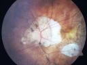
Myopic Macular Degeneration - Staphyloma - Recent Vision Loss Right Eye - Possible CNVMvaatamisi: 57983-year-old woman is a high myope with myopic macular degeneration. She had a retinal detachment repair in the right eye in 1968. I saw her in June of 2003. She had posterior colobomas and areas of pigment epithelial atrophy. She has noticed over the last six weeks substantial declining vision in the right eye. Prior to that, the vision in the two eyes was about the same. She uses Xalatan in the left eye and Alphagan in both eyes.
VISUAL ACUITY: Vision OD is 20/160, OS is 20/40juuni 28, 2011
|
|

Myopic Macular Degeneration - Staphyloma - Recent Vision Loss Right Eye - Possible CNVMvaatamisi: 59383-year-old woman is a high myope with myopic macular degeneration. She had a retinal detachment repair in the right eye in 1968. I saw her in June of 2003. She had posterior colobomas and areas of pigment epithelial atrophy. She has noticed over the last six weeks substantial declining vision in the right eye. Prior to that, the vision in the two eyes was about the same. She uses Xalatan in the left eye and Alphagan in both eyes.
VISUAL ACUITY: Vision OD is 20/160, OS is 20/40juuni 28, 2011
|
|

Myopic Macular Degeneration - Staphyloma - Recent Vision Loss Right Eye - Possible CNVMvaatamisi: 47183-year-old woman is a high myope with myopic macular degeneration. She had a retinal detachment repair in the right eye in 1968. I saw her in June of 2003. She had posterior colobomas and areas of pigment epithelial atrophy. She has noticed over the last six weeks substantial declining vision in the right eye. Prior to that, the vision in the two eyes was about the same. She uses Xalatan in the left eye and Alphagan in both eyes.
VISUAL ACUITY: Vision OD is 20/160, OS is 20/40juuni 28, 2011
|
|
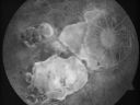
Myopic Macular Degeneration - Staphyloma - Recent Vision Loss Right Eye - Possible CNVMvaatamisi: 47883-year-old woman is a high myope with myopic macular degeneration. She had a retinal detachment repair in the right eye in 1968. I saw her in June of 2003. She had posterior colobomas and areas of pigment epithelial atrophy. She has noticed over the last six weeks substantial declining vision in the right eye. Prior to that, the vision in the two eyes was about the same. She uses Xalatan in the left eye and Alphagan in both eyes.
VISUAL ACUITY: Vision OD is 20/160, OS is 20/40juuni 28, 2011
|
|
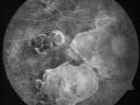
Myopic Macular Degeneration - Staphyloma - Recent Vision Loss Right Eye - Possible CNVMvaatamisi: 50583-year-old woman is a high myope with myopic macular degeneration. She had a retinal detachment repair in the right eye in 1968. I saw her in June of 2003. She had posterior colobomas and areas of pigment epithelial atrophy. She has noticed over the last six weeks substantial declining vision in the right eye. Prior to that, the vision in the two eyes was about the same. She uses Xalatan in the left eye and Alphagan in both eyes.
VISUAL ACUITY: Vision OD is 20/160, OS is 20/40juuni 28, 2011
|
|
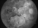
Myopic Macular Degeneration - Staphyloma - Recent Vision Loss Right Eye - Possible CNVMvaatamisi: 54983-year-old woman is a high myope with myopic macular degeneration. She had a retinal detachment repair in the right eye in 1968. I saw her in June of 2003. She had posterior colobomas and areas of pigment epithelial atrophy. She has noticed over the last six weeks substantial declining vision in the right eye. Prior to that, the vision in the two eyes was about the same. She uses Xalatan in the left eye and Alphagan in both eyes.
VISUAL ACUITY: Vision OD is 20/160, OS is 20/40juuni 28, 2011
|
|

Myopic Macular Degeneration - Staphyloma - Recent Vision Loss Right Eye - Possible CNVMvaatamisi: 50983-year-old woman is a high myope with myopic macular degeneration. She had a retinal detachment repair in the right eye in 1968. I saw her in June of 2003. She had posterior colobomas and areas of pigment epithelial atrophy. She has noticed over the last six weeks substantial declining vision in the right eye. Prior to that, the vision in the two eyes was about the same. She uses Xalatan in the left eye and Alphagan in both eyes.
VISUAL ACUITY: Vision OD is 20/160, OS is 20/40juuni 28, 2011
|
|

Myopic Macular Degeneration - Staphyloma - Recent Vision Loss Right Eye - Possible CNVMvaatamisi: 48183-year-old woman is a high myope with myopic macular degeneration. She had a retinal detachment repair in the right eye in 1968. I saw her in June of 2003. She had posterior colobomas and areas of pigment epithelial atrophy. She has noticed over the last six weeks substantial declining vision in the right eye. Prior to that, the vision in the two eyes was about the same. She uses Xalatan in the left eye and Alphagan in both eyes.
VISUAL ACUITY: Vision OD is 20/160, OS is 20/40juuni 28, 2011
|
|
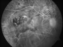
Myopic Macular Degeneration - Staphyloma - Recent Vision Loss Right Eye - Possible CNVMvaatamisi: 52283-year-old woman is a high myope with myopic macular degeneration. She had a retinal detachment repair in the right eye in 1968. I saw her in June of 2003. She had posterior colobomas and areas of pigment epithelial atrophy. She has noticed over the last six weeks substantial declining vision in the right eye. Prior to that, the vision in the two eyes was about the same. She uses Xalatan in the left eye and Alphagan in both eyes.
VISUAL ACUITY: Vision OD is 20/160, OS is 20/40juuni 28, 2011
|
|
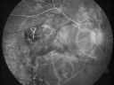
Myopic Macular Degeneration - Staphyloma - Recent Vision Loss Right Eye - Possible CNVMvaatamisi: 52583-year-old woman is a high myope with myopic macular degeneration. She had a retinal detachment repair in the right eye in 1968. I saw her in June of 2003. She had posterior colobomas and areas of pigment epithelial atrophy. She has noticed over the last six weeks substantial declining vision in the right eye. Prior to that, the vision in the two eyes was about the same. She uses Xalatan in the left eye and Alphagan in both eyes.
VISUAL ACUITY: Vision OD is 20/160, OS is 20/40juuni 28, 2011
|
|
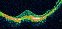
Myopic Macular Degeneration - Staphyloma - Recent Vision Loss Right Eye - Possible CNVMvaatamisi: 50383-year-old woman is a high myope with myopic macular degeneration. She had a retinal detachment repair in the right eye in 1968. I saw her in June of 2003. She had posterior colobomas and areas of pigment epithelial atrophy. She has noticed over the last six weeks substantial declining vision in the right eye. Prior to that, the vision in the two eyes was about the same. She uses Xalatan in the left eye and Alphagan in both eyes.
VISUAL ACUITY: Vision OD is 20/160, OS is 20/40juuni 28, 2011
|
|
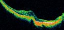
Myopic Macular Degeneration - Staphyloma - Recent Vision Loss Right Eye - Possible CNVMvaatamisi: 52383-year-old woman is a high myope with myopic macular degeneration. She had a retinal detachment repair in the right eye in 1968. I saw her in June of 2003. She had posterior colobomas and areas of pigment epithelial atrophy. She has noticed over the last six weeks substantial declining vision in the right eye. Prior to that, the vision in the two eyes was about the same. She uses Xalatan in the left eye and Alphagan in both eyes.
VISUAL ACUITY: Vision OD is 20/160, OS is 20/40juuni 28, 2011
|
|

Myopic Macular Degeneration - Staphyloma - Recent Vision Loss Right Eye - Possible CNVMvaatamisi: 73783-year-old woman is a high myope with myopic macular degeneration. She had a retinal detachment repair in the right eye in 1968. I saw her in June of 2003. She had posterior colobomas and areas of pigment epithelial atrophy. She has noticed over the last six weeks substantial declining vision in the right eye. Prior to that, the vision in the two eyes was about the same. She uses Xalatan in the left eye and Alphagan in both eyes.
VISUAL ACUITY: Vision OD is 20/160, OS is 20/40juuni 28, 2011
|
|
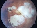
Myopic Macular Degeneration - Staphyloma - Recent Vision Loss Right Eye - Possible CNVMvaatamisi: 65583-year-old woman is a high myope with myopic macular degeneration. She had a retinal detachment repair in the right eye in 1968. I saw her in June of 2003. She had posterior colobomas and areas of pigment epithelial atrophy. She has noticed over the last six weeks substantial declining vision in the right eye. Prior to that, the vision in the two eyes was about the same. She uses Xalatan in the left eye and Alphagan in both eyes.
VISUAL ACUITY: Vision OD is 20/160, OS is 20/40juuni 28, 2011
|
|

Myopic Macular Degeneration - Staphyloma - Recent Vision Loss Right Eye - Possible CNVMvaatamisi: 67483-year-old woman is a high myope with myopic macular degeneration. She had a retinal detachment repair in the right eye in 1968. I saw her in June of 2003. She had posterior colobomas and areas of pigment epithelial atrophy. She has noticed over the last six weeks substantial declining vision in the right eye. Prior to that, the vision in the two eyes was about the same. She uses Xalatan in the left eye and Alphagan in both eyes.
VISUAL ACUITY: Vision OD is 20/160, OS is 20/40juuni 28, 2011
|
|

Macular Hemorrahge and Probable CNVM Right eye for 2 weeks VA 20/400 - 20 Year oldvaatamisi: 52220-year-old man noticed vision loss in the right eye about two weeks ago and if anything it seems to be getting worse. He is a high myope. VISUAL ACUITY: OD 20/400juuni 14, 2011
|
|
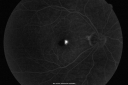
Macular Hemorrahge and Probable CNVM Right eye for 2 weeks VA 20/400 - 20 Year oldvaatamisi: 57220-year-old man noticed vision loss in the right eye about two weeks ago and if anything it seems to be getting worse. He is a high myope. VISUAL ACUITY: OD 20/400juuni 14, 2011
|
|

Macular Hemorrahge and Probable CNVM Right eye for 2 weeks VA 20/400 - 20 Year oldvaatamisi: 47120-year-old man noticed vision loss in the right eye about two weeks ago and if anything it seems to be getting worse. He is a high myope. VISUAL ACUITY: OD 20/400juuni 14, 2011
|
|

Macular Hemorrahge and Probable CNVM Right eye for 2 weeks VA 20/400 - 20 Year oldvaatamisi: 52820-year-old man noticed vision loss in the right eye about two weeks ago and if anything it seems to be getting worse. He is a high myope. VISUAL ACUITY: OD 20/400juuni 14, 2011
|
|

Macular Hemorrahge and Probable CNVM Right eye for 2 weeks VA 20/400 - 20 Year oldvaatamisi: 46720-year-old man noticed vision loss in the right eye about two weeks ago and if anything it seems to be getting worse. He is a high myope. VISUAL ACUITY: OD 20/400juuni 14, 2011
|
|

Macular Hemorrahge and Probable CNVM Right eye for 2 weeks VA 20/400 - 20 Year oldvaatamisi: 53620-year-old man noticed vision loss in the right eye about two weeks ago and if anything it seems to be getting worse. He is a high myope. VISUAL ACUITY: OD 20/400juuni 14, 2011
|
|
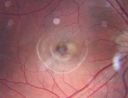
Macular Hemorrahge and Probable CNVM Right eye for 2 weeks VA 20/400 - 20 Year oldvaatamisi: 59720-year-old man noticed vision loss in the right eye about two weeks ago and if anything it seems to be getting worse. He is a high myope. VISUAL ACUITY: OD 20/400juuni 14, 2011
|
|

Macular Hemorrahge and Probable CNVM Right eye for 2 weeks VA 20/400 - 20 Year oldvaatamisi: 54620-year-old man noticed vision loss in the right eye about two weeks ago and if anything it seems to be getting worse. He is a high myope. VISUAL ACUITY: OD 20/400juuni 14, 2011
|
|

Myopic Choroidal Neovascular Membrane 54 year old woman 20/20vaatamisi: 63554-year-old woman has had problems in her vision in the right eye for about the last three months. Inferotemporally she sees a gray area, which is distorted. OD is 20/20, OS is 20/20nov 17, 2010
|
|

Myopic Choroidal Neovascular Membrane 54 year old woman 20/20vaatamisi: 63654-year-old woman has had problems in her vision in the right eye for about the last three months. Inferotemporally she sees a gray area, which is distorted. OD is 20/20, OS is 20/20nov 17, 2010
|
|

Myopic Choroidal Neovascular Membrane 54 year old woman 20/20vaatamisi: 68054-year-old woman has had problems in her vision in the right eye for about the last three months. Inferotemporally she sees a gray area, which is distorted. OD is 20/20, OS is 20/20nov 17, 2010
|
|

Myopic Choroidal Neovascular Membrane 54 year old woman 20/20vaatamisi: 60654-year-old woman has had problems in her vision in the right eye for about the last three months. Inferotemporally she sees a gray area, which is distorted. OD is 20/20, OS is 20/20nov 17, 2010
|
|

Myopic Choroidal Neovascular Membrane 54 year old woman 20/20vaatamisi: 59054-year-old woman has had problems in her vision in the right eye for about the last three months. Inferotemporally she sees a gray area, which is distorted. OD is 20/20, OS is 20/20nov 17, 2010
|
|

Myopic Choroidal Neovascular Membrane 54 year old woman 20/20vaatamisi: 63054-year-old woman has had problems in her vision in the right eye for about the last three months. Inferotemporally she sees a gray area, which is distorted. OD is 20/20, OS is 20/20nov 17, 2010
|
|

Myopic Choroidal Neovascular Membrane 54 year old woman 20/20vaatamisi: 69854-year-old woman has had problems in her vision in the right eye for about the last three months. Inferotemporally she sees a gray area, which is distorted. OD is 20/20, OS is 20/20nov 17, 2010
|
|

Myopic Choroidal Neovascular Membrane 54 year old woman 20/20vaatamisi: 68754-year-old woman has had problems in her vision in the right eye for about the last three months. Inferotemporally she sees a gray area, which is distorted. OD is 20/20, OS is 20/20nov 17, 2010
|
|

Myopic Choroidal Neovascular Membrane 54 year old woman 20/20vaatamisi: 89154-year-old woman has had problems in her vision in the right eye for about the last three months. Inferotemporally she sees a gray area, which is distorted. OD is 20/20, OS is 20/20nov 17, 2010
|
|

Myopic Choroidal Neovascular Membrane 54 year old woman 20/20vaatamisi: 76854-year-old woman has had problems in her vision in the right eye for about the last three months. Inferotemporally she sees a gray area, which is distorted. OD is 20/20, OS is 20/20nov 17, 2010
|
|

Myopic Choroidal Neovascular Membrane 54 year old woman 20/20vaatamisi: 106354-year-old woman has had problems in her vision in the right eye for about the last three months. Inferotemporally she sees a gray area, which is distorted. OD is 20/20, OS is 20/20nov 17, 2010
|
|

Myopic CNVM - Wet - rx Avastin for 1 yearvaatamisi: 114555-year-old woman OD is 20/40, OS is 20/20. There is a hyperpigmented disc diameter choroidal neovascular membrane with fluid touching the fovea.nov 06, 2010
|
|

Myopic CNVM - Wet - rx Avastin for 1 yearvaatamisi: 67455-year-old woman OD is 20/40, OS is 20/20. There is a hyperpigmented disc diameter choroidal neovascular membrane with fluid touching the fovea.nov 06, 2010
|
|

Myopic CNVM - Wet - rx Avastin for 1 yearvaatamisi: 64555-year-old woman OD is 20/40, OS is 20/20. There is a hyperpigmented disc diameter choroidal neovascular membrane with fluid touching the fovea.nov 06, 2010
|
|

Myopic CNVM - Wet - rx Avastin for 1 yearvaatamisi: 57855-year-old woman OD is 20/40, OS is 20/20. There is a hyperpigmented disc diameter choroidal neovascular membrane with fluid touching the fovea. nov 06, 2010
|
|

Myopic CNVM - Wet - rx Avastin for 1 yearvaatamisi: 64455-year-old woman OD is 20/40, OS is 20/20. There is a hyperpigmented disc diameter choroidal neovascular membrane with fluid touching the fovea. nov 06, 2010
|
|

Myopic CNVM - Wet - rx Avastin for 1 yearvaatamisi: 67955-year-old woman OD is 20/40, OS is 20/20. There is a hyperpigmented disc diameter choroidal neovascular membrane with fluid touching the fovea.nov 06, 2010
|
|

Myopic CNVM - Wet - rx Avastin for 1 yearvaatamisi: 62555-year-old woman OD is 20/40, OS is 20/20. There is a hyperpigmented disc diameter choroidal neovascular membrane with fluid touching the fovea.nov 06, 2010
|
|

Myopic CNVM - Wet - rx Avastin for 1 yearvaatamisi: 57955-year-old woman OD is 20/40, OS is 20/20. There is a hyperpigmented disc diameter choroidal neovascular membrane with fluid touching the fovea. nov 06, 2010
|
|

Myopic CNVM - Wet - rx Avastin for 1 yearvaatamisi: 68955-year-old woman OD is 20/40, OS is 20/20. There is a hyperpigmented disc diameter choroidal neovascular membrane with fluid touching the fovea.nov 06, 2010
|
|
|
|


