neueste Dateien - Retina Vascular Occlusions
|
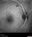
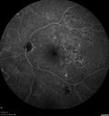
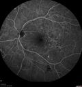
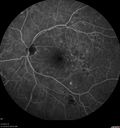
Branch Retinal Vein Occlusion with Edema - Failed avastin (bevacizumab) and did well with Eylea (Aflibercept)411 x angesehen67 year old female The vision in the left eye was good until about 4 months ago. Prior to that it was OK. The vision in the right eye is fine. She is not sure if she has had strokes from studies taken in December. She was, at that time, sent to the ER for high blood pressure. Â
VA OD: Dcc20/40 OS: 20/100
Four treatments with Avastin she was still 20/100 with edema. With two Eylea treatments she improved to 20/32 and has remained dry and with good vision23. April 2020
|
|
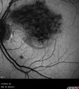
Branch Retinal Vein Occlusion with Edema - Failed avastin (bevacizumab) and did well with Eylea (Aflibercept)408 x angesehen67 year old female The vision in the left eye was good until about 4 months ago. Prior to that it was OK. The vision in the right eye is fine. She is not sure if she has had strokes from studies taken in December. She was, at that time, sent to the ER for high blood pressure. Â
VA OD: Dcc20/40 OS: 20/100
Four treatments with Avastin she was still 20/100 with edema. With two Eylea treatments she improved to 20/32 and has remained dry and with good vision23. April 2020
|
|
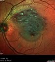
Branch Retinal Vein Occlusion with Edema - Failed avastin (bevacizumab) and did well with Eylea (Aflibercept)428 x angesehen67 year old female The vision in the left eye was good until about 4 months ago. Prior to that it was OK. The vision in the right eye is fine. She is not sure if she has had strokes from studies taken in December. She was, at that time, sent to the ER for high blood pressure. Â
VA OD: Dcc20/40 OS: 20/100
Four treatments with Avastin she was still 20/100 with edema. With two Eylea treatments she improved to 20/32 and has remained dry and with good vision23. April 2020
|
|
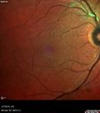
Branch Retinal Vein Occlusion with Edema - Failed avastin (bevacizumab) and did well with Eylea (Aflibercept)389 x angesehen67 year old female The vision in the left eye was good until about 4 months ago. Prior to that it was OK. The vision in the right eye is fine. She is not sure if she has had strokes from studies taken in December. She was, at that time, sent to the ER for high blood pressure. Â
VA OD: Dcc20/40 OS: 20/100
Four treatments with Avastin she was still 20/100 with edema. With two Eylea treatments she improved to 20/32 and has remained dry and with good vision23. April 2020
|
|
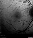
Branch Retinal Vein Occlusion with Edema - Failed avastin (bevacizumab) and did well with Eylea (Aflibercept)476 x angesehen67 year old female The vision in the left eye was good until about 4 months ago. Prior to that it was OK. The vision in the right eye is fine. She is not sure if she has had strokes from studies taken in December. She was, at that time, sent to the ER for high blood pressure. Â
VA OD: Dcc20/40 OS: 20/100
Four treatments with Avastin she was still 20/100 with edema. With two Eylea treatments she improved to 20/32 and has remained dry and with good vision23. April 2020
|
|

Branch Retinal Vein Occlusion with Edema - Failed avastin (bevacizumab) and did well with Eylea (Aflibercept)453 x angesehen67 year old female The vision in the left eye was good until about 4 months ago. Prior to that it was OK. The vision in the right eye is fine. She is not sure if she has had strokes from studies taken in December. She was, at that time, sent to the ER for high blood pressure. Â
VA OD: Dcc20/40 OS: 20/100
Four treatments with Avastin she was still 20/100 with edema. With two Eylea treatments she improved to 20/32 and has remained dry and with good vision23. April 2020
|
|
434 x angesehen23. April 2020
|
|
413 x angesehen23. April 2020
|
|
408 x angesehen23. April 2020
|
|
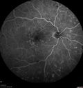
445 x angesehen23. April 2020
|
|
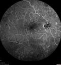
439 x angesehen23. April 2020
|
|
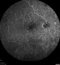
451 x angesehen23. April 2020
|
|
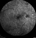
437 x angesehen23. April 2020
|
|
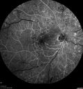
398 x angesehen23. April 2020
|
|
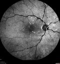
419 x angesehen23. April 2020
|
|
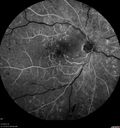
464 x angesehen23. April 2020
|
|
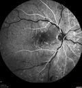
464 x angesehen23. April 2020
|
|
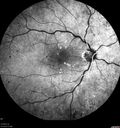
423 x angesehen23. April 2020
|
|
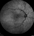
430 x angesehen23. April 2020
|
|
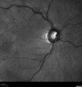
462 x angesehen23. April 2020
|
|
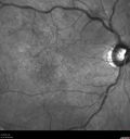
414 x angesehen23. April 2020
|
|

447 x angesehen23. April 2020
|
|
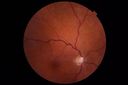
477 x angesehen23. April 2020
|
|
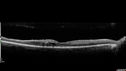
446 x angesehen23. April 2020
|
|
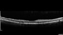
416 x angesehen23. April 2020
|
|

500 x angesehen23. April 2020
|
|
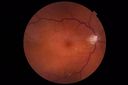
438 x angesehen23. April 2020
|
|
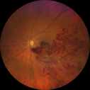
Acute Branch Retinal Vein Occlusion - 20/200501 x angesehen66 year old female with 3 weeks of vision loss in the left eye. VA 20/20 OD 20/200 OS11. Februar 2020
|
|
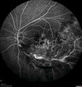
Acute Branch Retinal Vein Occlusion - 20/200448 x angesehen66 year old female with 3 weeks of vision loss in the left eye. VA 20/20 OD 20/200 OS11. Februar 2020
|
|
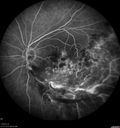
Acute Branch Retinal Vein Occlusion - 20/200476 x angesehen66 year old female with 3 weeks of vision loss in the left eye. VA 20/20 OD 20/200 OS11. Februar 2020
|
|
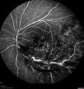
Acute Branch Retinal Vein Occlusion - 20/200423 x angesehen66 year old female with 3 weeks of vision loss in the left eye. VA 20/20 OD 20/200 OS11. Februar 2020
|
|

Acute Branch Retinal Vein Occlusion - 20/200405 x angesehen66 year old female with 3 weeks of vision loss in the left eye. VA 20/20 OD 20/200 OS11. Februar 2020
|
|
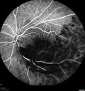
Acute Branch Retinal Vein Occlusion - 20/200426 x angesehen66 year old female with 3 weeks of vision loss in the left eye. VA 20/20 OD 20/200 OS11. Februar 2020
|
|
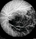
Acute Branch Retinal Vein Occlusion - 20/200472 x angesehen66 year old female with 3 weeks of vision loss in the left eye. VA 20/20 OD 20/200 OS11. Februar 2020
|
|
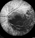
Acute Branch Retinal Vein Occlusion - 20/200469 x angesehen66 year old female with 3 weeks of vision loss in the left eye. VA 20/20 OD 20/200 OS11. Februar 2020
|
|
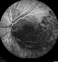
Acute Branch Retinal Vein Occlusion - 20/200436 x angesehen66 year old female with 3 weeks of vision loss in the left eye. VA 20/20 OD 20/200 OS11. Februar 2020
|
|
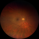
Acute Branch Retinal Vein Occlusion - 20/200493 x angesehen66 year old female with 3 weeks of vision loss in the left eye. VA 20/20 OD 20/200 OS11. Februar 2020
|
|
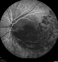
Acute Branch Retinal Vein Occlusion - 20/200440 x angesehen66 year old female with 3 weeks of vision loss in the left eye. VA 20/20 OD 20/200 OS11. Februar 2020
|
|

Acute Branch Retinal Vein Occlusion - 20/200464 x angesehen66 year old female with 3 weeks of vision loss in the left eye. VA 20/20 OD 20/200 OS11. Februar 2020
|
|
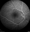
Acute Branch Retinal Vein Occlusion - 20/200425 x angesehen66 year old female with 3 weeks of vision loss in the left eye. VA 20/20 OD 20/200 OS11. Februar 2020
|
|
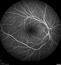
Acute Branch Retinal Vein Occlusion - 20/200464 x angesehen66 year old female with 3 weeks of vision loss in the left eye. VA 20/20 OD 20/200 OS11. Februar 2020
|
|
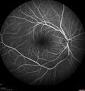
Acute Branch Retinal Vein Occlusion - 20/200460 x angesehen66 year old female with 3 weeks of vision loss in the left eye. VA 20/20 OD 20/200 OS11. Februar 2020
|
|
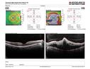
Acute Branch Retinal Vein Occlusion - 20/200436 x angesehen66 year old female with 3 weeks of vision loss in the left eye. VA 20/20 OD 20/200 OS11. Februar 2020
|
|

Acute Branch Retinal Vein Occlusion - 20/200475 x angesehen66 year old female with 3 weeks of vision loss in the left eye. VA 20/20 OD 20/200 OS11. Februar 2020
|
|
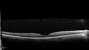
Acute Branch Retinal Vein Occlusion - 20/200423 x angesehen66 year old female with 3 weeks of vision loss in the left eye. VA 20/20 OD 20/200 OS11. Februar 2020
|
|
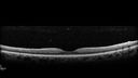
Acute Branch Retinal Vein Occlusion - 20/200459 x angesehen66 year old female with 3 weeks of vision loss in the left eye. VA 20/20 OD 20/200 OS11. Februar 2020
|
|

Central Retinal Vein Occlusion with Macular Edema566 x angesehen74 year old female The vision in the left eye has gotten worse over the last 3 weeks.
VA OD: Dcc20/25
VA OS: DccCF 3ft
IOP: TP: OD:16 OS:14
Vision improved to 20/40 in 3 months with monthly Aflibercept07. August 2019
|
|

Central Retinal Vein Occlusion with Macular Edema482 x angesehen74 year old female The vision in the left eye has gotten worse over the last 3 weeks.
VA OD: Dcc20/25
VA OS: DccCF 3ft
IOP: TP: OD:16 OS:14
Vision improved to 20/40 in 3 months with monthly Aflibercept07. August 2019
|
|

Central Retinal Vein Occlusion with Macular Edema545 x angesehen74 year old female The vision in the left eye has gotten worse over the last 3 weeks.
VA OD: Dcc20/25
VA OS: DccCF 3ft
IOP: TP: OD:16 OS:14
Vision improved to 20/40 in 3 months with monthly Aflibercept07. August 2019
|
|
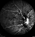
Central Retinal Vein Occlusion with Macular Edema596 x angesehen74 year old female The vision in the left eye has gotten worse over the last 3 weeks.
VA OD: Dcc20/25
VA OS: DccCF 3ft
IOP: TP: OD:16 OS:14
Vision improved to 20/40 in 3 months with monthly Aflibercept07. August 2019
|
|
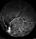
Central Retinal Vein Occlusion with Macular Edema558 x angesehen74 year old female The vision in the left eye has gotten worse over the last 3 weeks.
VA OD: Dcc20/25
VA OS: DccCF 3ft
IOP: TP: OD:16 OS:14
Vision improved to 20/40 in 3 months with monthly Aflibercept07. August 2019
|
|
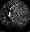
Central Retinal Vein Occlusion with Macular Edema562 x angesehen74 year old female The vision in the left eye has gotten worse over the last 3 weeks.
VA OD: Dcc20/25
VA OS: DccCF 3ft
IOP: TP: OD:16 OS:14
Vision improved to 20/40 in 3 months with monthly Aflibercept07. August 2019
|
|
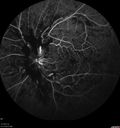
Central Retinal Vein Occlusion with Macular Edema457 x angesehen74 year old female The vision in the left eye has gotten worse over the last 3 weeks.
VA OD: Dcc20/25
VA OS: DccCF 3ft
IOP: TP: OD:16 OS:14
Vision improved to 20/40 in 3 months with monthly Aflibercept07. August 2019
|
|
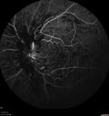
Central Retinal Vein Occlusion with Macular Edema494 x angesehen74 year old female The vision in the left eye has gotten worse over the last 3 weeks.
VA OD: Dcc20/25
VA OS: DccCF 3ft
IOP: TP: OD:16 OS:14
Vision improved to 20/40 in 3 months with monthly Aflibercept07. August 2019
|
|
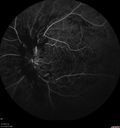
Central Retinal Vein Occlusion with Macular Edema465 x angesehen74 year old female The vision in the left eye has gotten worse over the last 3 weeks.
VA OD: Dcc20/25
VA OS: DccCF 3ft
IOP: TP: OD:16 OS:14
Vision improved to 20/40 in 3 months with monthly Aflibercept07. August 2019
|
|
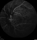
Central Retinal Vein Occlusion with Macular Edema527 x angesehen74 year old female The vision in the left eye has gotten worse over the last 3 weeks.
VA OD: Dcc20/25
VA OS: DccCF 3ft
IOP: TP: OD:16 OS:14
Vision improved to 20/40 in 3 months with monthly Aflibercept07. August 2019
|
|
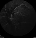
Central Retinal Vein Occlusion with Macular Edema474 x angesehen74 year old female The vision in the left eye has gotten worse over the last 3 weeks.
VA OD: Dcc20/25
VA OS: DccCF 3ft
IOP: TP: OD:16 OS:14
Vision improved to 20/40 in 3 months with monthly Aflibercept07. August 2019
|
|

Central Retinal Vein Occlusion with Macular Edema488 x angesehen74 year old female The vision in the left eye has gotten worse over the last 3 weeks.
VA OD: Dcc20/25
VA OS: DccCF 3ft
IOP: TP: OD:16 OS:14
Vision improved to 20/40 in 3 months with monthly Aflibercept07. August 2019
|
|
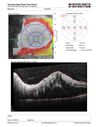
Central Retinal Vein Occlusion with Macular Edema600 x angesehen74 year old female The vision in the left eye has gotten worse over the last 3 weeks.
VA OD: Dcc20/25
VA OS: DccCF 3ft
IOP: TP: OD:16 OS:14
Vision improved to 20/40 in 3 months with monthly Aflibercept07. August 2019
|
|
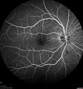
Central Retinal Vein Occlusion with Macular Edema526 x angesehen74 year old female The vision in the left eye has gotten worse over the last 3 weeks.
VA OD: Dcc20/25
VA OS: DccCF 3ft
IOP: TP: OD:16 OS:14
Vision improved to 20/40 in 3 months with monthly Aflibercept07. August 2019
|
|
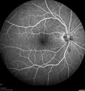
Central Retinal Vein Occlusion with Macular Edema474 x angesehen74 year old female The vision in the left eye has gotten worse over the last 3 weeks.
VA OD: Dcc20/25
VA OS: DccCF 3ft
IOP: TP: OD:16 OS:14
Vision improved to 20/40 in 3 months with monthly Aflibercept07. August 2019
|
|
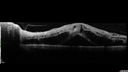
Central Retinal Vein Occlusion with Macular Edema546 x angesehen74 year old female The vision in the left eye has gotten worse over the last 3 weeks.
VA OD: Dcc20/25
VA OS: DccCF 3ft
IOP: TP: OD:16 OS:14
Vision improved to 20/40 in 3 months with monthly Aflibercept07. August 2019
|
|
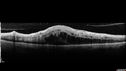
Central Retinal Vein Occlusion with Macular Edema486 x angesehen74 year old female The vision in the left eye has gotten worse over the last 3 weeks.
VA OD: Dcc20/25
VA OS: DccCF 3ft
IOP: TP: OD:16 OS:14
Vision improved to 20/40 in 3 months with monthly Aflibercept07. August 2019
|
|

Central Retinal Vein Occlusion with Macular Edema431 x angesehen74 year old female The vision in the left eye has gotten worse over the last 3 weeks.
VA OD: Dcc20/25
VA OS: DccCF 3ft
IOP: TP: OD:16 OS:14
Vision improved to 20/40 in 3 months with monthly Aflibercept07. August 2019
|
|

Central Retinal Vein Occlusion with Macular Edema452 x angesehen74 year old female The vision in the left eye has gotten worse over the last 3 weeks.
VA OD: Dcc20/25
VA OS: DccCF 3ft
IOP: TP: OD:16 OS:14
Vision improved to 20/40 in 3 months with monthly Aflibercept07. August 2019
|
|

Central Retinal Vein Occlusion with Macular Edema429 x angesehen74 year old female The vision in the left eye has gotten worse over the last 3 weeks.
VA OD: Dcc20/25
VA OS: DccCF 3ft
IOP: TP: OD:16 OS:14
Vision improved to 20/40 in 3 months with monthly Aflibercept07. August 2019
|
|

Central Retinal Vein Occlusion with Macular Edema498 x angesehen74 year old female The vision in the left eye has gotten worse over the last 3 weeks.
VA OD: Dcc20/25
VA OS: DccCF 3ft
IOP: TP: OD:16 OS:14
Vision improved to 20/40 in 3 months with monthly Aflibercept07. August 2019
|
|
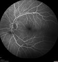
Subtle Branch Retinal Artery Occlusion440 x angesehen68 year old man with shadow in the left eye yesterday. IR and OCT show subtle retinal swelling. FA is normal07. August 2019
|
|

Subtle Branch Retinal Artery Occlusion450 x angesehen68 year old man with shadow in the left eye yesterday. IR and OCT show subtle retinal swelling. FA is normal07. August 2019
|
|

Subtle Branch Retinal Artery Occlusion514 x angesehen68 year old man with shadow in the left eye yesterday. IR and OCT show subtle retinal swelling. FA is normal07. August 2019
|
|
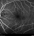
Subtle Branch Retinal Artery Occlusion479 x angesehen68 year old man with shadow in the left eye yesterday. IR and OCT show subtle retinal swelling. FA is normal07. August 2019
|
|

Subtle Branch Retinal Artery Occlusion498 x angesehen68 year old man with shadow in the left eye yesterday. IR and OCT show subtle retinal swelling. FA is normal07. August 2019
|
|

Subtle Branch Retinal Artery Occlusion423 x angesehen68 year old man with shadow in the left eye yesterday. IR and OCT show subtle retinal swelling. FA is normal07. August 2019
|
|
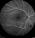
Subtle Branch Retinal Artery Occlusion495 x angesehen68 year old man with shadow in the left eye yesterday. IR and OCT show subtle retinal swelling. FA is normal07. August 2019
|
|

Subtle Branch Retinal Artery Occlusion507 x angesehen68 year old man with shadow in the left eye yesterday. IR and OCT show subtle retinal swelling. FA is normal07. August 2019
|
|

Subtle Branch Retinal Artery Occlusion500 x angesehen68 year old man with shadow in the left eye yesterday. IR and OCT show subtle retinal swelling. FA is normal07. August 2019
|
|
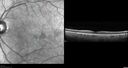
Subtle Branch Retinal Artery Occlusion573 x angesehen68 year old man with shadow in the left eye yesterday. IR and OCT show subtle retinal swelling. FA is normal07. August 2019
|
|

Subtle Branch Retinal Artery Occlusion492 x angesehen68 year old man with shadow in the left eye yesterday. IR and OCT show subtle retinal swelling. FA is normal07. August 2019
|
|

Subtle Branch Retinal Artery Occlusion502 x angesehen68 year old man with shadow in the left eye yesterday. IR and OCT show subtle retinal swelling. FA is normal07. August 2019
|
|

Subtle Branch Retinal Artery Occlusion593 x angesehen68 year old man with shadow in the left eye yesterday. IR and OCT show subtle retinal swelling. FA is normal07. August 2019
|
|
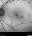
Subtle Branch Retinal Artery Occlusion468 x angesehen68 year old man with shadow in the left eye yesterday. IR and OCT show subtle retinal swelling. FA is normal07. August 2019
|
|
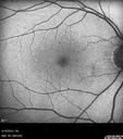
Subtle Branch Retinal Artery Occlusion495 x angesehen68 year old man with shadow in the left eye yesterday. IR and OCT show subtle retinal swelling. FA is normal07. August 2019
|
|
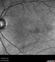
Subtle Branch Retinal Artery Occlusion533 x angesehen68 year old man with shadow in the left eye yesterday. IR and OCT show subtle retinal swelling. FA is normal07. August 2019
|
|

Subtle Branch Retinal Artery Occlusion493 x angesehen68 year old man with shadow in the left eye yesterday. IR and OCT show subtle retinal swelling. FA is normal07. August 2019
|
|

Subtle Branch Retinal Artery Occlusion483 x angesehen68 year old man with shadow in the left eye yesterday. IR and OCT show subtle retinal swelling. FA is normal07. August 2019
|
|

Subtle Branch Retinal Artery Occlusion559 x angesehen68 year old man with shadow in the left eye yesterday. IR and OCT show subtle retinal swelling. FA is normal07. August 2019
|
|
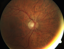
Subtle Branch Retinal Artery Occlusion628 x angesehen68 year old man with shadow in the left eye yesterday. IR and OCT show subtle retinal swelling. FA is normal07. August 2019
|
|

CRVO Shunt Vessels Right Eye - Optociliary Shunt Vessels768 x angesehenPrior CRVO right eye treated with injections13. Juni 2019
|
|

Sclerosed vein left eye735 x angesehen13. Juni 2019
|
|

Chronic BRAO with Hemorrhages - Sclerosed Vessel503 x angesehen95 year old female with blurred vision in both eyes in for a checkup. History of perpapillary CNVM in the left eye 2 years prior not treated. History of stroke with multiple emboli 1 year ago.
Vision 20/32 OD, 20/40 OS02. Juni 2019
|
|
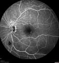
Chronic BRAO with Hemorrhages - Sclerosed Vessel444 x angesehen95 year old female with blurred vision in both eyes in for a checkup. History of perpapillary CNVM in the left eye 2 years prior not treated. History of stroke with multiple emboli 1 year ago.
Vision 20/32 OD, 20/40 OS02. Juni 2019
|
|

Chronic BRAO with Hemorrhages - Sclerosed Vessel463 x angesehen95 year old female with blurred vision in both eyes in for a checkup. History of perpapillary CNVM in the left eye 2 years prior not treated. History of stroke with multiple emboli 1 year ago.
Vision 20/32 OD, 20/40 OS02. Juni 2019
|
|
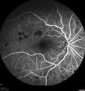
Chronic BRAO with Hemorrhages - Sclerosed Vessel471 x angesehen95 year old female with blurred vision in both eyes in for a checkup. History of perpapillary CNVM in the left eye 2 years prior not treated. History of stroke with multiple emboli 1 year ago.
Vision 20/32 OD, 20/40 OS02. Juni 2019
|
|
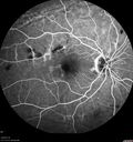
Chronic BRAO with Hemorrhages - Sclerosed Vessel448 x angesehen95 year old female with blurred vision in both eyes in for a checkup. History of perpapillary CNVM in the left eye 2 years prior not treated. History of stroke with multiple emboli 1 year ago.
Vision 20/32 OD, 20/40 OS02. Juni 2019
|
|
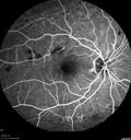
Chronic BRAO with Hemorrhages - Sclerosed Vessel458 x angesehen95 year old female with blurred vision in both eyes in for a checkup. History of perpapillary CNVM in the left eye 2 years prior not treated. History of stroke with multiple emboli 1 year ago.
Vision 20/32 OD, 20/40 OS02. Juni 2019
|
|

Chronic BRAO with Hemorrhages - Sclerosed Vessel502 x angesehen95 year old female with blurred vision in both eyes in for a checkup. History of perpapillary CNVM in the left eye 2 years prior not treated. History of stroke with multiple emboli 1 year ago.
Vision 20/32 OD, 20/40 OS02. Juni 2019
|
|

Chronic BRAO with Hemorrhages - Sclerosed Vessel436 x angesehen95 year old female with blurred vision in both eyes in for a checkup. History of perpapillary CNVM in the left eye 2 years prior not treated. History of stroke with multiple emboli 1 year ago.
Vision 20/32 OD, 20/40 OS02. Juni 2019
|
|

Chronic BRAO with Hemorrhages - Sclerosed Vessel426 x angesehen95 year old female with blurred vision in both eyes in for a checkup. History of perpapillary CNVM in the left eye 2 years prior not treated. History of stroke with multiple emboli 1 year ago.
Vision 20/32 OD, 20/40 OS02. Juni 2019
|
|
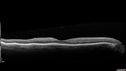
Chronic BRAO with Hemorrhages - Sclerosed Vessel414 x angesehen95 year old female with blurred vision in both eyes in for a checkup. History of perpapillary CNVM in the left eye 2 years prior not treated. History of stroke with multiple emboli 1 year ago.
Vision 20/32 OD, 20/40 OS02. Juni 2019
|
|
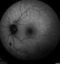
Chronic BRAO with Hemorrhages - Sclerosed Vessel485 x angesehen95 year old female with blurred vision in both eyes in for a checkup. History of perpapillary CNVM in the left eye 2 years prior not treated. History of stroke with multiple emboli 1 year ago.
Vision 20/32 OD, 20/40 OS02. Juni 2019
|
|
| 1845 Dateien auf 19 Seite(n) |
 |
|
|
|
|
8 | |
|
|
|
|


