Viimased lisandused - Retina Vascular Occlusions
|
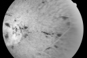
Central Retinal Vein Occlusion - New- and old Macular Scar from AMDvaatamisi: 48687-year-old woman with longstaning macular scars and new vision loss left eye for 2 months. OD: 20/200, OS 20/400. nov 28, 2010
|
|
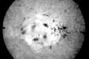
Central Retinal Vein Occlusion - New- and old Macular Scar from AMDvaatamisi: 47887-year-old woman with longstaning macular scars and new vision loss left eye for 2 months. OD: 20/200, OS 20/400. nov 28, 2010
|
|
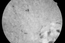
Central Retinal Vein Occlusion - New- and old Macular Scar from AMDvaatamisi: 50287-year-old woman with longstaning macular scars and new vision loss left eye for 2 months. OD: 20/200, OS 20/400. nov 28, 2010
|
|

Central Retinal Vein Occlusion - New- and old Macular Scar from AMDvaatamisi: 51487-year-old woman with longstaning macular scars and new vision loss left eye for 2 months. OD: 20/200, OS 20/400. nov 28, 2010
|
|
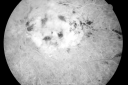
Central Retinal Vein Occlusion - New- and old Macular Scar from AMDvaatamisi: 44687-year-old woman with longstaning macular scars and new vision loss left eye for 2 months. OD: 20/200, OS 20/400. nov 28, 2010
|
|
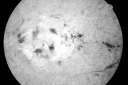
Central Retinal Vein Occlusion - New- and old Macular Scar from AMDvaatamisi: 49487-year-old woman with longstaning macular scars and new vision loss left eye for 2 months. OD: 20/200, OS 20/400. nov 28, 2010
|
|
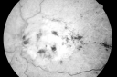
Central Retinal Vein Occlusion - New- and old Macular Scar from AMDvaatamisi: 53187-year-old woman with longstaning macular scars and new vision loss left eye for 2 months. OD: 20/200, OS 20/400. nov 28, 2010
|
|
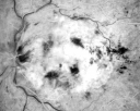
Central Retinal Vein Occlusion - New- and old Macular Scar from AMDvaatamisi: 50687-year-old woman with longstaning macular scars and new vision loss left eye for 2 months. OD: 20/200, OS 20/400. nov 28, 2010
|
|

Central Retinal Vein Occlusion - New- and old Macular Scar from AMDvaatamisi: 51487-year-old woman with longstaning macular scars and new vision loss left eye for 2 months. OD: 20/200, OS 20/400. nov 28, 2010
|
|
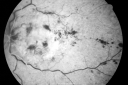
Central Retinal Vein Occlusion - New- and old Macular Scar from AMDvaatamisi: 49787-year-old woman with longstaning macular scars and new vision loss left eye for 2 months. OD: 20/200, OS 20/400. nov 28, 2010
|
|
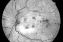
Central Retinal Vein Occlusion - New- and old Macular Scar from AMDvaatamisi: 49387-year-old woman with longstaning macular scars and new vision loss left eye for 2 months. OD: 20/200, OS 20/400. nov 28, 2010
|
|
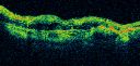
Central Retinal Vein Occlusion - New- and old Macular Scar from AMDvaatamisi: 48587-year-old woman with longstaning macular scars and new vision loss left eye for 2 months. OD: 20/200, OS 20/400. nov 28, 2010
|
|
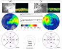
Central Retinal Vein Occlusion - New- and old Macular Scar from AMDvaatamisi: 56087-year-old woman with longstaning macular scars and new vision loss left eye for 2 months. OD: 20/200, OS 20/400. nov 28, 2010
|
|
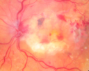
Central Retinal Vein Occlusion - New- and old Macular Scar from AMDvaatamisi: 62487-year-old woman with longstaning macular scars and new vision loss left eye for 2 months. OD: 20/200, OS 20/400. nov 28, 2010
|
|
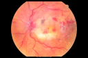
Central Retinal Vein Occlusion - New- and old Macular Scar from AMDvaatamisi: 64287-year-old woman with longstaning macular scars and new vision loss left eye for 2 months. OD: 20/200, OS 20/400. nov 28, 2010
|
|
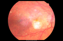
Central Retinal Vein Occlusion - New- and old Macular Scar from AMDvaatamisi: 65787-year-old woman with longstaning macular scars and new vision loss left eye for 2 months. OD: 20/200, OS 20/400. nov 28, 2010
|
|

Branch Retinal Vein Occlusion with Macular Edemavaatamisi: 688Central foveal thickness came to normal after laser and intravitreal Kenalognov 28, 2010
|
|
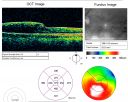
Branch Retinal Vein Occlusion with Macular Edemavaatamisi: 624Central foveal thickness came to normal after laser and intravitreal Kenalognov 28, 2010
|
|

Branch Retinal Vein Occlusion with Macular Edemavaatamisi: 550Central foveal thickness came to normal after laser and intravitreal Kenalognov 28, 2010
|
|
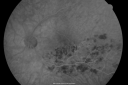
Branch Retinal Vein Occlusion with Macular Edemavaatamisi: 590Central foveal thickness came to normal after laser and intravitreal Kenalognov 28, 2010
|
|

Branch Retinal Vein Occlusion with Macular Edemavaatamisi: 555Central foveal thickness came to normal after laser and intravitreal Kenalognov 28, 2010
|
|

Branch Retinal Vein Occlusion with Macular Edemavaatamisi: 962Central foveal thickness came to normal after laser and intravitreal Kenalognov 28, 2010
|
|
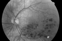
Branch Retinal Vein Occlusion with Macular Edemavaatamisi: 662Central foveal thickness came to normal after laser and intravitreal Kenalognov 28, 2010
|
|

Branch Retinal Vein Occlusion with Macular Edemavaatamisi: 784Central foveal thickness came to normal after laser and intravitreal Kenalognov 28, 2010
|
|

vaatamisi: 54249-year-old woman has central retinal vein occlusion right eye with fluctuating vision for a few month now with OD 20/100. IOP: OD 20nov 17, 2010
|
|
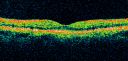
vaatamisi: 55449-year-old woman has central retinal vein occlusion right eye with fluctuating vision for a few month now with OD 20/100. IOP: OD 20nov 17, 2010
|
|
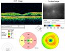
vaatamisi: 54249-year-old woman has central retinal vein occlusion right eye with fluctuating vision for a few month now with OD 20/100. IOP: OD 20nov 17, 2010
|
|
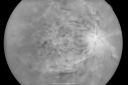
vaatamisi: 50649-year-old woman has central retinal vein occlusion right eye with fluctuating vision for a few month now with OD 20/100. IOP: OD 20nov 17, 2010
|
|
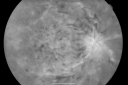
vaatamisi: 50149-year-old woman has central retinal vein occlusion right eye with fluctuating vision for a few month now with OD 20/100. IOP: OD 20nov 17, 2010
|
|
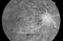
vaatamisi: 52749-year-old woman has central retinal vein occlusion right eye with fluctuating vision for a few month now with OD 20/100. IOP: OD 20nov 17, 2010
|
|
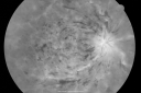
vaatamisi: 46949-year-old woman has central retinal vein occlusion right eye with fluctuating vision for a few month now with OD 20/100. IOP: OD 20nov 17, 2010
|
|
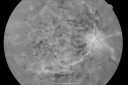
vaatamisi: 49449-year-old woman has central retinal vein occlusion right eye with fluctuating vision for a few month now with OD 20/100. IOP: OD 20nov 17, 2010
|
|
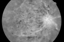
vaatamisi: 46249-year-old woman has central retinal vein occlusion right eye with fluctuating vision for a few month now with OD 20/100. IOP: OD 20nov 17, 2010
|
|
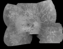
vaatamisi: 46649-year-old woman has central retinal vein occlusion right eye with fluctuating vision for a few month now with OD 20/100. IOP: OD 20nov 17, 2010
|
|
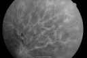
vaatamisi: 50149-year-old woman has central retinal vein occlusion right eye with fluctuating vision for a few month now with OD 20/100. IOP: OD 20nov 17, 2010
|
|
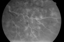
vaatamisi: 44349-year-old woman has central retinal vein occlusion right eye with fluctuating vision for a few month now with OD 20/100. IOP: OD 20nov 17, 2010
|
|
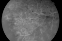
vaatamisi: 46949-year-old woman has central retinal vein occlusion right eye with fluctuating vision for a few month now with OD 20/100. IOP: OD 20nov 17, 2010
|
|
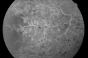
vaatamisi: 51449-year-old woman has central retinal vein occlusion right eye with fluctuating vision for a few month now with OD 20/100. IOP: OD 20nov 17, 2010
|
|
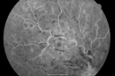
vaatamisi: 50249-year-old woman has central retinal vein occlusion right eye with fluctuating vision for a few month now with OD 20/100. IOP: OD 20nov 17, 2010
|
|
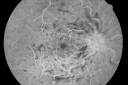
vaatamisi: 45949-year-old woman has central retinal vein occlusion right eye with fluctuating vision for a few month now with OD 20/100. IOP: OD 20nov 17, 2010
|
|
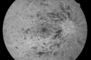
vaatamisi: 48949-year-old woman has central retinal vein occlusion right eye with fluctuating vision for a few month now with OD 20/100. IOP: OD 20nov 17, 2010
|
|
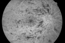
vaatamisi: 48049-year-old woman has central retinal vein occlusion right eye with fluctuating vision for a few month now with OD 20/100. IOP: OD 20nov 17, 2010
|
|
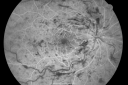
vaatamisi: 48949-year-old woman has central retinal vein occlusion right eye with fluctuating vision for a few month now with OD 20/100. IOP: OD 20nov 17, 2010
|
|

vaatamisi: 63049-year-old woman has central retinal vein occlusion right eye with fluctuating vision for a few month now with OD 20/100. IOP: OD 20nov 17, 2010
|
|
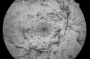
vaatamisi: 50749-year-old woman has central retinal vein occlusion right eye with fluctuating vision for a few month now with OD 20/100. IOP: OD 20nov 17, 2010
|
|
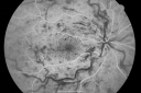
vaatamisi: 53149-year-old woman has central retinal vein occlusion right eye with fluctuating vision for a few month now with OD 20/100. IOP: OD 20nov 17, 2010
|
|
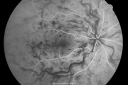
vaatamisi: 57449-year-old woman has central retinal vein occlusion right eye with fluctuating vision for a few month now with OD 20/100. IOP: OD 20nov 17, 2010
|
|
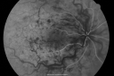
vaatamisi: 45049-year-old woman has central retinal vein occlusion right eye with fluctuating vision for a few month now with OD 20/100. IOP: OD 20nov 17, 2010
|
|
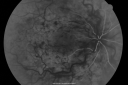
vaatamisi: 51049-year-old woman has central retinal vein occlusion right eye with fluctuating vision for a few month now with OD 20/100. IOP: OD 20nov 17, 2010
|
|
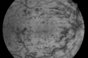
vaatamisi: 524nov 17, 2010
|
|
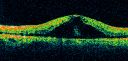
vaatamisi: 525nov 17, 2010
|
|
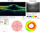
vaatamisi: 511nov 17, 2010
|
|
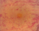
vaatamisi: 582nov 17, 2010
|
|
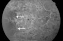
vaatamisi: 553nov 17, 2010
|
|
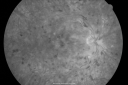
Central Retinal Vein Occlusion and Cystoid Macular Edema - Acute - Vision 20/50 vaatamisi: 60168-year-old man six weeks of vision loss right eye from CRVO and CME, vision right eye 20/50nov 11, 2010
|
|
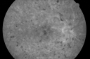
Central Retinal Vein Occlusion and Cystoid Macular Edema - Acute - Vision 20/50 vaatamisi: 57668-year-old man six weeks of vision loss right eye from CRVO and CME, vision right eye 20/50nov 11, 2010
|
|

Central Retinal Vein Occlusion and Cystoid Macular Edema - Acute - Vision 20/50 vaatamisi: 63368-year-old man six weeks of vision loss right eye from CRVO and CME, vision right eye 20/50nov 11, 2010
|
|

Central Retinal Vein Occlusion and Cystoid Macular Edema - Acute - Vision 20/50 vaatamisi: 57568-year-old man six weeks of vision loss right eye from CRVO and CME, vision right eye 20/50nov 11, 2010
|
|

Central Retinal Vein Occlusion and Cystoid Macular Edema - Acute - Vision 20/50 vaatamisi: 61568-year-old man six weeks of vision loss right eye from CRVO and CME, vision right eye 20/50nov 11, 2010
|
|

Central Retinal Vein Occlusion and Cystoid Macular Edema - Acute - Vision 20/50 vaatamisi: 61968-year-old man six weeks of vision loss right eye from CRVO and CME, vision right eye 20/50nov 11, 2010
|
|

Central Retinal Vein Occlusion and Cystoid Macular Edema - Acute - Vision 20/50 vaatamisi: 57668-year-old man six weeks of vision loss right eye from CRVO and CME, vision right eye 20/50nov 11, 2010
|
|

Central Retinal Vein Occlusion and Cystoid Macular Edema - Acute - Vision 20/50 vaatamisi: 75068-year-old man six weeks of vision loss right eye from CRVO and CME, vision right eye 20/50nov 11, 2010
|
|
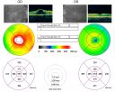
Central Retinal Vein Occlusion and Cystoid Macular Edema - Acute - Vision 20/50 vaatamisi: 61368-year-old man six weeks of vision loss right eye from CRVO and CME, vision right eye 20/50nov 11, 2010
|
|

Central Retinal Vein Occlusion and Cystoid Macular Edema - Acute - Vision 20/50 vaatamisi: 70668-year-old man six weeks of vision loss right eye from CRVO and CME, vision right eye 20/50nov 11, 2010
|
|

Central Retinal Vein Occlusion and Cystoid Macular Edema - Acute - Vision 20/50 vaatamisi: 82468-year-old man six weeks of vision loss right eye from CRVO and CME, vision right eye 20/50nov 11, 2010
|
|
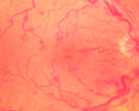
Central Retinal Vein Occlusion and Cystoid Macular Edema - Acute - Vision 20/50 vaatamisi: 80368-year-old man six weeks of vision loss right eye from CRVO and CME, vision right eye 20/50nov 11, 2010
|
|

Central Retinal Artery Occlusion - 2 months old by historyvaatamisi: 181977-year-old man was doing fine. His left eye was a little worse than the right eye, and then in the 2 months ago he had sudden severe vision loss in the right eye. Vision OD is 3/200; OS is 20/60. IOP: OD 30, OS 15.
OD: Vertical C/D ratio is 0.4. The macula is white from ischemia. The arteries are narrow.
Photos confirm clinical findings.
OCT SCAN: OCT scan of the right eye shows retinal thickening, consistent with macular edema associated with a central retinal artery occlusion. The left eye has severe inferior retinal thinning, consistent with an old hemicentral retinal artery occlusion in the left eye. The fovea in the left eye is a little bit thin but not too bad.
IMPRESSION: 1. CENTRAL RETINAL ARTERY OCCLUSION RIGHT EYE
2 OLD HEMICENTRAL RETINAL ARTERY OCCLUSION LEFT EYE
3 DIABETES WITHOUT RETINOPATHY
4 ELEVATED INTRAOCULAR PRESSURE RIGHT EYE STATUS POST INJECTION RIGHT EYE A WEEK AGO
DISCUSSION: I explained to the patient that the right eye unfortunately has had a central retinal artery and unfortunately we do not have anything to treat that. His vision will probably improve some over time. He needs to be watched though. It is possible he is developing rubeotic glaucoma, although it is probable the intraocular pressure is high from the injection last week. I asked him to restart the Alphagan, to stop the other two drops, and to return here in three weeks to make sure his pressure is more acceptable.
At this point, having lost almost all the retinal circulation in the right eye and half of the retinal circulation in the left eye, he only has half a retina in the left eye he is working off of. I told him it is very important he keep seeing you and anything that can be done to keep his blood pressure low and his cholesterol reasonable would be helpful to what remains of his retinal circulation.
nov 09, 2010
|
|

Central Retinal Artery Occlusion - 2 months old by historyvaatamisi: 86677-year-old man was doing fine. His left eye was a little worse than the right eye, and then in the 2 months ago he had sudden severe vision loss in the right eye. Vision OD is 3/200; OS is 20/60. IOP: OD 30, OS 15.
OD: Vertical C/D ratio is 0.4. The macula is white from ischemia. The arteries are narrow.
Photos confirm clinical findings.
OCT SCAN: OCT scan of the right eye shows retinal thickening, consistent with macular edema associated with a central retinal artery occlusion. The left eye has severe inferior retinal thinning, consistent with an old hemicentral retinal artery occlusion in the left eye. The fovea in the left eye is a little bit thin but not too bad.
IMPRESSION: 1. CENTRAL RETINAL ARTERY OCCLUSION RIGHT EYE
2 OLD HEMICENTRAL RETINAL ARTERY OCCLUSION LEFT EYE
3 DIABETES WITHOUT RETINOPATHY
4 ELEVATED INTRAOCULAR PRESSURE RIGHT EYE STATUS POST INJECTION RIGHT EYE A WEEK AGO
DISCUSSION: I explained to the patient that the right eye unfortunately has had a central retinal artery and unfortunately we do not have anything to treat that. His vision will probably improve some over time. He needs to be watched though. It is possible he is developing rubeotic glaucoma, although it is probable the intraocular pressure is high from the injection last week. I asked him to restart the Alphagan, to stop the other two drops, and to return here in three weeks to make sure his pressure is more acceptable.
At this point, having lost almost all the retinal circulation in the right eye and half of the retinal circulation in the left eye, he only has half a retina in the left eye he is working off of. I told him it is very important he keep seeing you and anything that can be done to keep his blood pressure low and his cholesterol reasonable would be helpful to what remains of his retinal circulation.
nov 09, 2010
|
|

Central Retinal Artery Occlusion - 2 months old by historyvaatamisi: 159377-year-old man was doing fine. His left eye was a little worse than the right eye, and then in the 2 months ago he had sudden severe vision loss in the right eye. Vision OD is 3/200; OS is 20/60. IOP: OD 30, OS 15.
OD: Vertical C/D ratio is 0.4. The macula is white from ischemia. The arteries are narrow.
Photos confirm clinical findings.
OCT SCAN: OCT scan of the right eye shows retinal thickening, consistent with macular edema associated with a central retinal artery occlusion. The left eye has severe inferior retinal thinning, consistent with an old hemicentral retinal artery occlusion in the left eye. The fovea in the left eye is a little bit thin but not too bad.
IMPRESSION: 1. CENTRAL RETINAL ARTERY OCCLUSION RIGHT EYE
2 OLD HEMICENTRAL RETINAL ARTERY OCCLUSION LEFT EYE
3 DIABETES WITHOUT RETINOPATHY
4 ELEVATED INTRAOCULAR PRESSURE RIGHT EYE STATUS POST INJECTION RIGHT EYE A WEEK AGO
DISCUSSION: I explained to the patient that the right eye unfortunately has had a central retinal artery and unfortunately we do not have anything to treat that. His vision will probably improve some over time. He needs to be watched though. It is possible he is developing rubeotic glaucoma, although it is probable the intraocular pressure is high from the injection last week. I asked him to restart the Alphagan, to stop the other two drops, and to return here in three weeks to make sure his pressure is more acceptable.
At this point, having lost almost all the retinal circulation in the right eye and half of the retinal circulation in the left eye, he only has half a retina in the left eye he is working off of. I told him it is very important he keep seeing you and anything that can be done to keep his blood pressure low and his cholesterol reasonable would be helpful to what remains of his retinal circulation.
nov 09, 2010
|
|

BRVO with collaterals and cystoid macular edemavaatamisi: 71066 Year Old Man with has had a branch retinal vein occlusion in the right eye with vision loss for the last few months. OD is 20/40, OS is 20/20. IOP: OD 18, OS 19. There is a superotemporal macular branch retinal vein occlusion with edema, hemorrhage, and exudate.
OCT SCAN: The right eye shows edema with a central foveal thickness of 354 microns FA: shows remodeling of the superotemporal vasculature. There are already venous-venous collaterals, suggesting the vein occlusion may be a little more than a few months old. There are multiple microaneurysms superotemporal to the fovea, and there is leakage in the late frames involving the fovea.
nov 06, 2010
|
|

BRVO with collaterals and cystoid macular edemavaatamisi: 74166 Year Old Man with has had a branch retinal vein occlusion in the right eye with vision loss for the last few months. OD is 20/40, OS is 20/20. IOP: OD 18, OS 19. There is a superotemporal macular branch retinal vein occlusion with edema, hemorrhage, and exudate.
OCT SCAN: The right eye shows edema with a central foveal thickness of 354 microns FA: shows remodeling of the superotemporal vasculature. There are already venous-venous collaterals, suggesting the vein occlusion may be a little more than a few months old. There are multiple microaneurysms superotemporal to the fovea, and there is leakage in the late frames involving the fovea.
nov 06, 2010
|
|

BRVO with collaterals and cystoid macular edemavaatamisi: 82766 Year Old Man with has had a branch retinal vein occlusion in the right eye with vision loss for the last few months. OD is 20/40, OS is 20/20. IOP: OD 18, OS 19. There is a superotemporal macular branch retinal vein occlusion with edema, hemorrhage, and exudate.
OCT SCAN: The right eye shows edema with a central foveal thickness of 354 microns FA: shows remodeling of the superotemporal vasculature. There are already venous-venous collaterals, suggesting the vein occlusion may be a little more than a few months old. There are multiple microaneurysms superotemporal to the fovea, and there is leakage in the late frames involving the fovea.
nov 06, 2010
|
|

BRVO with collaterals and cystoid macular edemavaatamisi: 93366 Year Old Man with has had a branch retinal vein occlusion in the right eye with vision loss for the last few months. OD is 20/40, OS is 20/20. IOP: OD 18, OS 19. There is a superotemporal macular branch retinal vein occlusion with edema, hemorrhage, and exudate.
OCT SCAN: The right eye shows edema with a central foveal thickness of 354 microns FA: shows remodeling of the superotemporal vasculature. There are already venous-venous collaterals, suggesting the vein occlusion may be a little more than a few months old. There are multiple microaneurysms superotemporal to the fovea, and there is leakage in the late frames involving the fovea.
nov 06, 2010
|
|

BRVO with collaterals and cystoid macular edemavaatamisi: 93666 Year Old Man with has had a branch retinal vein occlusion in the right eye with vision loss for the last few months. OD is 20/40, OS is 20/20. IOP: OD 18, OS 19. There is a superotemporal macular branch retinal vein occlusion with edema, hemorrhage, and exudate.
OCT SCAN: The right eye shows edema with a central foveal thickness of 354 microns FA: shows remodeling of the superotemporal vasculature. There are already venous-venous collaterals, suggesting the vein occlusion may be a little more than a few months old. There are multiple microaneurysms superotemporal to the fovea, and there is leakage in the late frames involving the fovea.
nov 06, 2010
|
|

BRVO with collaterals and cystoid macular edemavaatamisi: 80866 Year Old Man with has had a branch retinal vein occlusion in the right eye with vision loss for the last few months. OD is 20/40, OS is 20/20. IOP: OD 18, OS 19. There is a superotemporal macular branch retinal vein occlusion with edema, hemorrhage, and exudate.
OCT SCAN: The right eye shows edema with a central foveal thickness of 354 microns FA: shows remodeling of the superotemporal vasculature. There are already venous-venous collaterals, suggesting the vein occlusion may be a little more than a few months old. There are multiple microaneurysms superotemporal to the fovea, and there is leakage in the late frames involving the fovea.
nov 06, 2010
|
|

BRVO with collaterals and cystoid macular edemavaatamisi: 113966 Year Old Man with has had a branch retinal vein occlusion in the right eye with vision loss for the last few months. OD is 20/40, OS is 20/20. IOP: OD 18, OS 19. There is a superotemporal macular branch retinal vein occlusion with edema, hemorrhage, and exudate.
OCT SCAN: The right eye shows edema with a central foveal thickness of 354 microns FA: shows remodeling of the superotemporal vasculature. There are already venous-venous collaterals, suggesting the vein occlusion may be a little more than a few months old. There are multiple microaneurysms superotemporal to the fovea, and there is leakage in the late frames involving the fovea.
nov 06, 2010
|
|

BRVO with collaterals and cystoid macular edemavaatamisi: 81166 Year Old Man with has had a branch retinal vein occlusion in the right eye with vision loss for the last few months. OD is 20/40, OS is 20/20. IOP: OD 18, OS 19. There is a superotemporal macular branch retinal vein occlusion with edema, hemorrhage, and exudate.
OCT SCAN: The right eye shows edema with a central foveal thickness of 354 microns FA: shows remodeling of the superotemporal vasculature. There are already venous-venous collaterals, suggesting the vein occlusion may be a little more than a few months old. There are multiple microaneurysms superotemporal to the fovea, and there is leakage in the late frames involving the fovea.
nov 06, 2010
|
|
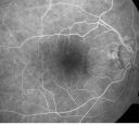
Old BRAO twig with plaquevaatamisi: 75181-year-old woman Her left eye is amblyopic. Her right eye was doing fine until about last Sunday, which is five days ago. She had a change in vision where for five minutes she saw a shadow in her side vision out of the right eye. She describes it as being more up into the left. It lasted for just a brief time and then went away. She has had atrial fibrillation since January of 2008. She is on Coumadin for that. She is diabetic for eleven years. OD is 20/25, OS is 20/60. IOP: OD 15, OS 18. nov 03, 2010
|
|
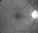
Old BRAO twig with plaquevaatamisi: 75481-year-old woman Her left eye is amblyopic. Her right eye was doing fine until about last Sunday, which is five days ago. She had a change in vision where for five minutes she saw a shadow in her side vision out of the right eye. She describes it as being more up into the left. It lasted for just a brief time and then went away. She has had atrial fibrillation since January of 2008. She is on Coumadin for that. She is diabetic for eleven years. OD is 20/25, OS is 20/60. IOP: OD 15, OS 18. nov 03, 2010
|
|

Old BRAO twig with plaquevaatamisi: 106581-year-old woman Her left eye is amblyopic. Her right eye was doing fine until about last Sunday, which is five days ago. She had a change in vision where for five minutes she saw a shadow in her side vision out of the right eye. She describes it as being more up into the left. It lasted for just a brief time and then went away. She has had atrial fibrillation since January of 2008. She is on Coumadin for that. She is diabetic for eleven years. OD is 20/25, OS is 20/60. IOP: OD 15, OS 18. nov 03, 2010
|
|

Old Branch Retinal Vein Occlusion- Severe CME - Pucker - Diabetic Late FAvaatamisi: 52161-year old man has diabetic retinopathy in the right eye. He also had a macular pucker and macular edema. I did a vitrectomy on September 5th. His vision was initially improving. His vision in the right eye seems to be a little more foggy.
VISUAL ACUITY: OD: 20/200okt 28, 2010
|
|

Old Branch Retinal Vein Occlusion- Severe CME - Pucker - Diabetic Early FAvaatamisi: 56461-year old man has diabetic retinopathy in the right eye. He also had a macular pucker and macular edema. I did a vitrectomy on September 5th. His vision was initially improving. His vision in the right eye seems to be a little more foggy.
VISUAL ACUITY: OD: 20/200okt 28, 2010
|
|

Old Branch Retinal Vein Occlusion- Severe CME - Pucker - Diabetic Early FAvaatamisi: 56361-year old man has diabetic retinopathy in the right eye. He also had a macular pucker and macular edema. I did a vitrectomy on September 5th. His vision was initially improving. His vision in the right eye seems to be a little more foggy.
VISUAL ACUITY: OD: 20/200okt 28, 2010
|
|

Old Branch Retinal Vein Occlusion- Severe CME - Pucker - Diabetic Map OCTvaatamisi: 81961-year old man has diabetic retinopathy in the right eye. He also had a macular pucker and macular edema. I did a vitrectomy on September 5th. His vision was initially improving. His vision in the right eye seems to be a little more foggy.
VISUAL ACUITY: OD: 20/200okt 28, 2010
|
|
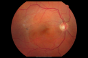
Old Branch Retinal Vein Occlusion- Severe CME - Pucker - Diabetic Fundus Photovaatamisi: 83461-year old man has diabetic retinopathy in the right eye. He also had a macular pucker and macular edema. I did a vitrectomy on September 5th. His vision was initially improving. His vision in the right eye seems to be a little more foggy.
VISUAL ACUITY: OD: 20/200okt 28, 2010
|
|

BRVO arrow showing obstruction point with collateral vessels - red free photovaatamisi: 87670 year old woman with 20/80 vision from a branch retinal vein occlusion and macular edema. She has had laser treatment and intravitreal steroids. Her vision with further laser improved to 20/50.okt 27, 2010
|
|

brvo cme with small serous macular detachment vaatamisi: 80670 year old woman with 20/80 vision from a branch retinal vein occlusion and macular edema. She has had laser treatment and intravitreal steroids. Her vision with further laser improved to 20/50.okt 27, 2010
|
|

branch retinal vein occlusion - fluorescein angiogramvaatamisi: 84470 year old woman with 20/80 vision from a branch retinal vein occlusion and macular edema. She has had laser treatment and intravitreal steroids. Her vision with further laser improved to 20/50.okt 27, 2010
|
|
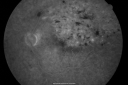
branch retinal vein occlusion - fluorescein angiogramvaatamisi: 70670 year old woman with 20/80 vision from a branch retinal vein occlusion and macular edema. She has had laser treatment and intravitreal steroids. Her vision with further laser improved to 20/50.okt 27, 2010
|
|

branch retinal vein occlusion - fluorescein angiogramvaatamisi: 68770 year old woman with 20/80 vision from a branch retinal vein occlusion and macular edema. She has had laser treatment and intravitreal steroids. Her vision with further laser improved to 20/50.okt 27, 2010
|
|

branch retinal vein occlusion - fluorescein angiogramvaatamisi: 63370 year old woman with 20/80 vision from a branch retinal vein occlusion and macular edema. She has had laser treatment and intravitreal steroids. Her vision with further laser improved to 20/50.okt 27, 2010
|
|

branch retinal vein occlusion - fluorescein angiogramvaatamisi: 75470 year old woman with 20/80 vision from a branch retinal vein occlusion and macular edema. She has had laser treatment and intravitreal steroids. Her vision with further laser improved to 20/50.okt 27, 2010
|
|
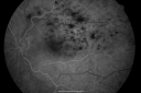
branch retinal vein occlusion - fluorescein angiogramvaatamisi: 64570 year old woman with 20/80 vision from a branch retinal vein occlusion and macular edema. She has had laser treatment and intravitreal steroids. Her vision with further laser improved to 20/50.okt 27, 2010
|
|

branch retinal vein occlusion - red free photovaatamisi: 91170 year old woman with 20/80 vision from a branch retinal vein occlusion and macular edema. She has had laser treatment and intravitreal steroids. Her vision with further laser improved to 20/50.okt 27, 2010
|
|

branch retinal vein occlusion - fundus photovaatamisi: 108370 year old woman with 20/80 vision from a branch retinal vein occlusion and macular edema. She has had laser treatment and intravitreal steroids. Her vision with further laser improved to 20/50.okt 27, 2010
|
|

branch retinal vein occlusion - oct line scanvaatamisi: 85170 year old woman with 20/80 vision from a branch retinal vein occlusion and macular edema. She has had laser treatment and intravitreal steroids. Her vision with further laser improved to 20/50.okt 27, 2010
|
|

branch retinal vein occlusion - oct mapvaatamisi: 97470 year old woman with 20/80 vision from a branch retinal vein occlusion and macular edema. She has had laser treatment and intravitreal steroids. Her vision with further laser improved to 20/50.okt 27, 2010
|
|

branch retinal vein occlusion - fluorescein angiogram - arrow shows obstruction point with collateral vesselsvaatamisi: 94270 year old woman with 20/80 vision from a branch retinal vein occlusion and macular edema. She has had laser treatment and intravitreal steroids. Her vision with further laser improved to 20/50.okt 27, 2010
|
|
|
|
|
|


