Last additions - Retina Vascular Occlusions
|
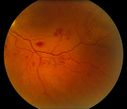
BRVO Inferiorly with Retinal Hemorrhages in the Left Eye361 viewsPatient comes in with complaint of "spot in vision", sort of a cloudy haze in the left eye. Patient's VA was 20/100-1 with no improvement with pinhole in the left eye. Fundus photos show multiple retinal hemorrhages scattered with a branch retinal vein occlusion inferiorily. Patient is also diabetic.Mar 12, 2013
|
|
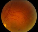
BRVO Inferiorly with Retinal Hemorrhages in the Left Eye433 viewsPatient comes in with complaint of "spot in vision", sort of a cloudy haze in the left eye. Patient's VA was 20/100-1 with no improvement with pinhole in the left eye. Fundus photos show multiple retinal hemorrhages scattered with a branch retinal vein occlusion inferiorily. Patient is also diabetic.Mar 12, 2013
|
|
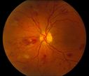
BRVO Inferiorly with Retinal Hemorrhages in the Left Eye465 viewsPatient comes in with complaint of "spot in vision", sort of a cloudy haze in the left eye. Patient's VA was 20/100-1 with no improvement with pinhole in the left eye. Fundus photos show multiple retinal hemorrhages scattered with a branch retinal vein occlusion inferiorily. Patient is also diabetic.Mar 12, 2013
|
|
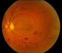
BRVO Inferiorly with Retinal Hemorrhages in the Left Eye539 viewsPatient comes in with complaint of "spot in vision", sort of a cloudy haze in the left eye. Patient's VA was 20/100-1 with no improvement with pinhole in the left eye. Fundus photos show multiple retinal hemorrhages scattered with a branch retinal vein occlusion inferiorily. Patient is also diabetic.Mar 12, 2013
|
|

BRVO Inferiorly with Retinal Hemorrhages in the Left Eye590 viewsPatient comes in with complaint of "spot in vision", sort of a cloudy haze in the left eye. Patient's VA was 20/100-1 with no improvement with pinhole in the left eye. Fundus photos show multiple retinal hemorrhages scattered with a branch retinal vein occlusion inferiorily. Patient is also diabetic.Mar 12, 2013
|
|

Ischemic HRVO389 views63 y/ o African American female with advanced glaucoma and longstanding loss of vision OD presents with acute vision loss OS. Ophthalmic history is significant for uncomplicated phaco/IOL OS 2 months prior to event. Medical history is significant for poorly controlled HTN. Pt characterizes vision loss as blurring centrally OS.Mar 05, 2013
|
|
345 viewsMar 05, 2013
|
|
296 viewsMar 05, 2013
|
|
370 viewsMar 05, 2013
|
|
363 viewsMar 05, 2013
|
|
350 viewsMar 05, 2013
|
|
370 viewsMar 05, 2013
|
|
357 viewsMar 05, 2013
|
|
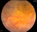
Central Retinal Vein Occlusion with Macular Edema and Retinal Hemorrhages 412 viewsFemale patient of 86-years of age presents a central retinal vein occlusion with macular edema in the right eye. Patient's VA was 20/70 in the right eye. Proceeded with laser treatment to the right eye.Jan 25, 2013
|
|
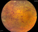
Central Retinal Vein Occlusion with Macular Edema and Retinal Hemorrhages 383 viewsFemale patient of 86-years of age presents a central retinal vein occlusion with macular edema in the right eye. Patient's VA was 20/70 in the right eye. Proceeded with laser treatment to the right eye.Jan 25, 2013
|
|
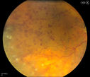
Central Retinal Vein Occlusion with Macular Edema and Retinal Hemorrhages 376 viewsFemale patient of 86-years of age presents a central retinal vein occlusion with macular edema in the right eye. Patient's VA was 20/70 in the right eye. Proceeded with laser treatment to the right eye.Jan 25, 2013
|
|
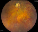
Central Retinal Vein Occlusion with Macular Edema and Retinal Hemorrhages 400 viewsFemale patient of 86-years of age presents a central retinal vein occlusion with macular edema in the right eye. Patient's VA was 20/70 in the right eye. Proceeded with laser treatment to the right eye.Jan 25, 2013
|
|
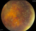
Central Retinal Vein Occlusion with Macular Edema and Retinal Hemorrhages 375 viewsFemale patient of 86-years of age presents a central retinal vein occlusion with macular edema in the right eye. Patient's VA was 20/70 in the right eye. Proceeded with laser treatment to the right eye.Jan 25, 2013
|
|
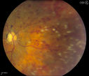
Central Retinal Vein Occlusion with Macular Edema and Retinal Hemorrhages 393 viewsFemale patient of 86-years of age presents a central retinal vein occlusion with macular edema in the right eye. Patient's VA was 20/70 in the right eye. Proceeded with laser treatment to the right eye.Jan 25, 2013
|
|
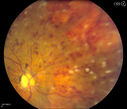
Central Retinal Vein Occlusion with Macular Edema and Retinal Hemorrhages 441 viewsFemale patient of 86-years of age presents a central retinal vein occlusion with macular edema in the right eye. Patient's VA was 20/70 in the right eye. Proceeded with laser treatment to the right eye.Jan 25, 2013
|
|
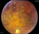
Central Retinal Vein Occlusion with Macular Edema and Retinal Hemorrhages 390 viewsFemale patient of 86-years of age presents a central retinal vein occlusion with macular edema in the right eye. Patient's VA was 20/70 in the right eye. Proceeded with laser treatment to the right eye.Jan 25, 2013
|
|
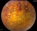
Central Retinal Vein Occlusion with Macular Edema and Retinal Hemorrhages 364 viewsFemale patient of 86-years of age presents a central retinal vein occlusion with macular edema in the right eye. Patient's VA was 20/70 in the right eye. Proceeded with laser treatment to the right eye.Jan 25, 2013
|
|
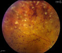
Central Retinal Vein Occlusion with Macular Edema and Retinal Hemorrhages 407 viewsFemale patient of 86-years of age presents a central retinal vein occlusion with macular edema in the right eye. Patient's VA was 20/70 in the right eye. Proceeded with laser treatment to the right eye.Jan 25, 2013
|
|
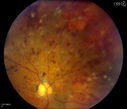
Central Retinal Vein Occlusion with Macular Edema and Retinal Hemorrhages 427 viewsFemale patient of 86-years of age presents a central retinal vein occlusion with macular edema in the right eye. Patient's VA was 20/70 in the right eye. Proceeded with laser treatment to the right eye.Jan 25, 2013
|
|
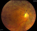
Central Retinal Vein Occlusion with Macular Edema and Retinal Hemorrhages 524 viewsFemale patient of 86-years of age presents a central retinal vein occlusion with macular edema in the right eye. Patient's VA was 20/70 in the right eye. Proceeded with laser treatment to the right eye.Jan 25, 2013
|
|
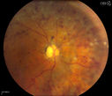
Central Retinal Vein Occlusion with Macular Edema and Retinal Hemorrhages 473 viewsFemale patient of 86-years of age presents a central retinal vein occlusion with macular edema in the right eye. Patient's VA was 20/70 in the right eye. Proceeded with laser treatment to the right eye.Jan 25, 2013
|
|
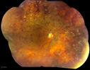
Central Retinal Vein Occlusion with Macular Edema and Retinal Hemorrhages 454 viewsFemale patient of 86-years of age presents a central retinal vein occlusion with macular edema in the right eye. Patient's VA was 20/70 in the right eye. Proceeded with laser treatment to the right eye.Jan 25, 2013
|
|
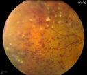
Central Retinal Vein Occlusion with Macular Edema and Retinal Hemorrhages 440 viewsFemale patient of 86-years of age presents a central retinal vein occlusion with macular edema in the right eye. Patient's VA was 20/70 in the right eye. Proceeded with laser treatment to the right eye.Jan 25, 2013
|
|
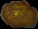
Combined Artery and Vein Occlusion561 viewsPatient comes in with decreased vision in both eyes. VA 20/200-OD, 20/60-OS. Fundus exam shows great amount of macular edema due to artery and vein occlusions. There is some neovascularization on the optic nerve in the right eye. Patient was treated with Eylea Injection in the left eye today and will return for Eylea Injection in the right eye. Jan 23, 2013
|
|
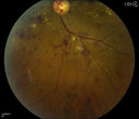
Combined Artery and Vein Occlusion376 viewsPatient comes in with decreased vision in both eyes. VA 20/200-OD, 20/60-OS. Fundus exam shows great amount of macular edema due to artery and vein occlusions. There is some neovascularization on the optic nerve in the right eye. Patient was treated with Eylea Injection in the left eye today and will return for Eylea Injection in the right eye.Jan 23, 2013
|
|
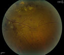
Combined Artery and Vein Occlusion382 viewsPatient comes in with decreased vision in both eyes. VA 20/200-OD, 20/60-OS. Fundus exam shows great amount of macular edema due to artery and vein occlusions. There is some neovascularization on the optic nerve in the right eye. Patient was treated with Eylea Injection in the left eye today and will return for Eylea Injection in the right eye.Jan 23, 2013
|
|
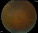
Combined Artery and Vein Occlusion382 viewsPatient comes in with decreased vision in both eyes. VA 20/200-OD, 20/60-OS. Fundus exam shows great amount of macular edema due to artery and vein occlusions. There is some neovascularization on the optic nerve in the right eye. Patient was treated with Eylea Injection in the left eye today and will return for Eylea Injection in the right eye.Jan 23, 2013
|
|
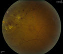
Combined Artery and Vein Occlusion392 viewsPatient comes in with decreased vision in both eyes. VA 20/200-OD, 20/60-OS. Fundus exam shows great amount of macular edema due to artery and vein occlusions. There is some neovascularization on the optic nerve in the right eye. Patient was treated with Eylea Injection in the left eye today and will return for Eylea Injection in the right eye.Jan 23, 2013
|
|
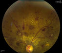
Combined Artery and Vein Occlusion410 viewsPatient comes in with decreased vision in both eyes. VA 20/200-OD, 20/60-OS. Fundus exam shows great amount of macular edema due to artery and vein occlusions. There is some neovascularization on the optic nerve in the right eye. Patient was treated with Eylea Injection in the left eye today and will return for Eylea Injection in the right eye.Jan 23, 2013
|
|
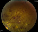
Combined Artery and Vein Occlusion391 viewsPatient comes in with decreased vision in both eyes. VA 20/200-OD, 20/60-OS. Fundus exam shows great amount of macular edema due to artery and vein occlusions. There is some neovascularization on the optic nerve in the right eye. Patient was treated with Eylea Injection in the left eye today and will return for Eylea Injection in the right eye.Jan 23, 2013
|
|
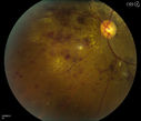
Combined Artery and Vein Occlusion394 viewsPatient comes in with decreased vision in both eyes. VA 20/200-OD, 20/60-OS. Fundus exam shows great amount of macular edema due to artery and vein occlusions. There is some neovascularization on the optic nerve in the right eye. Patient was treated with Eylea Injection in the left eye today and will return for Eylea Injection in the right eye.Jan 23, 2013
|
|
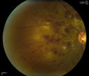
Combined Artery and Vein Occlusion428 viewsPatient comes in with decreased vision in both eyes. VA 20/200-OD, 20/60-OS. Fundus exam shows great amount of macular edema due to artery and vein occlusions. There is some neovascularization on the optic nerve in the right eye. Patient was treated with Eylea Injection in the left eye today and will return for Eylea Injection in the right eye.Jan 23, 2013
|
|
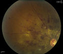
Combined Artery and Vein Occlusion377 viewsPatient comes in with decreased vision in both eyes. VA 20/200-OD, 20/60-OS. Fundus exam shows great amount of macular edema due to artery and vein occlusions. There is some neovascularization on the optic nerve in the right eye. Patient was treated with Eylea Injection in the left eye today and will return for Eylea Injection in the right eye.Jan 23, 2013
|
|
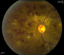
Combined Artery and Vein Occlusion340 viewsPatient comes in with decreased vision in both eyes. VA 20/200-OD, 20/60-OS. Fundus exam shows great amount of macular edema due to artery and vein occlusions. There is some neovascularization on the optic nerve in the right eye. Patient was treated with Eylea Injection in the left eye today and will return for Eylea Injection in the right eye.Jan 23, 2013
|
|
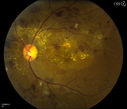
Combined Artery and Vein Occlusion430 viewsPatient comes in with decreased vision in both eyes. VA 20/200-OD, 20/60-OS. Fundus exam shows great amount of macular edema due to artery and vein occlusions. There is some neovascularization on the optic nerve in the right eye. Patient was treated with Eylea Injection in the left eye today and will return for Eylea Injection in the right eye.Jan 23, 2013
|
|
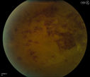
Combined Artery and Vein Occlusion389 viewsPatient comes in with decreased vision in both eyes. VA 20/200-OD, 20/60-OS. Fundus exam shows great amount of macular edema due to artery and vein occlusions. There is some neovascularization on the optic nerve in the right eye. Patient was treated with Eylea Injection in the left eye today and will return for Eylea Injection in the right eye.Jan 23, 2013
|
|
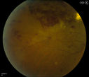
Combined Artery and Vein Occlusion434 viewsPatient comes in with decreased vision in both eyes. VA 20/200-OD, 20/60-OS. Fundus exam shows great amount of macular edema due to artery and vein occlusions. There is some neovascularization on the optic nerve in the right eye. Patient was treated with Eylea Injection in the left eye today and will return for Eylea Injection in the right eye.Jan 23, 2013
|
|

Combined Artery and Vein Occlusion394 viewsPatient comes in with decreased vision in both eyes. VA 20/200-OD, 20/60-OS. Fundus exam shows great amount of macular edema due to artery and vein occlusions. There is some neovascularization on the optic nerve in the right eye. Patient was treated with Eylea Injection in the left eye today and will return for Eylea Injection in the right eye.Jan 23, 2013
|
|
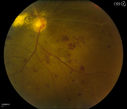
Combined Artery and Vein Occlusion489 viewsPatient comes in with decreased vision in both eyes. VA 20/200-OD, 20/60-OS. Fundus exam shows great amount of macular edema due to artery and vein occlusions. There is some neovascularization on the optic nerve in the right eye. Patient was treated with Eylea Injection in the left eye today and will return for Eylea Injection in the right eye.Jan 23, 2013
|
|
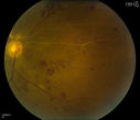
Combined Artery and Vein Occlusion479 viewsPatient comes in with decreased vision in both eyes. VA 20/200-OD, 20/60-OS. Fundus exam shows great amount of macular edema due to artery and vein occlusions. There is some neovascularization on the optic nerve in the right eye. Patient was treated with Eylea Injection in the left eye today and will return for Eylea Injection in the right eye.Jan 23, 2013
|
|
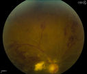
Combined Artery and Vein Occlusion409 viewsPatient comes in with decreased vision in both eyes. VA 20/200-OD, 20/60-OS. Fundus exam shows great amount of macular edema due to artery and vein occlusions. There is some neovascularization on the optic nerve in the right eye. Patient was treated with Eylea Injection in the left eye today and will return for Eylea Injection in the right eye.Jan 23, 2013
|
|
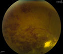
Combined Artery and Vein Occlusion495 viewsPatient comes in with decreased vision in both eyes. VA 20/200-OD, 20/60-OS. Fundus exam shows great amount of macular edema due to artery and vein occlusions. There is some neovascularization on the optic nerve in the right eye. Patient was treated with Eylea Injection in the left eye today and will return for Eylea Injection in the right eye.Jan 23, 2013
|
|

Combined Artery and Vein Occlusion384 viewsPatient comes in with decreased vision in both eyes. VA 20/200-OD, 20/60-OS. Fundus exam shows great amount of macular edema due to artery and vein occlusions. There is some neovascularization on the optic nerve in the right eye. Patient was treated with Eylea Injection in the left eye today and will return for Eylea Injection in the right eye.Jan 23, 2013
|
|

Combined Artery and Vein Occlusion415 viewsPatient comes in with decreased vision in both eyes. VA 20/200-OD, 20/60-OS. Fundus exam shows great amount of macular edema due to artery and vein occlusions. There is some neovascularization on the optic nerve in the right eye. Patient was treated with Eylea Injection in the left eye today and will return for Eylea Injection in the right eye.Jan 23, 2013
|
|
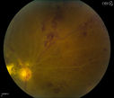
Combined Artery and Vein Occlusion431 viewsPatient comes in with decreased vision in both eyes. VA 20/200-OD, 20/60-OS. Fundus exam shows great amount of macular edema due to artery and vein occlusions. There is some neovascularization on the optic nerve in the right eye. Patient was treated with Eylea Injection in the left eye today and will return for Eylea Injection in the right eye.Jan 23, 2013
|
|
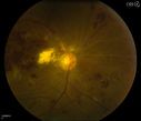
Combined Artery and Vein Occlusion407 viewsPatient comes in with decreased vision in both eyes. VA 20/200-OD, 20/60-OS. Fundus exam shows great amount of macular edema due to artery and vein occlusions. There is some neovascularization on the optic nerve in the right eye. Patient was treated with Eylea Injection in the left eye today and will return for Eylea Injection in the right eye.Jan 23, 2013
|
|
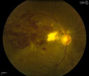
Combined Artery and Vein Occlusion483 viewsPatient comes in with decreased vision in both eyes. VA 20/200-OD, 20/60-OS. Fundus exam shows great amount of macular edema due to artery and vein occlusions. There is some neovascularization on the optic nerve in the right eye. Patient was treated with Eylea Injection in the left eye today and will return for Eylea Injection in the right eye.Jan 23, 2013
|
|
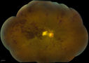
Combined Artery and Vein Occlusion491 viewsPatient comes in with decreased vision in both eyes. VA 20/200-OD, 20/60-OS. Fundus exam shows great amount of macular edema due to artery and vein occlusions. There is some neovascularization on the optic nerve in the right eye. Patient was treated with Eylea Injection in the left eye today and will return for Eylea Injection in the right eye.Jan 23, 2013
|
|
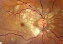
CRAO488 viewsCRAO Color MontageJan 11, 2013
|
|
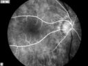
CRAO408 viewsCRAO in OD Tranisit Phase of FAJan 11, 2013
|
|

CRAO458 viewsCRAO in OD Tranisit Phase of FAJan 11, 2013
|
|
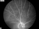
CRAO406 viewsCRAO in OD Late Phase of FAJan 11, 2013
|
|

CRAO465 viewsCRAO in OD of a 37 yr old Male, Transit PhaseJan 08, 2013
|
|

BRVO437 views50 yr old Female with a BRVO and ME in OSJan 08, 2013
|
|

Late674 viewsCorresponding FA for Cilioretinal artery occlusion with early venous stasis retinopathy Nov 06, 2012
|
|

48.2 seconds560 viewsCorresponding FA for Cilioretinal artery occlusion with early venous stasis retinopathy Nov 06, 2012
|
|

23.7 seconds414 viewsCorresponding FA for Cilioretinal artery occlusion with early venous stasis retinopathy Nov 06, 2012
|
|

14.5 seconds1055 viewsCorresponding FA for Cilioretinal artery occlusion with early venous stasis retinopathy Nov 06, 2012
|
|
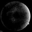
Corresponding FA504 viewsCorresponding FA for Cilioretinal artery occlusion with early venous stasis retinopathy - earlyNov 06, 2012
|
|

Central Retinal Vein Occlusion - Severe Optic Nerve Edema1054 views15 yr old male presents with sudden onset of painless decreased vision in right eye x 6 days gradually worsening. No history of Trauma OD 20/30 OS 20/25, Diagnosis CRVO with ? Papillophlebitis.Nov 06, 2012
|
|
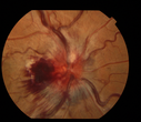
662 views15 yrs old male presents with sudden onset of painless decreased vision in right eye x 6 days gradually worsening. No history of Trauma OD 20/30 OS 20/25, Diagnosis CRVO with ? PapilophlebitisNov 06, 2012
|
|

Cilioretinal artery occlusion697 viewsCilioretinal artery occlusion with early venous stasis retinopathyNov 05, 2012
|
|
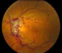
Central Retinal Vein Occlusion981 viewsNov 01, 2012
|
|

Central Retinal Vein Occlusion592 viewsNov 01, 2012
|
|

Central Retinal Artery Occlusion with Cilioretinal Sparing - Acute - Line scan good eye986 views 77-year-old man was doing fine until midnight of last night when he noticed sudden severe vision loss in the left eye. VISUAL ACUITY: OD 20/20, OS 2/200.Sep 05, 2012
|
|

Central Retinal Artery Occlusion with Cilioretinal Sparing - Acute - SD-OCT Line Scan Inner retinal edema1522 views 77-year-old man was doing fine until midnight of last night when he noticed sudden severe vision loss in the left eye. VISUAL ACUITY: OD 20/20, OS 2/200.Sep 05, 2012
|
|
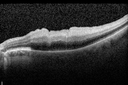
Central Retinal Artery Occlusion with Cilioretinal Sparing - Acute - SD-OCT Line Scan Inner retinal edema1174 views 77-year-old man was doing fine until midnight of last night when he noticed sudden severe vision loss in the left eye. VISUAL ACUITY: OD 20/20, OS 2/200.Sep 05, 2012
|
|
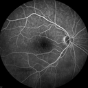
Central Retinal Artery Occlusion with Cilioretinal Sparing - Acute775 views 77-year-old man was doing fine until midnight of last night when he noticed sudden severe vision loss in the left eye. VISUAL ACUITY: OD 20/20, OS 2/200.Sep 05, 2012
|
|

Central Retinal Artery Occlusion with Cilioretinal Sparing - Acute688 views 77-year-old man was doing fine until midnight of last night when he noticed sudden severe vision loss in the left eye. VISUAL ACUITY: OD 20/20, OS 2/200.Sep 05, 2012
|
|
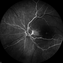
Central Retinal Artery Occlusion with Cilioretinal Sparing - Acute - FA Very Slow Filling CRA814 views 77-year-old man was doing fine until midnight of last night when he noticed sudden severe vision loss in the left eye. VISUAL ACUITY: OD 20/20, OS 2/200.Sep 05, 2012
|
|

Central Retinal Artery Occlusion with Cilioretinal Sparing - Acute - FA Very Slow Filling CRA829 views 77-year-old man was doing fine until midnight of last night when he noticed sudden severe vision loss in the left eye. VISUAL ACUITY: OD 20/20, OS 2/200.Sep 05, 2012
|
|

Central Retinal Artery Occlusion with Cilioretinal Sparing - Acute - FA Very Slow Filling CRA910 views 77-year-old man was doing fine until midnight of last night when he noticed sudden severe vision loss in the left eye. VISUAL ACUITY: OD 20/20, OS 2/200.Sep 05, 2012
|
|
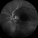
Central Retinal Artery Occlusion with Cilioretinal Sparing - Acute - FA Very Slow Filling CRA909 views 77-year-old man was doing fine until midnight of last night when he noticed sudden severe vision loss in the left eye. VISUAL ACUITY: OD 20/20, OS 2/200.Sep 05, 2012
|
|
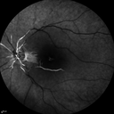
Central Retinal Artery Occlusion with Cilioretinal Sparing - Acute - FA Very Slow Filling CRA955 views 77-year-old man was doing fine until midnight of last night when he noticed sudden severe vision loss in the left eye. VISUAL ACUITY: OD 20/20, OS 2/200.Sep 05, 2012
|
|

Central Retinal Artery Occlusion with Cilioretinal Sparing - Acute - FA Very Slow Filling CRA1036 views 77-year-old man was doing fine until midnight of last night when he noticed sudden severe vision loss in the left eye. VISUAL ACUITY: OD 20/20, OS 2/200.Sep 05, 2012
|
|

Central Retinal Artery Occlusion with Cilioretinal Sparing - Acute916 views 77-year-old man was doing fine until midnight of last night when he noticed sudden severe vision loss in the left eye. VISUAL ACUITY: OD 20/20, OS 2/200.Sep 05, 2012
|
|

Central Retinal Artery Occlusion with Cilioretinal Sparing - Acute1207 views 77-year-old man was doing fine until midnight of last night when he noticed sudden severe vision loss in the left eye. VISUAL ACUITY: OD 20/20, OS 2/200.Sep 05, 2012
|
|
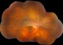
Montage of Hemi-Central Retinal Artery Occlusion723 viewsJul 06, 2012
|
|

Montage of Hemi-Central Retinal Artery Occlusion694 viewsJul 06, 2012
|
|

Classic BRVO884 viewsClassic branch retinal vein occlusionMay 08, 2012
|
|

Capillary Pruning570 viewsCapillary drop out secondary to proliferative diabetic retinopathyMay 08, 2012
|
|

Vasular Anomalies503 viewsThis patient shows vasculopathy seen after an old vein occlusion.May 08, 2012
|
|

648 viewsMay 07, 2012
|
|

747 viewsMay 07, 2012
|
|

774 viewsMay 07, 2012
|
|

CRVO399 views58 year old male with present CRVO OU. Visual Acuity 20/30 OD 20/200 OS. Pt. recently underwent surgery for cancer of the bladder and has other blood and hemoglobin issues. Pt sent for further blood work and see a neurologist. Apr 25, 2012
|
|

CRVO422 views58 year old male with present CRVO OU. Visual Acuity 20/30 OD 20/200 OS. Pt. recently underwent surgery for cancer of the bladder and has other blood and hemoglobin issues. Pt sent for further blood work and see a neurologist. Apr 25, 2012
|
|

CRVO514 views58 year old male with present CRVO OU. Visual Acuity 20/30 OD 20/200 OS. Pt. recently underwent surgery for cancer of the bladder and has other blood and hemoglobin issues. Pt sent for further blood work and see a neurologist. Apr 25, 2012
|
|
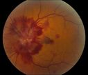
CRVO551 views58 year old male with present CRVO OU. Visual Acuity 20/30 OD 20/200 OS. Pt. recently underwent surgery for cancer of the bladder and has other blood and hemoglobin issues. Pt sent for further blood work and see a neurologist. Apr 25, 2012
|
|

Central Retinal Artery Occlusion1759 views60 year old male with LP vision due to extensive blood flow loss.Apr 25, 2012
|
|
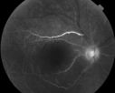
693 views60 year old male with LP vision due to extensive blood flow loss.Apr 25, 2012
|
|
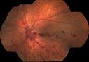
Hemi-retinal Vein Occlusion1598 views81 year old female seen in 2009 for a Hemi-retinal vein Occlusion OS with vision of 20/25. Apr 25, 2012
|
|

BRVO828 views42 year old female VA 20/25
Branch Retinal Vein Occlusion - Hot Spot on Vein at Occlusion SiteApr 19, 2012
|
|

Inferotemporal and Inferonasal BRAO1638 viewsBranch Retinal Artery Occlusions - MultipleApr 17, 2012
|
|

CVO438 viewsoct of a 27 year old male with crvo os VA 20/20-Jan 19, 2012
|
|
| 1798 files on 18 page(s) |
 |
|
|
|
|
|
14 | |
|
|
|


