Viimased lisandused - Kasutaja Tundmatu tegelane\ galerii
|

ARMD: Macular Degeneration with Subretinal Neovascular Membranevaatamisi: 649ARMD: Macular Degeneration with Subretinal Neovascular Membrane.
Upon presentation, this patient was found to have blots of subretinal hemorrhage extending into her FAZ, secondary to a subretinal neovascular membrane.
veebr 20, 2015
|
|

ERMvaatamisi: 631ERM: Epiretinal Membrane
Five years prior to presentation this patient was struck in the eye with a basketball, suffering a vitreal hemorrhage. He underwent vitrectomy and cataract surgery. He later developed a faint ERM. On recent presentation he complained of increased blurring and a floater in his O.D. Examination determined that the ERM had become more prominent.
veebr 20, 2015
|
|

Branch Retinal Artery Occlusion with Hollenhorst Plaquevaatamisi: 507This patient was found to have retinal edema along the superotemporal arcade secondary to Branch Retinal Artery Occlusion. A Hollenhorst plaque is visible at the first bifurcation of central artery.jaan 20, 2015
|
|

Calcification of the Choroidvaatamisi: 476CALCIFICATION OF THE CHOROID
Most patients with calcification of the choroid have it as an incidental finding. However, as a few have parathyroid abnormalities, this patient was screened for a serum calcium, that returned as normal.jaan 20, 2015
|
|

CHOROIDAL MELANOMA Stereo Pairvaatamisi: 450This patient was seen approximately five years ago with a suspicious choroidal nodule. The patient did not return for scheduled follow up appointments. When referred back to the practice, the nodule had erupted through Bruchs’ membrane to assume a brawny mushroom cloud configuration which is visible when the stereo images are merged.dets 22, 2014
|
|

CRAO WITH SPARING OF CILIORETINAL ARTERYvaatamisi: 591This patient was found to have retinal edema from a Central Retinal Artery Occlusion with sparing of the cilioretinal artery. There were no discernible Hollenhorst plaques dets 22, 2014
|
|

Ahmed Valvevaatamisi: 510AHMED VALVE: This 57 year old diabetic has end stage glaucoma. An Ahmed valve was inserted into her right anterior chamber to help control her IOP.nov 17, 2014
|
|

Choroiditisvaatamisi: 478CHOROIDITIS: This 46 year old female patient was found to have a peripheral sector of old chorioretinal inflammation accompanied by low grade oozing, intraretinal telangietatic vessels and hard exudates. There was no active bleeding or overlying vitritis.nov 17, 2014
|
|
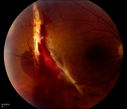
Choroidal Rupture with Subretinal Hemorrhagevaatamisi: 637This patient was punched in the eye with a fist, resulting in a choroidal rupture.okt 14, 2014
|
|

Actively Bleeding NVEvaatamisi: 528This male patient was referred for a diabetic examination. Upon dilated exam, he started to hemorrhage from a frond of NVE in the superior arcade. These six photographs follow the flow of the hemorrhage through the vitreous to the inferior arcade.okt 14, 2014
|
|

Papilledemavaatamisi: 464sept 04, 2014
|
|

Acute Multifocal Placoid Pigment Epitheliopathy (AMPPE) related scarringvaatamisi: 1321sept 04, 2014
|
|

Nonproliferative Diabetic Retinopathy with Circinate Hard Exudatesvaatamisi: 1515aug 14, 2014
|
|

Choriodal Rupture with Hemorrhagevaatamisi: 1126aug 14, 2014
|
|

August;Acute Ischemic Optic Neuropathy with Hemorrhagevaatamisi: 587aug 14, 2014
|
|
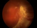
Diabetic Tractional Detachmentvaatamisi: 468juuni 06, 2014
|
|

Branch Retinal Artery Occlusion with Hollenhorst Plaquevaatamisi: 617juuni 06, 2014
|
|

Retinal Tear Surrounded By Laser Bordervaatamisi: 981mai 16, 2014
|
|

Papilledemavaatamisi: 441mai 16, 2014
|
|

Central Retinal Vein Occlusion with Disc Collateralsvaatamisi: 1153apr 04, 2014
|
|

Epiretinal Membrane with Flecks of Hemorrhagevaatamisi: 1239apr 01, 2014
|
|

Corneal Neovascularizationvaatamisi: 486apr 01, 2014
|
|

Idiopathic Intracranial Hypertension O.S.vaatamisi: 553märts 03, 2014
|
|
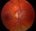
Idiopathic Intracranial Hypertension O.D.vaatamisi: 521märts 03, 2014
|
|

Epiretinal Membrane Stereo Pairvaatamisi: 544märts 03, 2014
|
|
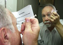
Intraocular Self injectionvaatamisi: 972The treatment of wet macular degeneration was revolutionized in late 2013 when Geneneron introduced its innovative and inexpensive take-home kit. Now, millions of patients around the world treat themselves on a PRN basis, using instructions written in 10 different languages.
April Fool!veebr 18, 2014
|
|
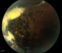
Traumatic Chorioretinal Scarvaatamisi: 752veebr 18, 2014
|
|

Choroidal Rupture Secondary to Ocular Traumavaatamisi: 945veebr 18, 2014
|
|

Molteno Drainage Tubevaatamisi: 1058jaan 07, 2014
|
|

Macular Hole Photograph with superimposed OCTvaatamisi: 430jaan 07, 2014
|
|

ERM with Pseudoholevaatamisi: 873dets 13, 2013
|
|

Central Retinal Venous Occlusion with Papilledema, Intraretinal Hemorrhage and Macular Edemavaatamisi: 943dets 13, 2013
|
|
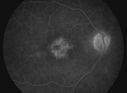
Pseudophakic Cystoid Macular Edema with Disc Hyperfluorescencevaatamisi: 867dets 13, 2013
|
|

Central Retinal Venous Occlusion with Papilledema, Irtraretinal Hemorrhage and Vascular Tortuosityvaatamisi: 812okt 22, 2013
|
|

Epiretinal Membranevaatamisi: 804okt 22, 2013
|
|
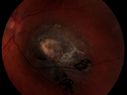
Elevated Nevusvaatamisi: 717okt 22, 2013
|
|

Diabetic Retinopathy with Hard Exudatesvaatamisi: 587sept 10, 2013
|
|

Stargardt's Dystrophy - Silent Choroid - Fluorscein Angiogramvaatamisi: 1048sept 10, 2013
|
|

Pseudophakic Cystoid Macular Edema FA late framevaatamisi: 1098aug 16, 2013
|
|

Epiretinal Membrane with Pseudoholevaatamisi: 1194aug 16, 2013
|
|

Retinoschisisvaatamisi: 930aug 16, 2013
|
|

Melanomavaatamisi: 781juuli 01, 2013
|
|

Retinal Detachmentvaatamisi: 385juuli 01, 2013
|
|

Acute Multifocal Placoid Pigment Epitheliopathyvaatamisi: 907juuni 28, 2013
|
|
Superior Retinal Detachmentvaatamisi: 408juuni 28, 2013
|
|
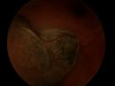
Melanomavaatamisi: 787mai 03, 2013
|
|

Ocular Trauma with Retinal Detachmentvaatamisi: 910Patient suffered blunt force trauma to the eye resulting total retinal detachment. The retina is visible in the pupillary space.mai 03, 2013
|
|

Iris Neovascularization secondary to Central Retinal Artery Occlusionvaatamisi: 1393apr 04, 2013
|
|

Eales Disease - Fluorescein Angiogramvaatamisi: 1462apr 04, 2013
|
|
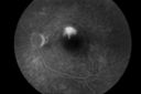
Central Serous Choroidopathy with Smoke Stack and Mushroom Cloud - Fluorescein Angiogramvaatamisi: 1285apr 04, 2013
|
|

Choroidal Rupture Secondary to Ocular Traumavaatamisi: 1170apr 04, 2013
|
|

Nonprolific Diabetic Retinopathy with Circinate Ringvaatamisi: 1013Nonprolific Diabetic Retinopathy with Circinate Ringmärts 12, 2013
|
|

Proliferative Diabetic Retinopathy with Tractional Retinal Detachmentvaatamisi: 1052Proliferative Diabetic Retinopathy with Tractional Retinal Detachmentmärts 12, 2013
|
|

Optic Nerve Pitvaatamisi: 1417Optic Nerve Pitmärts 12, 2013
|
|

Cavernous Hemangiomavaatamisi: 1084jaan 04, 2013
|
|
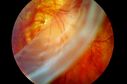
Giant Retinal Tearvaatamisi: 972jaan 04, 2013
|
|

Macular Hole with Subretinal Hemorrhage Secondary to Ocular Traumavaatamisi: 1161dets 07, 2012
|
|
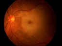
Cilio-Retinal Artery Occlusion with "Box Carring" and Cherry Red Spotvaatamisi: 1858Cilioretinal Artery Occlusion with Cherry Red Spotdets 07, 2012
|
|

Retinal Tearvaatamisi: 1097dets 07, 2012
|
|

Branch Retinal Artery Occlusion with Hollenhorst Plaquesvaatamisi: 585Branch Retinal Artery Occlusion with Hollenhorst Plaquesnov 06, 2012
|
|
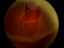
Retinal Detachment Secondary to Irregular Retinal Tearvaatamisi: 1041Retinal Detachment with Irregular Retinal Tearnov 06, 2012
|
|
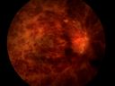
Central Retinal Vein Occlusionvaatamisi: 532Central Retinal Vein Occlusionnov 06, 2012
|
|
Choroidal Rupture Secondary to Ocular Traumavaatamisi: 548Choroidal Rupture Secondary to Ocular Traumanov 06, 2012
|
|
|
|
|


