Last additions - Chronic CSR - treated with Photodynamic Laser (Visudyne) PDT 66-year-old woman has central serous retinopathy in the right eye. She has had some retinal fluid there for some time. Her vision unfortunately is declining.
VISUAL ACUITY: OD 20/125, OS 20/25. IOP: 14 OD, 13 OS. There is 2+ nuclear sclerosis in both eyes.
EXTENDED OPHTHALMOSCOPY:
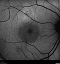
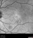
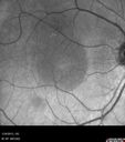
OD: Vertical C/D ratio is 0.2. There is a serous macular detachment centrally.
OS: Vertical C/D ratio is 0.2. The macula has some irregularities inferiorly.
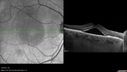
SPECTRALIS-SD-OCT SCAN: The OCT scan of the right eye shows persistent subretinal fluid and some increase in choroidal thickness. There is a pigment epithelial detachment just superior to the fovea. The left eye shows a normal macular contour.
COLOR PHOTOS: The color photos do show a serous macular detachment in the right eye. The left eye has a pigment spot inferior to the fovea.
FLUORESCEIN ANGIOGRAPHY: Fluorescein angiography shows a leaky spot superior to the fovea. In the left eye there is a leaky spot inferior to the fovea.
INDOCYANINE GREEN ANGIOGRAPHY: The indocyanine green angiogram in the right eye shows a leaky spot just superior to the fovea limited to the same area shown on the fluorescein angiogram. The left eye has a leaky spot inferior to the fovea.
IMPRESSION:
1. CENTRAL SEROUS RETINOPATHY – BOTH EYES
2. SEROUS MACULAR DETACHMENT – RIGHT EYE
DISCUSSION: I explained to the patient with photodynamic laser there is an excellent chance of drying the macula in the right eye and improving her vision.
|

Chronic Central Serous Retinopathy 1 month after Photodynamic Therapy (Visudyne)668 viewsSerous Retinal Detachment is Gone (Absorbed)Apr 25, 2013
|
|
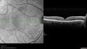
Chronic Central Serous Retinopathy 1 month after Photodynamic Therapy (Visudyne)621 viewsSerous Retinal Detachment is Gone (Absorbed)Apr 25, 2013
|
|

Chronic Central Serous Retinopathy - Indocyanine Green Angiogram 520 viewsGood EyeApr 25, 2013
|
|
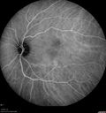
Chronic Central Serous Retinopathy - Indocyanine Green Angiogram 594 viewsGood EyeApr 25, 2013
|
|
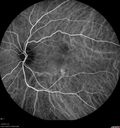
Chronic Central Serous Retinopathy - Indocyanine Green Angiogram 617 viewsGood EyeApr 25, 2013
|
|
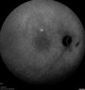
Chronic Central Serous Retinopathy - Indocyanine Green Angiogram 575 viewsApr 25, 2013
|
|
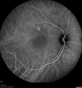
Chronic Central Serous Retinopathy - Indocyanine Green Angiogram 556 viewsApr 25, 2013
|
|
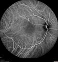
Chronic Central Serous Retinopathy - Indocyanine Green Angiogram 656 viewsApr 25, 2013
|
|
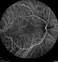
Chronic Central Serous Retinopathy - Indocyanine Green Angiogram 581 viewsApr 25, 2013
|
|
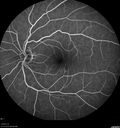
Chronic Central Serous Retinopathy - Fluorescein Angiogram540 viewsApr 25, 2013
|
|
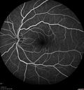
Chronic Central Serous Retinopathy - Fluorescein Angiogram560 viewsApr 25, 2013
|
|
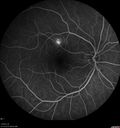
Chronic Central Serous Retinopathy - Fluorescein Angiogram656 viewsApr 25, 2013
|
|
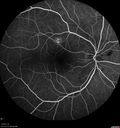
Chronic Central Serous Retinopathy - Fluorescein Angiogram559 viewsApr 25, 2013
|
|
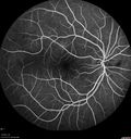
Chronic Central Serous Retinopathy - Fluorescein Angiogram544 viewsApr 25, 2013
|
|

Chronic Central Serous Retinopathy - Fluorescein Angiogram548 viewsApr 25, 2013
|
|

Chronic Central Serous Retinopathy - SD-OCT594 viewsApr 25, 2013
|
|
Chronic Central Serous Retinopathy - SD-OCT598 viewsApr 25, 2013
|
|

Chronic Central Serous Retinopathy - FAF538 viewsApr 25, 2013
|
|
Chronic Central Serous Retinopathy - FAF731 viewsApr 25, 2013
|
|
Chronic Central Serous Retinopathy - IR537 viewsApr 25, 2013
|
|
Chronic Central Serous Retinopathy - IR534 viewsApr 25, 2013
|
|
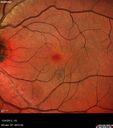
Chronic Central Serous Retinopathy - False Color598 viewsApr 25, 2013
|
|
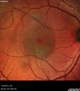
Chronic Central Serous Retinopathy - False Color530 viewsApr 25, 2013
|
|
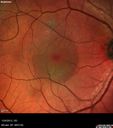
Chronic Central Serous Retinopathy - False Color608 viewsApr 25, 2013
|
|
|
|
66-year-old woman has central serous retinopathy in the right eye. She has had some retinal fluid there for some time. Her vision unfortunately is declining.
VISUAL ACUITY: OD 20/125, OS 20/25. IOP: 14 OD, 13 OS. There is 2+ nuclear sclerosis in both eyes.
EXTENDED OPHTHALMOSCOPY:
OD: Vertical C/D ratio is 0.2. There is a serous macular detachment centrally.
OS: Vertical C/D ratio is 0.2. The macula has some irregularities inferiorly.
SPECTRALIS-SD-OCT SCAN: The OCT scan of the right eye shows persistent subretinal fluid and some increase in choroidal thickness. There is a pigment epithelial detachment just superior to the fovea. The left eye shows a normal macular contour.
COLOR PHOTOS: The color photos do show a serous macular detachment in the right eye. The left eye has a pigment spot inferior to the fovea.
FLUORESCEIN ANGIOGRAPHY: Fluorescein angiography shows a leaky spot superior to the fovea. In the left eye there is a leaky spot inferior to the fovea.
INDOCYANINE GREEN ANGIOGRAPHY: The indocyanine green angiogram in the right eye shows a leaky spot just superior to the fovea limited to the same area shown on the fluorescein angiogram. The left eye has a leaky spot inferior to the fovea.
IMPRESSION:
1. CENTRAL SEROUS RETINOPATHY – BOTH EYES
2. SEROUS MACULAR DETACHMENT – RIGHT EYE
DISCUSSION: I explained to the patient with photodynamic laser there is an excellent chance of drying the macula in the right eye and improving her vision.


