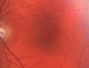
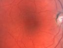
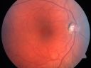
Last additions - Pigmented Macula Mother and Daughter 12-year-old was seen in the office on 4/29/2011. She was in for an evaluation. Her vision is perfectly fine. She sees fine in bright lights and she sees fine in dim lights. She was noticing when she was reading, the images seemed to jump around a little bit. She had a dilated macular exam and the macula look abnormal. Interestingly, her mother, who is with her at this visit, was told about a year ago that her maculae are abnormal too. Her mother’s vision is fine. In the family tree, there are no known retinal dystrophies. They are both healthy.
VISUAL ACUITY: Her vision is 20/20 in both eyes. IOP: OD 14, OS 15. The color vision is normal. The Amsler grid is normal. The pupil response is normal.
SLIT LAMP EXAM: Normal with clear lenses.
EXTENDED OPHTHALMOSCOPY:
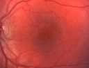
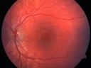
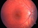
OD: Vertical C/D ratio is 0.2. There is no posterior vitreous separation. There is minimal hyperpigmentation to the macula with some granularity of the macular pigment.
OS: Vertical C/D ratio is 0.2. There is no posterior vitreous separation. There is some granularity to the macula. There is no evidence of any drusen though nor is there any pigment atrophy.
PHOTOGRAPHS: Photos confirm clinical findings.
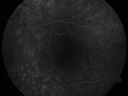
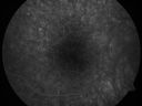
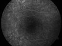
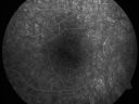
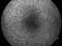
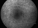
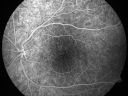
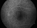
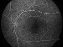
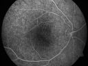
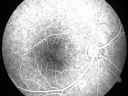
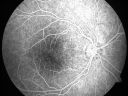
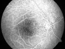
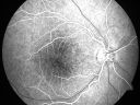
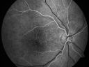
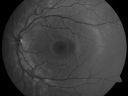
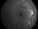
FLUORESCEIN ANGIOGRAPHY: The FA shows some irregular hyperfluorescence in the macula, which is mostly of a transmission than a blockage suggesting that there is some loss of pigmentation of the retinal pigment epithelium.
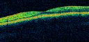
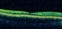
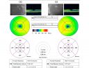
OCT SCAN: The OCT scans are normal.
I checked the mother’s eyes and they look exactly the same.
1. IRREGULAR MACULAR DEPIGMENTATION – BOTH EYES
2. POSSIBLE VARIANT OF CUTICULAR OR LAMINAR DRUSEN
DISCUSSION: I explained to the patient and her mother that the retinas are abnormal but they are exactly the same. The fact that the mother has good vision suggests that whatever she has is benign. The closest disease I can link it to is cuticular drusen, which is autosomal dominant but generally manifest with more pinpoint areas of depigmentation whereas their areas are more diffuse. They are both lightly pigmented but this is not consistent with albinism. There is not any foveal aplasia nor is there is any loss on the angiogram of the foveal avascular zone.
|
Pigmented Maculae - Mother and Daughter - Normal Visual Acuity774 viewsMothers macular photosApr 30, 2011
|
|
Pigmented Maculae - Mother and Daughter - Normal Visual Acuity657 viewsMothers macular photosApr 30, 2011
|
|

Pigmented Maculae - Mother and Daughter - Normal Visual Acuity636 viewsMothers macular photosApr 30, 2011
|
|
Pigmented Maculae - Mother and Daughter - Normal Visual Acuity579 viewsMothers macular photosApr 30, 2011
|
|
Pigmented Maculae - Mother and Daughter - Normal Visual Acuity580 views12-year-old had a dilated macular exam and the macula look abnormal. Interestingly, her mother, who is with her at this visit, was told about a year ago that her maculae are abnormal too. Her mother’s vision is fine. In the family tree, there are no known retinal dystrophies. They are both healthy.
VISUAL ACUITY: Her vision is 20/20 in both eyes.Apr 30, 2011
|
|

Pigmented Maculae - Mother and Daughter - Normal Visual Acuity568 views12-year-old had a dilated macular exam and the macula look abnormal. Interestingly, her mother, who is with her at this visit, was told about a year ago that her maculae are abnormal too. Her mother’s vision is fine. In the family tree, there are no known retinal dystrophies. They are both healthy.
VISUAL ACUITY: Her vision is 20/20 in both eyes.Apr 30, 2011
|
|

Pigmented Maculae - Mother and Daughter - Normal Visual Acuity536 views12-year-old had a dilated macular exam and the macula look abnormal. Interestingly, her mother, who is with her at this visit, was told about a year ago that her maculae are abnormal too. Her mother’s vision is fine. In the family tree, there are no known retinal dystrophies. They are both healthy.
VISUAL ACUITY: Her vision is 20/20 in both eyes.Apr 30, 2011
|
|
Pigmented Maculae - Mother and Daughter - Normal Visual Acuity551 views12-year-old had a dilated macular exam and the macula look abnormal. Interestingly, her mother, who is with her at this visit, was told about a year ago that her maculae are abnormal too. Her mother’s vision is fine. In the family tree, there are no known retinal dystrophies. They are both healthy.
VISUAL ACUITY: Her vision is 20/20 in both eyes.Apr 30, 2011
|
|
Pigmented Maculae - Mother and Daughter - Normal Visual Acuity571 views12-year-old had a dilated macular exam and the macula look abnormal. Interestingly, her mother, who is with her at this visit, was told about a year ago that her maculae are abnormal too. Her mother’s vision is fine. In the family tree, there are no known retinal dystrophies. They are both healthy.
VISUAL ACUITY: Her vision is 20/20 in both eyes.Apr 30, 2011
|
|
Pigmented Maculae - Mother and Daughter - Normal Visual Acuity565 views12-year-old had a dilated macular exam and the macula look abnormal. Interestingly, her mother, who is with her at this visit, was told about a year ago that her maculae are abnormal too. Her mother’s vision is fine. In the family tree, there are no known retinal dystrophies. They are both healthy.
VISUAL ACUITY: Her vision is 20/20 in both eyes.Apr 30, 2011
|
|
Pigmented Maculae - Mother and Daughter - Normal Visual Acuity565 views12-year-old had a dilated macular exam and the macula look abnormal. Interestingly, her mother, who is with her at this visit, was told about a year ago that her maculae are abnormal too. Her mother’s vision is fine. In the family tree, there are no known retinal dystrophies. They are both healthy.
VISUAL ACUITY: Her vision is 20/20 in both eyes.Apr 30, 2011
|
|
Pigmented Maculae - Mother and Daughter - Normal Visual Acuity548 views12-year-old had a dilated macular exam and the macula look abnormal. Interestingly, her mother, who is with her at this visit, was told about a year ago that her maculae are abnormal too. Her mother’s vision is fine. In the family tree, there are no known retinal dystrophies. They are both healthy.
VISUAL ACUITY: Her vision is 20/20 in both eyes.Apr 30, 2011
|
|
Pigmented Maculae - Mother and Daughter - Normal Visual Acuity511 views12-year-old had a dilated macular exam and the macula look abnormal. Interestingly, her mother, who is with her at this visit, was told about a year ago that her maculae are abnormal too. Her mother’s vision is fine. In the family tree, there are no known retinal dystrophies. They are both healthy.
VISUAL ACUITY: Her vision is 20/20 in both eyes.Apr 30, 2011
|
|
Pigmented Maculae - Mother and Daughter - Normal Visual Acuity538 views12-year-old had a dilated macular exam and the macula look abnormal. Interestingly, her mother, who is with her at this visit, was told about a year ago that her maculae are abnormal too. Her mother’s vision is fine. In the family tree, there are no known retinal dystrophies. They are both healthy.
VISUAL ACUITY: Her vision is 20/20 in both eyes.Apr 30, 2011
|
|
Pigmented Maculae - Mother and Daughter - Normal Visual Acuity555 views12-year-old had a dilated macular exam and the macula look abnormal. Interestingly, her mother, who is with her at this visit, was told about a year ago that her maculae are abnormal too. Her mother’s vision is fine. In the family tree, there are no known retinal dystrophies. They are both healthy.
VISUAL ACUITY: Her vision is 20/20 in both eyes.Apr 30, 2011
|
|
Pigmented Maculae - Mother and Daughter - Normal Visual Acuity510 views12-year-old had a dilated macular exam and the macula look abnormal. Interestingly, her mother, who is with her at this visit, was told about a year ago that her maculae are abnormal too. Her mother’s vision is fine. In the family tree, there are no known retinal dystrophies. They are both healthy.
VISUAL ACUITY: Her vision is 20/20 in both eyes.Apr 30, 2011
|
|
Pigmented Maculae - Mother and Daughter - Normal Visual Acuity530 views12-year-old had a dilated macular exam and the macula look abnormal. Interestingly, her mother, who is with her at this visit, was told about a year ago that her maculae are abnormal too. Her mother’s vision is fine. In the family tree, there are no known retinal dystrophies. They are both healthy.
VISUAL ACUITY: Her vision is 20/20 in both eyes.Apr 30, 2011
|
|
Pigmented Maculae - Mother and Daughter - Normal Visual Acuity706 views12-year-old had a dilated macular exam and the macula look abnormal. Interestingly, her mother, who is with her at this visit, was told about a year ago that her maculae are abnormal too. Her mother’s vision is fine. In the family tree, there are no known retinal dystrophies. They are both healthy.
VISUAL ACUITY: Her vision is 20/20 in both eyes.Apr 30, 2011
|
|
Pigmented Maculae - Mother and Daughter - Normal Visual Acuity617 views12-year-old had a dilated macular exam and the macula look abnormal. Interestingly, her mother, who is with her at this visit, was told about a year ago that her maculae are abnormal too. Her mother’s vision is fine. In the family tree, there are no known retinal dystrophies. They are both healthy.
VISUAL ACUITY: Her vision is 20/20 in both eyes.Apr 30, 2011
|
|
Pigmented Maculae - Mother and Daughter - Normal Visual Acuity629 views12-year-old had a dilated macular exam and the macula look abnormal. Interestingly, her mother, who is with her at this visit, was told about a year ago that her maculae are abnormal too. Her mother’s vision is fine. In the family tree, there are no known retinal dystrophies. They are both healthy.
VISUAL ACUITY: Her vision is 20/20 in both eyes.Apr 30, 2011
|
|
Pigmented Maculae - Mother and Daughter - Normal Visual Acuity626 views12-year-old had a dilated macular exam and the macula look abnormal. Interestingly, her mother, who is with her at this visit, was told about a year ago that her maculae are abnormal too. Her mother’s vision is fine. In the family tree, there are no known retinal dystrophies. They are both healthy.
VISUAL ACUITY: Her vision is 20/20 in both eyes.Apr 30, 2011
|
|
Pigmented Maculae - Mother and Daughter - Normal Visual Acuity615 views12-year-old had a dilated macular exam and the macula look abnormal. Interestingly, her mother, who is with her at this visit, was told about a year ago that her maculae are abnormal too. Her mother’s vision is fine. In the family tree, there are no known retinal dystrophies. They are both healthy.
VISUAL ACUITY: Her vision is 20/20 in both eyes.Apr 30, 2011
|
|
Pigmented Maculae - Mother and Daughter - Normal Visual Acuity594 views12-year-old had a dilated macular exam and the macula look abnormal. Interestingly, her mother, who is with her at this visit, was told about a year ago that her maculae are abnormal too. Her mother’s vision is fine. In the family tree, there are no known retinal dystrophies. They are both healthy.
VISUAL ACUITY: Her vision is 20/20 in both eyes.Apr 30, 2011
|
|
Pigmented Maculae - Mother and Daughter - Normal Visual Acuity597 views12-year-old had a dilated macular exam and the macula look abnormal. Interestingly, her mother, who is with her at this visit, was told about a year ago that her maculae are abnormal too. Her mother’s vision is fine. In the family tree, there are no known retinal dystrophies. They are both healthy.
VISUAL ACUITY: Her vision is 20/20 in both eyes.Apr 30, 2011
|
|
Pigmented Maculae - Mother and Daughter - Normal Visual Acuity624 views12-year-old had a dilated macular exam and the macula look abnormal. Interestingly, her mother, who is with her at this visit, was told about a year ago that her maculae are abnormal too. Her mother’s vision is fine. In the family tree, there are no known retinal dystrophies. They are both healthy.
VISUAL ACUITY: Her vision is 20/20 in both eyes.Apr 30, 2011
|
|
628 viewsApr 30, 2011
|
|
642 viewsApr 30, 2011
|
|

638 viewsApr 30, 2011
|
|
713 viewsApr 30, 2011
|
|
879 viewsApr 30, 2011
|
|
|
|
|
|
12-year-old was seen in the office on 4/29/2011. She was in for an evaluation. Her vision is perfectly fine. She sees fine in bright lights and she sees fine in dim lights. She was noticing when she was reading, the images seemed to jump around a little bit. She had a dilated macular exam and the macula look abnormal. Interestingly, her mother, who is with her at this visit, was told about a year ago that her maculae are abnormal too. Her mother’s vision is fine. In the family tree, there are no known retinal dystrophies. They are both healthy.
VISUAL ACUITY: Her vision is 20/20 in both eyes. IOP: OD 14, OS 15. The color vision is normal. The Amsler grid is normal. The pupil response is normal.
SLIT LAMP EXAM: Normal with clear lenses.
EXTENDED OPHTHALMOSCOPY:
OD: Vertical C/D ratio is 0.2. There is no posterior vitreous separation. There is minimal hyperpigmentation to the macula with some granularity of the macular pigment.
OS: Vertical C/D ratio is 0.2. There is no posterior vitreous separation. There is some granularity to the macula. There is no evidence of any drusen though nor is there any pigment atrophy.
PHOTOGRAPHS: Photos confirm clinical findings.
FLUORESCEIN ANGIOGRAPHY: The FA shows some irregular hyperfluorescence in the macula, which is mostly of a transmission than a blockage suggesting that there is some loss of pigmentation of the retinal pigment epithelium.
OCT SCAN: The OCT scans are normal.
I checked the mother’s eyes and they look exactly the same.
1. IRREGULAR MACULAR DEPIGMENTATION – BOTH EYES
2. POSSIBLE VARIANT OF CUTICULAR OR LAMINAR DRUSEN
DISCUSSION: I explained to the patient and her mother that the retinas are abnormal but they are exactly the same. The fact that the mother has good vision suggests that whatever she has is benign. The closest disease I can link it to is cuticular drusen, which is autosomal dominant but generally manifest with more pinpoint areas of depigmentation whereas their areas are more diffuse. They are both lightly pigmented but this is not consistent with albinism. There is not any foveal aplasia nor is there is any loss on the angiogram of the foveal avascular zone.


