Retina Gallery ~ Full Sized Retina Images

Library of Free, Non-Copyrighted Retina Images and Videos

Choisissez votre langue:

Derniers ajouts - MacTel with symptomatic blind spots - non-neovascular

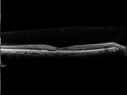
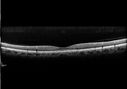
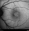
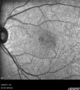
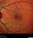
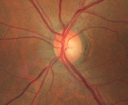
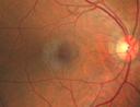
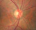
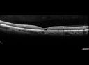
55 year old man The patient has been having trouble with his vision off and on for about 6 years. He was treated for glaucoma for a time but has been off the medications for about 3 years. He feels like something has changed in his left eye. When he goes to read he cannot read properly out of the left eye because letters disappear. Also the left eye will sometime hurt. VA 20/20 OD; 20/50 OS. Images show prominent temporal gray area and OCT scans show significant outer retinal atrophy especially in the left eye.

mactel_raco_020617_14.jpg
Macular Telangiectasis with Symptomatic Blind Spotsvu 402 fois55 year old man with non-neovascular MacTel. VA 20/20 right eye 20/50 left eye. The color images show gray areas temporally. The FAF shows decreased macular pigment. The OCT shows outer retinal atrophy.06 Août 2017
mactel_raco_020617_13.jpg
Macular Telangiectasis with Symptomatic Blind Spotsvu 383 fois55 year old man with non-neovascular MacTel. VA 20/20 right eye 20/50 left eye. The color images show gray areas temporally. The FAF shows decreased macular pigment. The OCT shows outer retinal atrophy.06 Août 2017
mactel_raco_020617_12.jpg
Macular Telangiectasis with Symptomatic Blind Spotsvu 378 fois55 year old man with non-neovascular MacTel. VA 20/20 right eye 20/50 left eye. The color images show gray areas temporally. The FAF shows decreased macular pigment. The OCT shows outer retinal atrophy.06 Août 2017
mactel_raco_020617_11.jpg
Macular Telangiectasis with Symptomatic Blind Spotsvu 374 fois55 year old man with non-neovascular MacTel. VA 20/20 right eye 20/50 left eye. The color images show gray areas temporally. The FAF shows decreased macular pigment. The OCT shows outer retinal atrophy.06 Août 2017
mactel_raco_020617_10.jpg
Macular Telangiectasis with Symptomatic Blind Spotsvu 377 fois55 year old man with non-neovascular MacTel. VA 20/20 right eye 20/50 left eye. The color images show gray areas temporally. The FAF shows decreased macular pigment. The OCT shows outer retinal atrophy.06 Août 2017
mactel_raco_020617_09.jpg
Macular Telangiectasis with Symptomatic Blind Spotsvu 381 fois55 year old man with non-neovascular MacTel. VA 20/20 right eye 20/50 left eye. The color images show gray areas temporally. The FAF shows decreased macular pigment. The OCT shows outer retinal atrophy.06 Août 2017
mactel_raco_020617_08.jpg
Macular Telangiectasis with Symptomatic Blind Spotsvu 444 fois55 year old man with non-neovascular MacTel. VA 20/20 right eye 20/50 left eye. The color images show gray areas temporally. The FAF shows decreased macular pigment. The OCT shows outer retinal atrophy.06 Août 2017
mactel_raco_020617_07.jpg
Macular Telangiectasis with Symptomatic Blind Spotsvu 402 fois55 year old man with non-neovascular MacTel. VA 20/20 right eye 20/50 left eye. The color images show gray areas temporally. The FAF shows decreased macular pigment. The OCT shows outer retinal atrophy.06 Août 2017
mactel_raco_020617_06.jpg
Macular Telangiectasis with Symptomatic Blind Spotsvu 437 fois55 year old man with non-neovascular MacTel. VA 20/20 right eye 20/50 left eye. The color images show gray areas temporally. The FAF shows decreased macular pigment. The OCT shows outer retinal atrophy.06 Août 2017
mactel_raco_020617_05.jpg
Macular Telangiectasis with Symptomatic Blind Spotsvu 534 fois55 year old man with non-neovascular MacTel. VA 20/20 right eye 20/50 left eye. The color images show gray areas temporally. The FAF shows decreased macular pigment. The OCT shows outer retinal atrophy.06 Août 2017
mactel_raco_020617_04.png
Macular Telangiectasis with Symptomatic Blind Spotsvu 363 fois55 year old man with non-neovascular MacTel. VA 20/20 right eye 20/50 left eye. The color images show gray areas temporally. The FAF shows decreased macular pigment. The OCT shows outer retinal atrophy.06 Août 2017
mactel_raco_020617_01.png
Macular Telangiectasis with Symptomatic Blind Spotsvu 401 fois55 year old man with non-neovascular MacTel. VA 20/20 right eye 20/50 left eye. The color images show gray areas temporally. The FAF shows decreased macular pigment. The OCT shows outer retinal atrophy.06 Août 2017
mactel_raco_020617_02.png
Macular Telangiectasis with Symptomatic Blind Spotsvu 414 fois55 year old man with non-neovascular MacTel. VA 20/20 right eye 20/50 left eye. The color images show gray areas temporally. The FAF shows decreased macular pigment. The OCT shows outer retinal atrophy.06 Août 2017
     
13 photos sur 1 page(s)

55 year old man The patient has been having trouble with his vision off and on for about 6 years. He was treated for glaucoma for a time but has been off the medications for about 3 years. He feels like something has changed in his left eye. When he goes to read he cannot read properly out of the left eye because letters disappear. Also the left eye will sometime hurt. VA 20/20 OD; 20/50 OS. Images show prominent temporal gray area and OCT scans show significant outer retinal atrophy especially in the left eye.