Retina Gallery ~ Full Sized Retina Images

Library of Free, Non-Copyrighted Retina Images and Videos

Wähle Sprache:


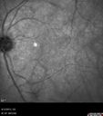
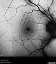
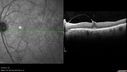
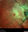
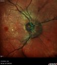
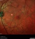
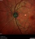
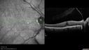
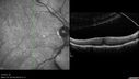
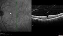
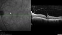
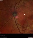
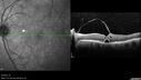
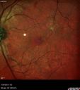
81 y.o woman with chronic macular pucker in the right eye who progressed from vitreo-macular traction to full thickness macular hole and then spontaneously closed macular hole. On her initial visit VA OD: cc20/32 PHNI NccJ1 OS: cc20/32 Anterior Right Eye Left Eye • Lens Centered PC IOL. Open PC. Centered PC IOL. 1+ PCO. OD has macular pucker and OS vitreomacular traction on initial visit
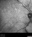
Abort_betu1_081213_01.jpg
Macular Hole Left Eye - progression from VMT - Stage II macular hole - Spontaneously Closed Macular Hole612 x angesehen00000
(0 Bewertungen)
Abort_betu1_081213_02.jpg
Macular Hole Left Eye - progression from VMT - Stage II macular hole - Spontaneously Closed Macular Hole548 x angesehen00000
(0 Bewertungen)
Abort_betu1_081213_03.jpg
Macular Hole Left Eye - progression from VMT - Stage II macular hole - Spontaneously Closed Macular Hole508 x angesehen00000
(0 Bewertungen)
Abort_betu1_081213_04.jpg
Macular Hole Left Eye - progression from VMT - Stage II macular hole - Spontaneously Closed Macular Hole534 x angesehen00000
(0 Bewertungen)
Abort_betu1_081213_05.jpg
Macular Hole Left Eye - progression from VMT - Stage II macular hole - Spontaneously Closed Macular Hole523 x angesehen00000
(0 Bewertungen)
Abort_betu1_081213_07.jpg
Macular Hole Left Eye - progression from VMT - Stage II macular hole - Spontaneously Closed Macular Hole538 x angesehenAsymptomatic VMT OS with 20/30 VA00000
(0 Bewertungen)
Abort_betu2_111113_08.jpg
Macular Hole Left Eye - progression from VMT - Stage II macular hole - Spontaneously Closed Macular Hole484 x angesehen00000
(0 Bewertungen)
Abort_betu2_111113_09.jpg
Macular Hole Left Eye - progression from VMT - Stage II macular hole - Spontaneously Closed Macular Hole540 x angesehenVision better left eye VMT has released the fovea (was 20/25)00000
(0 Bewertungen)
Abort_betu2_111113_10.jpg
Macular Hole Left Eye - progression from VMT - Stage II macular hole - Spontaneously Closed Macular Hole528 x angesehenVision better left eye VMT has released the fovea (was 20/25)00000
(0 Bewertungen)
Abort_betu3_021214_12.jpg
Macular Hole Left Eye - progression from VMT - Stage II macular hole - Spontaneously Closed Macular Hole534 x angesehenPucker False Color00000
(0 Bewertungen)
Abort_betu3_021214_13.jpg
Macular Hole Left Eye - progression from VMT - Stage II macular hole - Spontaneously Closed Macular Hole494 x angesehen00000
(0 Bewertungen)
Abort_betu3_021214_14.jpg
Macular Hole Left Eye - progression from VMT - Stage II macular hole - Spontaneously Closed Macular Hole522 x angesehenVision has dropped to 20/50 and there is a full thickness macular hole00000
(0 Bewertungen)
Abort_betu3_021214_15.jpg
Macular Hole Left Eye - progression from VMT - Stage II macular hole - Spontaneously Closed Macular Hole456 x angesehenVision has dropped to 20/50 and there is a full thickness macular hole00000
(0 Bewertungen)
Abort_betu3_021214_16.jpg
Macular Hole Left Eye - progression from VMT - Stage II macular hole - Spontaneously Closed Macular Hole495 x angesehen00000
(0 Bewertungen)
Abort_betu3_021214_17.jpg
Macular Hole Left Eye - progression from VMT - Stage II macular hole - Spontaneously Closed Macular Hole468 x angesehenPucker in fellow eye is stable00000
(0 Bewertungen)
Abort_betu3_021214_18.jpg
Macular Hole Left Eye - progression from VMT - Stage II macular hole - Spontaneously Closed Macular Hole489 x angesehenVision has dropped to 20/50 and there is a full thickness macular hole00000
(0 Bewertungen)
Abort_betu3_021214_19.jpg
Macular Hole Left Eye - progression from VMT - Stage II macular hole - Spontaneously Closed Macular Hole611 x angesehenVision has dropped to 20/50 and there is a full thickness macular hole00000
(0 Bewertungen)
Abort_betu4_031314_22.jpg
Macular Hole Left Eye - progression from VMT - Stage II macular hole - Spontaneously Closed Macular Hole538 x angesehen00000
(0 Bewertungen)
Abort_betu4_031314_23.jpg
Macular Hole Left Eye - progression from VMT - Stage II macular hole - Spontaneously Closed Macular Hole626 x angesehen00000
(0 Bewertungen)
Abort_betu4_031314_24.jpg
Macular Hole Left Eye - progression from VMT - Stage II macular hole - Spontaneously Closed Macular Hole613 x angesehen00000
(0 Bewertungen)
Abort_betu4_031314_25.jpg
Macular Hole Left Eye - progression from VMT - Stage II macular hole - Spontaneously Closed Macular Hole584 x angesehenMacular hole is closing and vision has improved to 20/4000000
(0 Bewertungen)
Abort_betu4_031314_26.jpg
Macular Hole Left Eye - progression from VMT - Stage II macular hole - Spontaneously Closed Macular Hole555 x angesehenMacular hole is closing and vision has improved to 20/4000000
(0 Bewertungen)
Abort_betu5_042414_28.jpg
Macular Hole Left Eye - progression from VMT - Stage II macular hole - Spontaneously Closed Macular Hole537 x angesehenMacular hole is closing and vision has improved to 20/4000000
(0 Bewertungen)
Abort_betu5_042414_29.jpg
Macular Hole Left Eye - progression from VMT - Stage II macular hole - Spontaneously Closed Macular Hole567 x angesehenMacular hole is closing and vision has improved to 20/4000000
(0 Bewertungen)
Abort_betu6_072414_31.jpg
Macular Hole Left Eye - progression from VMT - Stage II macular hole - Spontaneously Closed Macular Hole668 x angesehenVision has improved to 20/40 and the hole is closed00000
(0 Bewertungen)
Abort_betu6_072414_32.jpg
Macular Hole Left Eye - progression from VMT - Stage II macular hole - Spontaneously Closed Macular Hole633 x angesehenVision has improved to 20/40 and the hole is closed00000
(0 Bewertungen)
Abort_betu6_072414_33.jpg
Macular Hole Left Eye - progression from VMT - Stage II macular hole - Spontaneously Closed Macular Hole577 x angesehenVision has improved to 20/40 and the hole is closed00000
(0 Bewertungen)
 
27 Dateien auf 1 Seite(n)

81 y.o woman with chronic macular pucker in the right eye who progressed from vitreo-macular traction to full thickness macular hole and then spontaneously closed macular hole. On her initial visit VA OD: cc20/32 PHNI NccJ1 OS: cc20/32 Anterior Right Eye Left Eye • Lens Centered PC IOL. Open PC. Centered PC IOL. 1+ PCO. OD has macular pucker and OS vitreomacular traction on initial visit