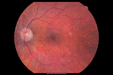
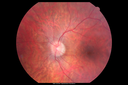
28 y.o man with more flashes and floaters in both eyes starting a few months ago. There are a few big floaters and a lot of little ones in both eyes. The flashes he is seeing used to start when he was exposed to bright light. The last few weeks the flashes have been constant and all over. They come and go. They can form in an arc below fixation. It used to be more like a ring. Now it is more like an L shape more to the left and below center. The flashes do not come with headaches. He has occipital migraines. The lights have been more constant than they used to be. There is a lot of junk in his eye. The patient has less than normal night vision and side vision is a little bit hazy VA OD: cc20/25 OS: cc20/25 IOP: TP OD: 15 OS: 15 Findings OD: Lens: Clear. Findings OS: Lens: Clear. Posterior Segment: Findings OD: Nerve: Pink. Sharp. CDR-V 0.1. Vitreous: Posterior Vitreous Detachment. Retinal Vessels: Attenuated Vessels. Macula: No Hemorrhage. No Subretinal Fluid. No Edema. Periphery: Attached 360 Degrees. Paving Stone Degeneration (Inferior). Findings OS: Nerve: Pink. Sharp. CDR-V 0.1. Vitreous: Posterior Vitreous Detachment. Retinal Vessels: Attenuated Vessels. Macula: No Hemorrhage. No Subretinal Fluid. No Edema. Periphery: Attached 360 Degrees. Paving Stone Degeneration (Inferior). OCT Macula: Findings OD: Outer Retinal Atrophy (Extrafoveal). Findings OS: Outer Retinal Atrophy (Extrafoveal). Fundus Photos (Infrared): Findings OD: Findings OS: Fundus Photos (Autofluorescence): Findings OD: White ring around the macula typical for RP. Findings OS: White ring around the macula typical for RP. Imp/Plan: 1. Retinitis Pigmentosa OU (sine pigmente). I explained to the patient that his findings are typical for RP. I'm going to see about getting him genetically tested either through UF or directly through Carver Lab. |
|||||||
|
|
|
|
||||
|
|
|
|
||||
|
|
|
|
||||
|
|
||||||
|
|||||||
28 y.o man with more flashes and floaters in both eyes starting a few months ago. There are a few big floaters and a lot of little ones in both eyes. The flashes he is seeing used to start when he was exposed to bright light. The last few weeks the flashes have been constant and all over. They come and go. They can form in an arc below fixation. It used to be more like a ring. Now it is more like an L shape more to the left and below center. The flashes do not come with headaches. He has occipital migraines. The lights have been more constant than they used to be. There is a lot of junk in his eye. The patient has less than normal night vision and side vision is a little bit hazy VA OD: cc20/25 OS: cc20/25 IOP: TP OD: 15 OS: 15 Findings OD: Lens: Clear. Findings OS: Lens: Clear. Posterior Segment: Findings OD: Nerve: Pink. Sharp. CDR-V 0.1. Vitreous: Posterior Vitreous Detachment. Retinal Vessels: Attenuated Vessels. Macula: No Hemorrhage. No Subretinal Fluid. No Edema. Periphery: Attached 360 Degrees. Paving Stone Degeneration (Inferior). Findings OS: Nerve: Pink. Sharp. CDR-V 0.1. Vitreous: Posterior Vitreous Detachment. Retinal Vessels: Attenuated Vessels. Macula: No Hemorrhage. No Subretinal Fluid. No Edema. Periphery: Attached 360 Degrees. Paving Stone Degeneration (Inferior). OCT Macula: Findings OD: Outer Retinal Atrophy (Extrafoveal). Findings OS: Outer Retinal Atrophy (Extrafoveal). Fundus Photos (Infrared): Findings OD: Findings OS: Fundus Photos (Autofluorescence): Findings OD: White ring around the macula typical for RP. Findings OS: White ring around the macula typical for RP. Imp/Plan: 1. Retinitis Pigmentosa OU (sine pigmente). I explained to the patient that his findings are typical for RP. I'm going to see about getting him genetically tested either through UF or directly through Carver Lab.