CC: Decreased Vision OS>OD. Activity Affected: reading. Context Onset/Aggravation: on Amsler grid. Other: lines seemed straighter yesterday. Pertinent Negatives: No Floaters. Course: variable, some
days better than others. Extended HPI: Pt reports that the left eye is bothering him "a lot" for the past few weeks. He characterizes the vision loss as "distortion."
VA OD: cc20/40 OS: cc20/50 PHNI IOP: TP OD: 18 OS: 12
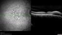
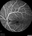
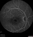
DISCUSSION: Macular Edema OS > OD: The macular edema involves
the center of the macula in the left eye and is likely contributing to the patient’s vision
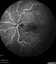
loss. The etiology of the macular edema is not clear. The right eye has good vision and
no leakage at all on angiography so I will obsere the right side while I treat the left.
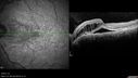
Serous Macular Detachment OS: The subretinal fluid in the macula is probably
responsible for some of the patients vision loss. The etiology of the edema in the left eye is unclear and vexing. There is no
subretinal leakage on exam. I am going to try topical anti-inflammatories for a month
and see if he improves before considering more aggressive intravitreal therapy. There is
also a chance that he has a hypertensive etiology to his problem and I recommended
optimizing his systemic status.
|
|
|
|
|
|
|
|
|
|
|
|
|
|
|
|
|
|
CC: Decreased Vision OS>OD. Activity Affected: reading. Context Onset/Aggravation: on Amsler grid. Other: lines seemed straighter yesterday. Pertinent Negatives: No Floaters. Course: variable, some
days better than others. Extended HPI: Pt reports that the left eye is bothering him "a lot" for the past few weeks. He characterizes the vision loss as "distortion."
VA OD: cc20/40 OS: cc20/50 PHNI IOP: TP OD: 18 OS: 12
DISCUSSION: Macular Edema OS > OD: The macular edema involves
the center of the macula in the left eye and is likely contributing to the patient’s vision
loss. The etiology of the macular edema is not clear. The right eye has good vision and
no leakage at all on angiography so I will obsere the right side while I treat the left.
Serous Macular Detachment OS: The subretinal fluid in the macula is probably
responsible for some of the patients vision loss. The etiology of the edema in the left eye is unclear and vexing. There is no
subretinal leakage on exam. I am going to try topical anti-inflammatories for a month
and see if he improves before considering more aggressive intravitreal therapy. There is
also a chance that he has a hypertensive etiology to his problem and I recommended
optimizing his systemic status.