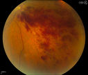
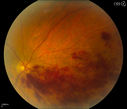
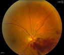
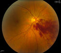
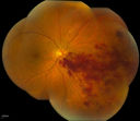
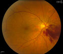
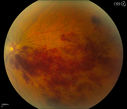
Branch Retinal Vein Occlusionvu 500 foisPatient's VA was 20/40, right eye. Fundus exam shows branch retinal vein occlusion nasally to the optic nerve in the right eye. (0 votes)
Branch Retinal Vein Occlusionvu 494 foisPatient's VA was 20/40, right eye. Fundus exam shows branch retinal vein occlusion nasally to the optic nerve in the right eye. (0 votes)
Branch Retinal Vein Occlusionvu 429 foisPatient's VA was 20/40, right eye. Fundus exam shows branch retinal vein occlusion nasally to the optic nerve in the right eye. (0 votes)
Branch Retinal Vein Occlusionvu 404 foisPatient's VA was 20/40, right eye. Fundus exam shows branch retinal vein occlusion nasally to the optic nerve in the right eye. (0 votes)
Branch Retinal Vein Occlusionvu 478 foisPatient's VA was 20/40, right eye. Fundus exam shows branch retinal vein occlusion nasally to the optic nerve in the right eye. (0 votes)
Branch Retinal Vein Occlusionvu 468 foisPatient's VA was 20/40, right eye. Fundus exam shows branch retinal vein occlusion nasally to the optic nerve in the right eye. (0 votes)
Branch Retinal Vein Occlusionvu 431 foisPatient's VA was 20/40, right eye. Fundus exam shows branch retinal vein occlusion nasally to the optic nerve in the right eye. (0 votes)
Branch Retinal Vein Occlusionvu 463 foisPatient's VA was 20/40, right eye. Fundus exam shows branch retinal vein occlusion nasally to the optic nerve in the right eye. (0 votes)