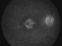
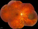
024 December 2013 - Best Image Finalist - Winner - Dislocated IOL - Jason Calhoun - Photographer - Mayo Clinic Jacksonville
Pseudophakic Cystoid Macular Edema with Disc Hyperfluorescencevu 874 fois (1 votes)
Central Retinal Venous Occlusion with Papilledema, Intraretinal Hemorrhage and Macular Edemavu 949 fois (1 votes)
ERM with Pseudoholevu 881 fois (1 votes)
Optic Nerve Tumorvu 1037 foisYoung male patient with a tumor present on the optic nerve in the right eye. VA is hand motion in the right eye (1 votes)
Subluxed IOL with TIDvu 1195 foisPatient comes in with blurred vision in the left eye. Slit lamp exam shows dislocated IOL, inferior, left eye. PXF material on the implant inferiorly. (2 votes)
Giant Papillary Conjunctivitisvu 1078 foisPatient wears soft contact lenses complained of irritation when the SCL would move. Inverted eyelid in both eyes and there was papillary +2 underneath the eyelid. (2 votes)
Prominent Corneal Nervesvu 1245 foisSlit lamp exam shows prominent corneal nerves present at 8-10-o'clock and 4-6-o'clock. Baseline work to rule out Fuch's dystrophy and recurrent corneal erosion. (2 votes)
Diabetic Retinopathy with CMEvu 1289 foisSecond opinion for diabetic retinopathy. VA is 20/30, right eye, 20/50, left eye. Patient has mild swelling in the macula of the right eye. No evidence of CME but does have a moderate cataract in the left eye. (2 votes)