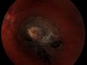
022 October 2013 - Best Image Finalist - Winner - Marissa Scherf - Unilateral Hemorrhagic Retinopathy - New England Retina Associates
Unilateral Hemorrhagic Retinopathy1402 x angesehenA 53 yr-old male was referred for a Macular Aneurysm. The FA should no apparent Aneurysm, nor did it show any evident VO or Edema. The patient is in the Navy but hadn't gone on any deep sea dives or any air flights.
The cause of the hemorrhages is unknown, the following ARVO abstract from 2012 identifies similar cases found in females.
Clinical Features Of Unilateral Hemorrhagic Retinopathy: A New Retinal Entity?
Presentation Start/End Time: Wednesday, May 09, 2012, 1:45 PM - 3:30 PM
Session 466 (4 Bewertungen)
Rhegmatogenous Retinal Detachment872 x angesehenPatient comes in for second opinion on RD, right eye. VA is NLP in the right eye. Fundus photography shows inferior retinal detachment with holes and subretinal fibrosis. No further surgery is suggested at this time. (1 Bewertungen)
Elevated Nevus726 x angesehen (0 Bewertungen)
Epiretinal Membrane808 x angesehen (0 Bewertungen)
Central Retinal Venous Occlusion with Papilledema, Irtraretinal Hemorrhage and Vascular Tortuosity816 x angesehen (0 Bewertungen)
Papilledema unspecified 1096 x angesehen (0 Bewertungen)
Embolic CRAO color847 x angesehen (0 Bewertungen)
Myopic Degeneration with RPE Loss1168 x angesehenPatient comes in for follow up for glaucoma. VA is 20/30 in both eyes. Fundus photography shows the RPE loss due to myopic degeneration, nasally, inferiority in the left eye. (1 Bewertungen)