014 February 2013 Best Image Award Finalists - Choroidal Melanoma with Radiation Retinopathy - Jason Calhoun - Photographer - Mayo Clinic Jacksonville
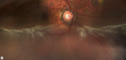
Choroidal Melanoma with Radiation Retinopathyvu 1272 foisPatient comes with follow up on Choroidal Melanoma, Right Eye that was treated back in June of 2009 with Radioactive Implant. Vein Occlusion is also present with VA - Hand Motion. Hemorrhages visible with hard exudates from the Radiation Retinopathy. (2 votes)
Nonproliferative Diabetic Retinopathyvu 8330 fois65-year old female with diabetes. Has had cataract surgery in the left eye with VA 20/25. She has had laser in the past. Fundus examination shows microaneurysms with retinal hemorrahages and exudates in the left eye. (1 votes)
RDvu 819 foisInferior RD (0 votes)
RD with PVRvu 1462 foisRD with Proliferative Vitreoretinopathy (1 votes)
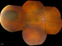
Hemangiomavu 991 foisFP Composite of Hemangioma in OD (0 votes)
Uveal Choroidal Melanomavu 1001 foisPatient comes in for evaluation on a Choroidal Melanoma in the right eye. VA was 20/25 in both eyes. The melanoma is in the temporal aspect of the right eye. It measured at 0.7mm elevated after doing a BSCAN Ultrasound. (1 votes)
Gonioscopy, Blood in the Anterior Chamber from Hyphemavu 924 foisPatient comes in with blunt trauma to the right eye due to a BB gun incident. Patient was present with a hyphema at 8-o'clock about 1mm thick. Gonioscopy photos were then taken to show blood from the hyphema entered into the anterior chamber. Patient had no angle recession in the right eye. (1 votes)
Corneal Abrasion with Foreign Body Present vu 961 foisER patient comes in with corneal abrasion in the left eye which was getting worst. VA 20/25. Slit lamp exam showed corneal abrasion superiorly at 2-o'clock. Flipped eyelid and foreign body appeared on the upper tarsal plate. Foreign body was removed. (1 votes)
Corneal Abrasion with Foreign Body Present vu 1113 foisER patient comes in with corneal abrasion in the left eye which was getting worst. VA 20/25. Slit lamp exam showed corneal abrasion superiorly at 2-o'clock. Flipped eyelid and foreign body appeared on the upper tarsal plate. Foreign body was removed. (1 votes)