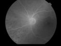
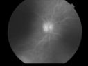
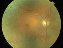
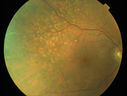
80 Year Old Female Patient came in 1 year ago with a retinal whitening in her eye and uveitis. Her uveitis work-up was negative, and because of the retinal whitening and the uveitis, I was concerned about a possible occult acute retinal necrosis case. I put her on Valtrex, but the eyes did not get any better and the retinal whitening continued. Then later, I became concerned about a possible primary ocular lymphoma. She had an extensive cancer evaluation that was all negative. She has had MRI scans and ultimately I did a vitrectomy on both eyes. I did the right eye on May 30th of 2012 and flow cytometry and cytology was negative. That eye cleared nicely, but then, because of increasing uveitis in the left eye, I did a vitrectomy in the left eye on December 19th of 2012. That sample I sent to the lab in Miami and that result was similarly negative. I am going to send you all of the results so you have them when you see her. At this point, it has been unclear whether she had uveitis, infectious retinitis, or possibly primary ocular lymphoma.
When I saw her in my office on November 27th of 2012, she had a white subretinal mass growing in the right eye and the left eye had vitreous cells. After the vitrectomy in the left eye, I started rechecking both eyes and the white subretinal mass in the right eye had doubled in size. The right eye now looks very much like it has primary ocular lymphoma.
I appreciate you seeing her and, if possible, performing some sort of a biopsy of that mass, which has now grown to the point where I think a fine-needle aspiration biopsy would be possible to help secure the diagnosis.
++++++++++++ Visit 2/5/2013. 80-year-old woman has possible ocular lymphoma. She has had two vitreous biopsies, one in one eye and one in the other eye, and the tests have been negative. Recently she is manifesting increasing anemia. She also has glaucoma. She is taking Pred Forte in both eyes and Combigan in both eyes.
VISUAL ACUITY: Vision OD is 20/40, OS is 20/160. IOP: OD 22, OS 24. The eyes are quiet with a posterior chamber intraocular lens in good position in both eyes.
EXTENDED OPHTHALMOSCOPY:
OD: Vertical C/D ratio is 0.4. The white mass in the macula is much bigger than it was when I saw her in November. It is growing on the superior half of the macula, as well as just nasal to the optic nerve. It has about doubled in size. There are no significant vitreous cells.
OS: Vertical C/D ratio is 0.4. There are patchy retinal hemorrhages.
FUNDUS PHOTOGRAPHY – COLOR AND AUTOFLUORESCENCE: Color and autofluorescence photographs do show the mass to be much bigger.
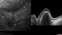
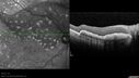
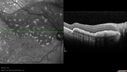
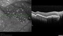
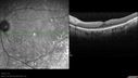
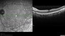
SPECTRALIS SD-OCT SCAN: The OCT scans over that area show the probable tumor to be about 1.5 mm high.
IMPRESSION:
1. PROBABLE OCULAR LYMPHOMA – BOTH EYES
DISCUSSION: I explained to the patient, given the growth of the lesion and the absence of inflammation around it, I think at this point it is likely ocular lymphoma.
++++++++++ VISIT - 1/4/2013. 80-year-old woman had a vitreous biopsy in the left eye with a fairly full vitrectomy back on December 19th, of 2012. Her vitreous was sent down to Miami where it was carefully analyzed in the pathology lab. The flow cytometry was normal. Her B cell testing was normal. The T cell testing did show two small peaks. I contacted the pathologist about that result and it is not uncommon in cerebrospinal fluid and vitreous samples, because there are so few cells, to see some peaks on the T cells. Generally that is not considered indicative of a T cell lymphoma. There is no such thing as a primary intraocular T cell lymphoma. The primary intraocular lymphomas are B cells and the findings overall were consistent with intraocular inflammation. She did have extensive blood testing by
Dr. Friedberg when this all started a few years ago and that was all negative. I have not repeated the blood tests though. The vision in the left eye is better since the vitrectomy.
VISUAL ACUITY: Vision OD is 20/25; OS is 20/100, PH is 20/40. IOP: OS 18. The left eye is quiet in the anterior chamber. The posterior chamber intraocular lens is in good position.
EXTENDED OPHTHALMOSCOPY:
OS: Vertical C/D ratio is 0.2. There is peripheral retinal hemorrhaging, less than on last visit. The retina is everywhere attached.
IMPRESSION:
1. NEGATIVE VITREOUS BIOPSY FOR INTRAOCULAR LYMPHOMA
2. PANUVEITIS – BOTH EYES
DISCUSSION: I explained to the patient, given all testing, it looks at the moment like she has an inflammatory panuveitis, and I am going to rerun the initial blood tests to see if anything comes up positive. Inflammatory and infectious diseases sometimes infect the posterior pole.
|
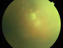
Uveitis and Retinal Whitening - 04-27-12 - Prior to PPV OD - Possible Primary Ocular Lymphoma vu 869 fois
(0 votes)
|
|

Uveitis and Retinal Whitening - 04-27-12 - Prior to PPV OD - Possible Primary Ocular Lymphoma vu 733 fois
(0 votes)
|
|
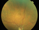
Uveitis and Retinal Whitening - 04-27-12 - Prior to PPV OD - Possible Primary Ocular Lymphoma vu 696 fois
(0 votes)
|
|
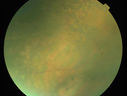
Uveitis and Retinal Whitening - 04-27-12 - Prior to PPV OD - Possible Primary Ocular Lymphoma vu 709 fois
(0 votes)
|
|

Uveitis and Retinal Whitening - 04-27-12 - Prior to PPV OD - Possible Primary Ocular Lymphoma vu 816 fois
(0 votes)
|
|
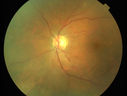
Uveitis and Retinal Whitening - 04-27-12 - Prior to PPV OD - Possible Primary Ocular Lymphoma vu 764 fois
(0 votes)
|
|
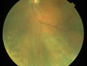
Uveitis and Retinal Whitening - 04-27-12 - Prior to PPV OD - Possible Primary Ocular Lymphoma vu 659 fois
(0 votes)
|
|
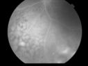
Uveitis and Retinal Whitening - 04-27-12 - Prior to PPV OD - Possible Primary Ocular Lymphoma vu 681 fois
(0 votes)
|
|
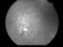
Uveitis and Retinal Whitening - 04-27-12 - Prior to PPV OD - Possible Primary Ocular Lymphoma vu 671 fois
(0 votes)
|
|

Uveitis and Retinal Whitening - 04-27-12 - Prior to PPV OD - Possible Primary Ocular Lymphoma vu 581 fois
(0 votes)
|
|
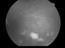
Uveitis and Retinal Whitening - 04-27-12 - Prior to PPV OD - Possible Primary Ocular Lymphoma vu 610 fois
(0 votes)
|
|

Uveitis and Retinal Whitening - 04-27-12 - Prior to PPV OD - Possible Primary Ocular Lymphoma vu 644 fois
(0 votes)
|
|

Uveitis and Retinal Whitening - 04-27-12 - Prior to PPV OD - Possible Primary Ocular Lymphoma vu 572 fois
(0 votes)
|
|
Uveitis and Retinal Whitening - 04-27-12 - Prior to PPV OD - Possible Primary Ocular Lymphoma vu 621 fois
(0 votes)
|
|
Uveitis and Retinal Whitening - 04-27-12 - Prior to PPV OD - Possible Primary Ocular Lymphoma vu 639 fois
(0 votes)
|
|
Primary Ocular Lymphoma - 051812 vu 591 fois
(0 votes)
|
|

Primary Ocular Lymphoma - 051812 vu 563 fois
(0 votes)
|
|
Primary Ocular Lymphoma - 051812 vu 575 fois
(0 votes)
|
|
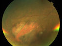
Primary Ocular Lymphoma - 051812 vu 582 fois
(0 votes)
|
|

Primary Ocular Lymphoma - 051812 vu 540 fois
(0 votes)
|
|
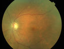
Primary Ocular Lymphoma - 051812 vu 573 fois
(0 votes)
|
|

Primary Ocular Lymphoma - 051812 vu 583 fois
(0 votes)
|
|
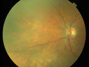
Primary Ocular Lymphoma - 051812 vu 563 fois
(0 votes)
|
|
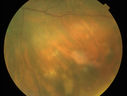
Primary Ocular Lymphoma - 051812 vu 604 fois
(0 votes)
|
|
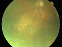
Primary Ocular Lymphoma - 051812 vu 595 fois
(0 votes)
|
|

Primary Ocular Lymphoma - White Mass Left Eye - 112712 vu 673 fois
(0 votes)
|
|

Primary Ocular Lymphoma - White Mass Left Eye - 112712 vu 863 fois
(0 votes)
|
|

Primary Ocular Lymphoma - White Mass Left Eye - 112712 vu 652 fois
(0 votes)
|
|
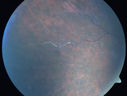
Primary Ocular Lymphoma - White Mass Left Eye - 112712 vu 818 fois
(0 votes)
|
|

Primary Ocular Lymphoma - White Mass Left Eye - 112712 vu 751 fois
(0 votes)
|
|
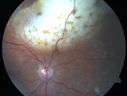
Primary Ocular Lymphoma - White Mass Left Eye - 112712 vu 736 fois
(0 votes)
|
|

Primary Ocular Lymphoma - White Mass Left Eye - 112712 vu 796 fois
(0 votes)
|
|

Primary Ocular Lymphoma - White Mass Left Eye - 112712 vu 699 fois
(0 votes)
|
|

Primary Ocular Lymphoma - White Mass Left Eye - 112712 vu 759 fois
(0 votes)
|
|
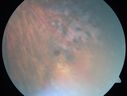
Primary Ocular Lymphoma - White Mass Left Eye - 112712 vu 618 fois
(0 votes)
|
|

Primary Ocular Lymphoma - White Mass Left Eye - 112712 vu 624 fois
(0 votes)
|
|

Primary Ocular Lymphoma - White Mass Left Eye - 112712 vu 637 fois
(0 votes)
|
|

Primary Ocular Lymphoma - White Mass Left Eye - 112712 vu 764 fois
(0 votes)
|
|

Primary Ocular Lymphoma - White Mass Left Eye - 112712 vu 758 fois
(0 votes)
|
|
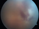
Primary Ocular Lymphoma - White Mass Left Eye - 112712 vu 720 fois
(0 votes)
|
|

Primary Ocular Lymphoma - White Mass Left Eye - 112712 vu 752 fois
(0 votes)
|
|
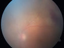
Primary Ocular Lymphoma - White Mass Left Eye - 112712 vu 709 fois
(0 votes)
|
|

Primary Ocular Lymphoma - White Mass Left Eye - 112712 vu 668 fois
(0 votes)
|
|

Primary Ocular Lymphoma - White Mass Left Eye - 112712 vu 566 fois
(0 votes)
|
|
Primary Ocular Lymphoma - White Mass Left Eye - 112712 vu 553 fois
(0 votes)
|
|

Primary Ocular Lymphoma - White Mass Left Eye - 112712 vu 607 fois
(0 votes)
|
|
Primary Ocular Lymphoma - White Mass Left Eye - 112712 vu 575 fois
(0 votes)
|
|
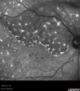
Primary Ocular Lymphoma - 020513 vu 594 fois
(0 votes)
|
|

Primary Ocular Lymphoma - 020513 vu 642 fois
(0 votes)
|
|
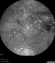
Primary Ocular Lymphoma - 020513 vu 620 fois
(0 votes)
|
|

Primary Ocular Lymphoma - 020513 vu 571 fois
(0 votes)
|
|
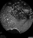
Primary Ocular Lymphoma - 020513 - FUndus Autofluorescencevu 710 fois
(0 votes)
|
|

Primary Ocular Lymphoma - 020513 - FUndus Autofluorescencevu 702 fois
(0 votes)
|
|
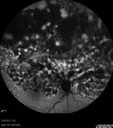
Primary Ocular Lymphoma - 020513 - FUndus Autofluorescencevu 694 fois
(0 votes)
|
|

Primary Ocular Lymphoma - 020513 - FUndus Autofluorescencevu 576 fois
(0 votes)
|
|

Primary Ocular Lymphoma - 020513 - FUndus Autofluorescencevu 737 fois
(0 votes)
|
|
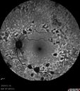
Primary Ocular Lymphoma - 020513 - FUndus Autofluorescencevu 820 fois
(0 votes)
|
|
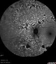
Primary Ocular Lymphoma - 020513 - FUndus Autofluorescencevu 791 fois
(0 votes)
|
|
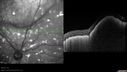
Primary Ocular Lymphoma - 020513 - SD OCTvu 658 fois
(0 votes)
|
|
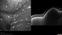
Primary Ocular Lymphoma - 020513 - SD OCTvu 703 fois
(0 votes)
|
|
Primary Ocular Lymphoma - 020513 - SD OCTvu 820 fois
(0 votes)
|
|
Primary Ocular Lymphoma - 020513 - SD OCTvu 697 foisVIDEO TAKES TIME TO LOAD
(0 votes)
|
|
Primary Ocular Lymphoma - 020513 - SD OCTvu 630 foisVIDEO TAKES TIME TO LOAD
(0 votes)
|
|
Primary Ocular Lymphoma - 020513 - SD OCT - SD OCTvu 1011 fois
(0 votes)
|
|
Primary Ocular Lymphoma - 020513 - SD OCTvu 933 fois
(0 votes)
|
|
Primary Ocular Lymphoma - 020513 - SD OCTvu 673 foisVIDEO TAKES TIME TO LOAD
(0 votes)
|
|

Primary Ocular Lymphoma - Lesion has grown in size over 3 monthsvu 803 fois
(0 votes)
|
|
|
|
|
80 Year Old Female Patient came in 1 year ago with a retinal whitening in her eye and uveitis. Her uveitis work-up was negative, and because of the retinal whitening and the uveitis, I was concerned about a possible occult acute retinal necrosis case. I put her on Valtrex, but the eyes did not get any better and the retinal whitening continued. Then later, I became concerned about a possible primary ocular lymphoma. She had an extensive cancer evaluation that was all negative. She has had MRI scans and ultimately I did a vitrectomy on both eyes. I did the right eye on May 30th of 2012 and flow cytometry and cytology was negative. That eye cleared nicely, but then, because of increasing uveitis in the left eye, I did a vitrectomy in the left eye on December 19th of 2012. That sample I sent to the lab in Miami and that result was similarly negative. I am going to send you all of the results so you have them when you see her. At this point, it has been unclear whether she had uveitis, infectious retinitis, or possibly primary ocular lymphoma.
When I saw her in my office on November 27th of 2012, she had a white subretinal mass growing in the right eye and the left eye had vitreous cells. After the vitrectomy in the left eye, I started rechecking both eyes and the white subretinal mass in the right eye had doubled in size. The right eye now looks very much like it has primary ocular lymphoma.
I appreciate you seeing her and, if possible, performing some sort of a biopsy of that mass, which has now grown to the point where I think a fine-needle aspiration biopsy would be possible to help secure the diagnosis.
++++++++++++ Visit 2/5/2013. 80-year-old woman has possible ocular lymphoma. She has had two vitreous biopsies, one in one eye and one in the other eye, and the tests have been negative. Recently she is manifesting increasing anemia. She also has glaucoma. She is taking Pred Forte in both eyes and Combigan in both eyes.
VISUAL ACUITY: Vision OD is 20/40, OS is 20/160. IOP: OD 22, OS 24. The eyes are quiet with a posterior chamber intraocular lens in good position in both eyes.
EXTENDED OPHTHALMOSCOPY:
OD: Vertical C/D ratio is 0.4. The white mass in the macula is much bigger than it was when I saw her in November. It is growing on the superior half of the macula, as well as just nasal to the optic nerve. It has about doubled in size. There are no significant vitreous cells.
OS: Vertical C/D ratio is 0.4. There are patchy retinal hemorrhages.
FUNDUS PHOTOGRAPHY – COLOR AND AUTOFLUORESCENCE: Color and autofluorescence photographs do show the mass to be much bigger.
SPECTRALIS SD-OCT SCAN: The OCT scans over that area show the probable tumor to be about 1.5 mm high.
IMPRESSION:
1. PROBABLE OCULAR LYMPHOMA – BOTH EYES
DISCUSSION: I explained to the patient, given the growth of the lesion and the absence of inflammation around it, I think at this point it is likely ocular lymphoma.
++++++++++ VISIT - 1/4/2013. 80-year-old woman had a vitreous biopsy in the left eye with a fairly full vitrectomy back on December 19th, of 2012. Her vitreous was sent down to Miami where it was carefully analyzed in the pathology lab. The flow cytometry was normal. Her B cell testing was normal. The T cell testing did show two small peaks. I contacted the pathologist about that result and it is not uncommon in cerebrospinal fluid and vitreous samples, because there are so few cells, to see some peaks on the T cells. Generally that is not considered indicative of a T cell lymphoma. There is no such thing as a primary intraocular T cell lymphoma. The primary intraocular lymphomas are B cells and the findings overall were consistent with intraocular inflammation. She did have extensive blood testing by
Dr. Friedberg when this all started a few years ago and that was all negative. I have not repeated the blood tests though. The vision in the left eye is better since the vitrectomy.
VISUAL ACUITY: Vision OD is 20/25; OS is 20/100, PH is 20/40. IOP: OS 18. The left eye is quiet in the anterior chamber. The posterior chamber intraocular lens is in good position.
EXTENDED OPHTHALMOSCOPY:
OS: Vertical C/D ratio is 0.2. There is peripheral retinal hemorrhaging, less than on last visit. The retina is everywhere attached.
IMPRESSION:
1. NEGATIVE VITREOUS BIOPSY FOR INTRAOCULAR LYMPHOMA
2. PANUVEITIS – BOTH EYES
DISCUSSION: I explained to the patient, given all testing, it looks at the moment like she has an inflammatory panuveitis, and I am going to rerun the initial blood tests to see if anything comes up positive. Inflammatory and infectious diseases sometimes infect the posterior pole.


