Occult Macular Dystophy - 9 year follow-up
|
|
45-year-old man was seen in the office on 9/14/2010. He did not wear glasses until he was older, mostly in high school. He remembers his vision correcting to be pretty good and even without his glasses he was able to drive until when he was younger. He has had a slight decline, as far as he can tell. He does have a family history of eye problems. His mother and his maternal grandmother each had vision loss at a relatively young age. His mother was in her twenties or thirties, and the vision loss got to the point where she was actually tested for a possible brain tumor. His grandmother lost vision in her fifties. Other than that, everyone in the family tree sees fine. His is a high myope, but
with his glasses and contacts, his best corrected vision is limited.
VISUAL ACUITY: Vision OD is 20/80, PH 20/70; OS is 20/80, PH 20/70. IOP: 11 in both eyes.
SLIT LAMP EXAM: Normal with clear lenses.
COLOR VISION: His color vision is normal with 13 out of 14 correct in each eye. There is no afferent pupillary defect.
NERVE FIBER LAYER SCANS: Normal.
EXTENDED OPHTHALMOSCOPY:
OD: Vertical C/D ratio is 0.3. The macula and periphery look healthy.
OS: Vertical C/D ratio is 0.3. The macula and periphery look healthy.
OCT SCAN: The OCT scan shows some retinal thinning centrally, with particular loss of the photoreceptor cell bodies around the very center of the fovea.
PHOTOGRAPHS: Photos confirm clinical findings.
FLUORESCEIN ANGIOGRAPHY: The FA is entirely normal.
IMPRESSION:
1. Occult Maculopathy - New Entity
DISCUSSION: I explained to the patient at this moment, based on the foveal findings, it may be that he has a very subtle macular dystrophy, although the atypical thing in him is that most people with isolated macular dystrophy either have pigment epithelial changes which are evident on angiography or color vision changes which are evident on color vision testing, neither of which are a problem with him.
I recently attended a Gass Society meeting in Chicago and a case was presented and discussed, which matched this case nearly exactly, and the diagnosis that was coined at the time was occult maculopathy. His syndrome does not seem to be associated with anything. In his case, he has a family history suggesting there may be some sort of genetic issue. I may have his case published with some other ones with similar findings, but at this point I do not think there is any particular treatment that would be worth considering
|

Occult Macular Dystrophy - vu 589 fois53 year old man. Vision is better at 20/50 OU
(0 votes)
|
|

Occult Macular Dystrophy - 9 Years later vu 523 fois53 year old man. Vision is better at 20/50 OU
(0 votes)
|
|

Occult Macular Dystrophy - 9 Years later vu 496 fois53 year old man. Vision is better at 20/50 OU
(0 votes)
|
|

Occult Macular Dystrophy - 9 Years later vu 704 fois53 year old man. Vision is better at 20/50 OU
(0 votes)
|
|

Occult Macular Dystrophy - 9 Years later vu 533 fois53 year old man. Vision is better at 20/50 OU
(0 votes)
|
|

Occult Macular Dystrophy - 9 Years later vu 583 fois53 year old man. Vision is better at 20/50 OU
(0 votes)
|
|

Occult Macular Dystrophy - 9 Years later vu 514 fois53 year old man. Vision is better at 20/50 OU
(0 votes)
|
|

Occult Macular Dystrophy - 9 Years later vu 571 fois53 year old man. Vision is better at 20/50 OU
(0 votes)
|
|

Occult Macular Dystrophy - 9 Years later vu 578 fois53 year old man. Vision is better at 20/50 OU
(0 votes)
|
|

Occult Macular Dystrophy - 9 Years later vu 531 fois53 year old man. Vision is better at 20/50 OU
(0 votes)
|
|

Occult Macular Dystrophy - 9 Years later vu 479 fois53 year old man. Vision is better at 20/50 OU
(0 votes)
|
|

Occult Macular Dystrophy - 9 Years later vu 503 fois53 year old man. Vision is better at 20/50 OU
(0 votes)
|
|

Occult Macular Dystrophy - 9 Years later vu 676 fois53 year old man. Vision is better at 20/50 OU
(0 votes)
|
|

Occult Macular Dystrophy - 9 Years later vu 739 fois53 year old man. Vision is better at 20/50 OU
(0 votes)
|
|

Occult Maculopathy - Thin Fovea on OCT and Normal Color VA, Photos, FA VA 20/80 OUvu 1186 fois45-year-old man his mother and his maternal grandmother each had vision loss at a relatively young age. Retina OD
(0 votes)
|
|

Occult Maculopathy - Thin Fovea on OCT and Normal Color VA, Photos, FA VA 20/80 OUvu 843 fois45-year-old man his mother and his maternal grandmother each had vision loss at a relatively young age. Macula OD
(0 votes)
|
|

Occult Maculopathy - Thin Fovea on OCT and Normal Color VA, Photos, FA VA 20/80 OUvu 935 fois45-year-old man his mother and his maternal grandmother each had vision loss at a relatively young age. Retina OS normal
(0 votes)
|
|

Occult Maculopathy - Thin Fovea on OCT and Normal Color VA, Photos, FA VA 20/80 OUvu 808 fois45-year-old man his mother and his maternal grandmother each had vision loss at a relatively young age. Macula OS looks normal
(0 votes)
|
|

Occult Maculopathy - Thin Fovea on OCT and Normal Color VA, Photos, FA VA 20/80 OUvu 807 fois45-year-old man his mother and his maternal grandmother each had vision loss at a relatively young age. Red Free Photo
(0 votes)
|
|

Occult Maculopathy - Thin Fovea on OCT and Normal Color VA, Photos, FA VA 20/80 OUvu 729 fois45-year-old man his mother and his maternal grandmother each had vision loss at a relatively young age. Red Free Photo - Normal
(0 votes)
|
|
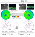
Occult Maculopathy - Thin Fovea on OCT and Normal Color VA, Photos, FA VA 20/80 OUvu 1081 fois45-year-old man his mother and his maternal grandmother each had vision loss at a relatively young age. OCT MAP shows foveal thinning.
(0 votes)
|
|

Occult Maculopathy - Thin Fovea on OCT and Normal Color VA, Photos, FA VA 20/80 OUvu 1020 fois45-year-old man his mother and his maternal grandmother each had vision loss at a relatively young age. LINE SCAN SHOW FOVEAL ATROPHY
(0 votes)
|
|

Occult Maculopathy - Thin Fovea on OCT and Normal Color VA, Photos, FA VA 20/80 OUvu 873 fois45-year-old man his mother and his maternal grandmother each had vision loss at a relatively young age. Line scan shows foveal atrophy.
(0 votes)
|
|

Occult Maculopathy - Thin Fovea on OCT and Normal Color VA, Photos, FA VA 20/80 OUvu 1299 fois45-year-old man OCT scan shows foveal thinning
(0 votes)
|
|
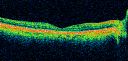
Occult Maculopathy - Thin Fovea on OCT and Normal Color VA, Photos, FA VA 20/80 OUvu 972 foisOCT Scan Right Eye shows foveal thinning. OD is 20/80, PH 20/70; OS is 20/80, PH 20/70. His color vision is normal with 13 out of 14 correct in each eye. There is no afferent pupillary defect.
FLUORESCEIN ANGIOGRAPHY: The FA is entirely normal.
(0 votes)
|
|

Occult Maculopathy - Thin Fovea on OCT and Normal Color VA, Photos, FA VA 20/80 OUvu 817 fois45-year-old man Normal FA
(0 votes)
|
|
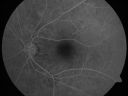
Occult Maculopathy - Thin Fovea on OCT and Normal Color VA, Photos, FA VA 20/80 OUvu 769 fois45-year-old man The FA is entirely normal.
(0 votes)
|
|
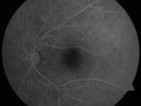
Occult Maculopathy - Thin Fovea on OCT and Normal Color VA, Photos, FA VA 20/80 OUvu 755 fois45-year-old man The FA is entirely normal.
(0 votes)
|
|
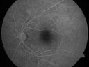
Occult Maculopathy - Thin Fovea on OCT and Normal Color VA, Photos, FA VA 20/80 OUvu 689 fois45-year-old man Normal FA
(0 votes)
|
|

Occult Maculopathy - Thin Fovea on OCT and Normal Color VA, Photos, FA VA 20/80 OUvu 751 fois45-year-old man Normal FA
(0 votes)
|
|
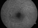
Occult Maculopathy - Thin Fovea on OCT and Normal Color VA, Photos, FA VA 20/80 OUvu 836 fois45-year-old man Normal FA
(0 votes)
|
|

Occult Maculopathy - Thin Fovea on OCT and Normal Color VA, Photos, FA VA 20/80 OUvu 978 fois45-year-old man Normal FA
(0 votes)
|
|

Occult Maculopathy - Thin Fovea on OCT and Normal Color VA, Photos, FA VA 20/80 OUvu 799 fois45-year-old man his mother and his maternal grandmother each had vision loss at a relatively young age. Normal FA.
(0 votes)
|
|
|
|
|
|
|
45-year-old man was seen in the office on 9/14/2010. He did not wear glasses until he was older, mostly in high school. He remembers his vision correcting to be pretty good and even without his glasses he was able to drive until when he was younger. He has had a slight decline, as far as he can tell. He does have a family history of eye problems. His mother and his maternal grandmother each had vision loss at a relatively young age. His mother was in her twenties or thirties, and the vision loss got to the point where she was actually tested for a possible brain tumor. His grandmother lost vision in her fifties. Other than that, everyone in the family tree sees fine. His is a high myope, but
with his glasses and contacts, his best corrected vision is limited.
VISUAL ACUITY: Vision OD is 20/80, PH 20/70; OS is 20/80, PH 20/70. IOP: 11 in both eyes.
SLIT LAMP EXAM: Normal with clear lenses.
COLOR VISION: His color vision is normal with 13 out of 14 correct in each eye. There is no afferent pupillary defect.
NERVE FIBER LAYER SCANS: Normal.
EXTENDED OPHTHALMOSCOPY:
OD: Vertical C/D ratio is 0.3. The macula and periphery look healthy.
OS: Vertical C/D ratio is 0.3. The macula and periphery look healthy.
OCT SCAN: The OCT scan shows some retinal thinning centrally, with particular loss of the photoreceptor cell bodies around the very center of the fovea.
PHOTOGRAPHS: Photos confirm clinical findings.
FLUORESCEIN ANGIOGRAPHY: The FA is entirely normal.
IMPRESSION:
1. Occult Maculopathy - New Entity
DISCUSSION: I explained to the patient at this moment, based on the foveal findings, it may be that he has a very subtle macular dystrophy, although the atypical thing in him is that most people with isolated macular dystrophy either have pigment epithelial changes which are evident on angiography or color vision changes which are evident on color vision testing, neither of which are a problem with him.
I recently attended a Gass Society meeting in Chicago and a case was presented and discussed, which matched this case nearly exactly, and the diagnosis that was coined at the time was occult maculopathy. His syndrome does not seem to be associated with anything. In his case, he has a family history suggesting there may be some sort of genetic issue. I may have his case published with some other ones with similar findings, but at this point I do not think there is any particular treatment that would be worth considering


