Retinitis Pigmentosa - Vascular Narrowing
|
|
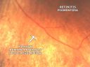
This pleasant 55-year-old man was seen in the office on 6/17/08. He noticed his vision declining recently, over the last few weeks, particularly in the right eye. He said previous to that the vision was not normal but was pretty good. He has been getting along nicely driving. He had a metal injury a long time ago in his right eye that was treated in the emergency room. Surgery was not needed for this injury. He does not have a significant family history of retinal diseases, although most family members do wear glasses. He does smoke cigarettes, but he is going to try to quit.
VISUAL ACUITY: Vision OD is 20/100, OS is 20/50. IOP: OD 15, OS 13. Color vision OD 7 out of 10 correction, OS 8 out of 10 correct.
AMSLER GRID: Normal OU.
SLIT LAMP EXAM: There is 3+ nuclear sclerosis in both eyes.
EXTENDED OPHTHALMOSCOPY:
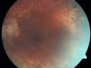
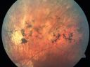
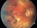
OD: Vertical C/D ratio is 0.2. There is a posterior vitreous separation with 2+ vitreous cells. There is 2+ cystoid macular edema. There is peripapillary atrophy, chorioretinal scarring, and bone spicules 360° at the equator, touching the macula.
OS: Vertical C/D ratio is 0.2. There is a posterior vitreous separation and 2+ macular edema. Again, there is peripapillary atrophy and there are bone spicule pigment irregularities 360° at the equator.
OCT SCAN: There is macular thickening in both eyes with intraretinal cysts.
FLUORESCEIN ANGIOGRAPHY: Photos confirm clinical findings. FA shows hyperfluorescence in the periphery 360° around the macula, consistent with retinitis pigmentosa. The fovea has had minimal staining with fluorescein in the late frames.
IMPRESSION:
1. POSSIBLE RETINITIS PIGMENTOSA – BOTH EYES
2. POSSIBLE CHORIORETINAL INFLAMMATORY INFECTIOUS CHORIORETINOPATHY – BOTH EYES
3. MACULAR EDEMA – BOTH EYES
DISCUSSION: I explained to the patient he does have macular edema in both eyes, limiting his vision. I put him on Xibrom and Econopred drops to see if that will eliminate the macular edema. If it is from retinitis pigmentosa, sometimes Acetazolamide will help.
Since he has a new diagnosis of retinitis pigmentosa and since this is atypical in that the symptoms are presenting so late in life, I am going to obtain blood tests to rule out possible infectious or inflammatory etiologies. He does not have any health problems that point to any specific diagnosis except for arthritis. He will return for check in 6-8 weeks, sooner should he notice any problems.
|
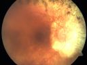
Retinitis Pigmentosa Newly Diagnosed 55 Year Oldvista 810 veces55-year-old noticed his vision declining recently, over the last few weeks, particularly in the right eye. He said previous to that the vision was not normal but was pretty good. OD is 20/100, OS is 20/50
OCT SCAN: There is macular thickening in both eyes with intraretinal cysts.
(0 votos)
|
|

Retinitis Pigmentosa Newly Diagnosed 55 Year Oldvista 618 veces55-year-old noticed his vision declining recently, over the last few weeks, particularly in the right eye. He said previous to that the vision was not normal but was pretty good. OD is 20/100, OS is 20/50
OCT SCAN: There is macular thickening in both eyes with intraretinal cysts.
(0 votos)
|
|
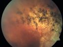
Retinitis Pigmentosa Newly Diagnosed 55 Year Oldvista 612 veces55-year-old noticed his vision declining recently, over the last few weeks, particularly in the right eye. He said previous to that the vision was not normal but was pretty good. OD is 20/100, OS is 20/50
OCT SCAN: There is macular thickening in both eyes with intraretinal cysts.
(0 votos)
|
|
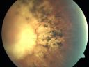
Retinitis Pigmentosa Newly Diagnosed 55 Year Oldvista 568 veces55-year-old noticed his vision declining recently, over the last few weeks, particularly in the right eye. He said previous to that the vision was not normal but was pretty good. OD is 20/100, OS is 20/50
OCT SCAN: There is macular thickening in both eyes with intraretinal cysts.
(0 votos)
|
|

Retinitis Pigmentosa Newly Diagnosed 55 Year Oldvista 605 veces55-year-old noticed his vision declining recently, over the last few weeks, particularly in the right eye. He said previous to that the vision was not normal but was pretty good. OD is 20/100, OS is 20/50
OCT SCAN: There is macular thickening in both eyes with intraretinal cysts.
(0 votos)
|
|

Retinitis Pigmentosa Newly Diagnosed 55 Year Oldvista 661 veces55-year-old noticed his vision declining recently, over the last few weeks, particularly in the right eye. He said previous to that the vision was not normal but was pretty good. OD is 20/100, OS is 20/50
OCT SCAN: There is macular thickening in both eyes with intraretinal cysts.
(0 votos)
|
|

Retinitis Pigmentosa Newly Diagnosed 55 Year Oldvista 594 veces55-year-old noticed his vision declining recently, over the last few weeks, particularly in the right eye. He said previous to that the vision was not normal but was pretty good. OD is 20/100, OS is 20/50
OCT SCAN: There is macular thickening in both eyes with intraretinal cysts.
(0 votos)
|
|
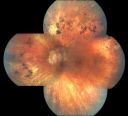
Retinitis Pigmentosa Newly Diagnosed 55 Year Oldvista 599 veces55-year-old noticed his vision declining recently, over the last few weeks, particularly in the right eye. He said previous to that the vision was not normal but was pretty good. OD is 20/100, OS is 20/50
OCT SCAN: There is macular thickening in both eyes with intraretinal cysts.
(0 votos)
|
|
Retinitis Pigmentosa Newly Diagnosed 55 Year Oldvista 660 veces55-year-old noticed his vision declining recently, over the last few weeks, particularly in the right eye. He said previous to that the vision was not normal but was pretty good. OD is 20/100, OS is 20/50
OCT SCAN: There is macular thickening in both eyes with intraretinal cysts.
(0 votos)
|
|
Retinitis Pigmentosa Newly Diagnosed 55 Year Oldvista 532 veces55-year-old noticed his vision declining recently, over the last few weeks, particularly in the right eye. He said previous to that the vision was not normal but was pretty good. OD is 20/100, OS is 20/50
OCT SCAN: There is macular thickening in both eyes with intraretinal cysts.
(0 votos)
|
|
Retinitis Pigmentosa Newly Diagnosed 55 Year Oldvista 664 veces55-year-old noticed his vision declining recently, over the last few weeks, particularly in the right eye. He said previous to that the vision was not normal but was pretty good. OD is 20/100, OS is 20/50
OCT SCAN: There is macular thickening in both eyes with intraretinal cysts.
(0 votos)
|
|

Retinitis Pigmentosa Newly Diagnosed 55 Year Oldvista 631 veces55-year-old noticed his vision declining recently, over the last few weeks, particularly in the right eye. He said previous to that the vision was not normal but was pretty good. OD is 20/100, OS is 20/50
OCT SCAN: There is macular thickening in both eyes with intraretinal cysts.
(0 votos)
|
|
Retinitis Pigmentosa Newly Diagnosed 55 Year Oldvista 654 veces55-year-old noticed his vision declining recently, over the last few weeks, particularly in the right eye. He said previous to that the vision was not normal but was pretty good. OD is 20/100, OS is 20/50
OCT SCAN: There is macular thickening in both eyes with intraretinal cysts.
(0 votos)
|
|
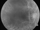
Retinitis Pigmentosa Newly Diagnosed 55 Year Oldvista 649 veces55-year-old noticed his vision declining recently, over the last few weeks, particularly in the right eye. He said previous to that the vision was not normal but was pretty good. OD is 20/100, OS is 20/50
OCT SCAN: There is macular thickening in both eyes with intraretinal cysts.
(0 votos)
|
|
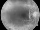
Retinitis Pigmentosa Newly Diagnosed 55 Year Oldvista 551 veces55-year-old noticed his vision declining recently, over the last few weeks, particularly in the right eye. He said previous to that the vision was not normal but was pretty good. OD is 20/100, OS is 20/50
OCT SCAN: There is macular thickening in both eyes with intraretinal cysts.
(0 votos)
|
|
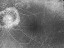
Retinitis Pigmentosa Newly Diagnosed 55 Year Oldvista 525 veces55-year-old noticed his vision declining recently, over the last few weeks, particularly in the right eye. He said previous to that the vision was not normal but was pretty good. OD is 20/100, OS is 20/50
OCT SCAN: There is macular thickening in both eyes with intraretinal cysts.
(0 votos)
|
|

Retinitis Pigmentosa Newly Diagnosed 55 Year Oldvista 490 veces55-year-old noticed his vision declining recently, over the last few weeks, particularly in the right eye. He said previous to that the vision was not normal but was pretty good. OD is 20/100, OS is 20/50
OCT SCAN: There is macular thickening in both eyes with intraretinal cysts.
(0 votos)
|
|

Retinitis Pigmentosa Newly Diagnosed 55 Year Oldvista 541 veces55-year-old noticed his vision declining recently, over the last few weeks, particularly in the right eye. He said previous to that the vision was not normal but was pretty good. OD is 20/100, OS is 20/50
OCT SCAN: There is macular thickening in both eyes with intraretinal cysts.
(0 votos)
|
|
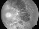
Retinitis Pigmentosa Newly Diagnosed 55 Year Oldvista 519 veces55-year-old noticed his vision declining recently, over the last few weeks, particularly in the right eye. He said previous to that the vision was not normal but was pretty good. OD is 20/100, OS is 20/50
OCT SCAN: There is macular thickening in both eyes with intraretinal cysts.
(0 votos)
|
|
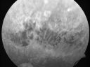
Retinitis Pigmentosa Newly Diagnosed 55 Year Oldvista 574 veces55-year-old noticed his vision declining recently, over the last few weeks, particularly in the right eye. He said previous to that the vision was not normal but was pretty good. OD is 20/100, OS is 20/50
OCT SCAN: There is macular thickening in both eyes with intraretinal cysts.
(0 votos)
|
|
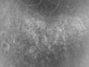
Retinitis Pigmentosa Newly Diagnosed 55 Year Oldvista 597 veces55-year-old noticed his vision declining recently, over the last few weeks, particularly in the right eye. He said previous to that the vision was not normal but was pretty good. OD is 20/100, OS is 20/50
OCT SCAN: There is macular thickening in both eyes with intraretinal cysts.
(0 votos)
|
|
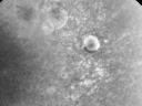
Retinitis Pigmentosa Newly Diagnosed 55 Year Oldvista 567 veces55-year-old noticed his vision declining recently, over the last few weeks, particularly in the right eye. He said previous to that the vision was not normal but was pretty good. OD is 20/100, OS is 20/50
OCT SCAN: There is macular thickening in both eyes with intraretinal cysts.
(0 votos)
|
|
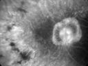
Retinitis Pigmentosa Newly Diagnosed 55 Year Oldvista 638 veces55-year-old noticed his vision declining recently, over the last few weeks, particularly in the right eye. He said previous to that the vision was not normal but was pretty good. OD is 20/100, OS is 20/50
OCT SCAN: There is macular thickening in both eyes with intraretinal cysts.
(0 votos)
|
|
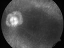
Retinitis Pigmentosa Newly Diagnosed 55 Year Oldvista 604 veces55-year-old noticed his vision declining recently, over the last few weeks, particularly in the right eye. He said previous to that the vision was not normal but was pretty good. OD is 20/100, OS is 20/50
OCT SCAN: There is macular thickening in both eyes with intraretinal cysts.
(0 votos)
|
|
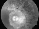
Retinitis Pigmentosa Newly Diagnosed 55 Year Oldvista 579 veces55-year-old noticed his vision declining recently, over the last few weeks, particularly in the right eye. He said previous to that the vision was not normal but was pretty good. OD is 20/100, OS is 20/50
OCT SCAN: There is macular thickening in both eyes with intraretinal cysts.
(0 votos)
|
|
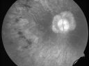
Retinitis Pigmentosa Newly Diagnosed 55 Year Oldvista 535 veces55-year-old noticed his vision declining recently, over the last few weeks, particularly in the right eye. He said previous to that the vision was not normal but was pretty good. OD is 20/100, OS is 20/50
OCT SCAN: There is macular thickening in both eyes with intraretinal cysts.
(0 votos)
|
|
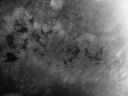
Retinitis Pigmentosa Newly Diagnosed 55 Year Oldvista 615 veces55-year-old noticed his vision declining recently, over the last few weeks, particularly in the right eye. He said previous to that the vision was not normal but was pretty good. OD is 20/100, OS is 20/50
OCT SCAN: There is macular thickening in both eyes with intraretinal cysts.
(0 votos)
|
|
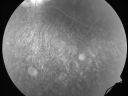
Retinitis Pigmentosa Newly Diagnosed 55 Year Oldvista 641 veces55-year-old noticed his vision declining recently, over the last few weeks, particularly in the right eye. He said previous to that the vision was not normal but was pretty good. OD is 20/100, OS is 20/50
OCT SCAN: There is macular thickening in both eyes with intraretinal cysts.
(0 votos)
|
|

Retinitis Pigmentosa Newly Diagnosed 55 Year Oldvista 730 veces55-year-old noticed his vision declining recently, over the last few weeks, particularly in the right eye. He said previous to that the vision was not normal but was pretty good. OD is 20/100, OS is 20/50
OCT SCAN: There is macular thickening in both eyes with intraretinal cysts.
(0 votos)
|
|
|
|
|
| 29 archivos en 1 página(s) |
|
This pleasant 55-year-old man was seen in the office on 6/17/08. He noticed his vision declining recently, over the last few weeks, particularly in the right eye. He said previous to that the vision was not normal but was pretty good. He has been getting along nicely driving. He had a metal injury a long time ago in his right eye that was treated in the emergency room. Surgery was not needed for this injury. He does not have a significant family history of retinal diseases, although most family members do wear glasses. He does smoke cigarettes, but he is going to try to quit.
VISUAL ACUITY: Vision OD is 20/100, OS is 20/50. IOP: OD 15, OS 13. Color vision OD 7 out of 10 correction, OS 8 out of 10 correct.
AMSLER GRID: Normal OU.
SLIT LAMP EXAM: There is 3+ nuclear sclerosis in both eyes.
EXTENDED OPHTHALMOSCOPY:
OD: Vertical C/D ratio is 0.2. There is a posterior vitreous separation with 2+ vitreous cells. There is 2+ cystoid macular edema. There is peripapillary atrophy, chorioretinal scarring, and bone spicules 360° at the equator, touching the macula.
OS: Vertical C/D ratio is 0.2. There is a posterior vitreous separation and 2+ macular edema. Again, there is peripapillary atrophy and there are bone spicule pigment irregularities 360° at the equator.
OCT SCAN: There is macular thickening in both eyes with intraretinal cysts.
FLUORESCEIN ANGIOGRAPHY: Photos confirm clinical findings. FA shows hyperfluorescence in the periphery 360° around the macula, consistent with retinitis pigmentosa. The fovea has had minimal staining with fluorescein in the late frames.
IMPRESSION:
1. POSSIBLE RETINITIS PIGMENTOSA – BOTH EYES
2. POSSIBLE CHORIORETINAL INFLAMMATORY INFECTIOUS CHORIORETINOPATHY – BOTH EYES
3. MACULAR EDEMA – BOTH EYES
DISCUSSION: I explained to the patient he does have macular edema in both eyes, limiting his vision. I put him on Xibrom and Econopred drops to see if that will eliminate the macular edema. If it is from retinitis pigmentosa, sometimes Acetazolamide will help.
Since he has a new diagnosis of retinitis pigmentosa and since this is atypical in that the symptoms are presenting so late in life, I am going to obtain blood tests to rule out possible infectious or inflammatory etiologies. He does not have any health problems that point to any specific diagnosis except for arthritis. He will return for check in 6-8 weeks, sooner should he notice any problems.


