26-year-old man who I saw in the office on June 25, 2009. He lived in Knoxville, Tennessee and was diagnosed there with congenital achromatopsia. He does have family members who also have this disorder. He says he sees better in dim lighting than in bright lighting. He believes as a child he might have had electroretinography testing. He has not had any testing done lately.
VISUAL ACUITY: OD 20/200, OS 20/200. IOP: OD 17, OS 18.
SLIT EXAMINATION: Normal with clear lenses.
EXTENDED OPHTHALMOSCOPY:
OD: Vertical C/D ratio is 0.1. There is no posterior vitreous separation. The macula looks normal.
OS: Vertical C/D ratio is 0.1. There is no posterior vitreous separation. The macula and periphery look normal.
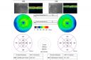
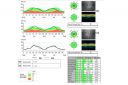
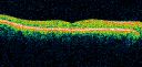
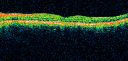
OCT SCAN: The OCT scan shows a normal foveal contour with normal retinal architecture. Photos confirm clinical findings.
FLUORESCEIN ANGIOGRAPHY: Fluorescein angiography shows a very minimal patchy hyperfluorescence in the macula, which is not significantly different than normal.
IMPRESSION:
1. PROBABLE CONGENITAL ACHROMATOPSIA
DISCUSSION: I explained to the patient that I do not see any reason why any other diagnosis would fit his particular condition. Of course electroretinography would be diagnostic in a case such as this and I suggested he continue using dim lightning and sun glasses, which he is already doing.
|

Congenital Achromatopsia 1016 views26-year-old man was diagnosed with congenital achromatopsia by ERG as a child. He has family members who also have this disorder. OD 20/200, OS 20/200.
(0 votes)
|
|
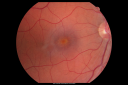
Congenital Achromatopsia 902 views26-year-old man was diagnosed with congenital achromatopsia by ERG as a child. He has family members who also have this disorder. OD 20/200, OS 20/200.
(0 votes)
|
|
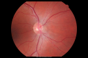
Congenital Achromatopsia 673 views26-year-old man was diagnosed with congenital achromatopsia by ERG as a child. He has family members who also have this disorder. OD 20/200, OS 20/200.
(0 votes)
|
|
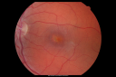
Congenital Achromatopsia 898 views26-year-old man was diagnosed with congenital achromatopsia by ERG as a child. He has family members who also have this disorder. OD 20/200, OS 20/200.
(0 votes)
|
|
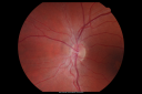
Congenital Achromatopsia 659 views26-year-old man was diagnosed with congenital achromatopsia by ERG as a child. He has family members who also have this disorder. OD 20/200, OS 20/200.
(0 votes)
|
|
Congenital Achromatopsia 679 views26-year-old man was diagnosed with congenital achromatopsia by ERG as a child. He has family members who also have this disorder. OD 20/200, OS 20/200.
(0 votes)
|
|
Congenital Achromatopsia 581 views26-year-old man was diagnosed with congenital achromatopsia by ERG as a child. He has family members who also have this disorder. OD 20/200, OS 20/200.
(0 votes)
|
|
Congenital Achromatopsia 553 views26-year-old man was diagnosed with congenital achromatopsia by ERG as a child. He has family members who also have this disorder. OD 20/200, OS 20/200.
(0 votes)
|
|
Congenital Achromatopsia 703 views26-year-old man was diagnosed with congenital achromatopsia by ERG as a child. He has family members who also have this disorder. OD 20/200, OS 20/200.
(0 votes)
|
|
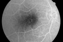
Congenital Achromatopsia 689 views26-year-old man was diagnosed with congenital achromatopsia by ERG as a child. He has family members who also have this disorder. OD 20/200, OS 20/200.
(0 votes)
|
|
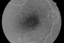
Congenital Achromatopsia 591 views26-year-old man was diagnosed with congenital achromatopsia by ERG as a child. He has family members who also have this disorder. OD 20/200, OS 20/200.
(0 votes)
|
|
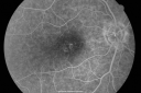
Congenital Achromatopsia 550 views26-year-old man was diagnosed with congenital achromatopsia by ERG as a child. He has family members who also have this disorder. OD 20/200, OS 20/200.
(0 votes)
|
|
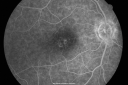
Congenital Achromatopsia 580 views26-year-old man was diagnosed with congenital achromatopsia by ERG as a child. He has family members who also have this disorder. OD 20/200, OS 20/200.
(0 votes)
|
|
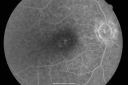
Congenital Achromatopsia 618 views26-year-old man was diagnosed with congenital achromatopsia by ERG as a child. He has family members who also have this disorder. OD 20/200, OS 20/200.
(0 votes)
|
|
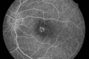
Congenital Achromatopsia 672 views26-year-old man was diagnosed with congenital achromatopsia by ERG as a child. He has family members who also have this disorder. OD 20/200, OS 20/200.
(0 votes)
|
|
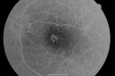
Congenital Achromatopsia 622 views26-year-old man was diagnosed with congenital achromatopsia by ERG as a child. He has family members who also have this disorder. OD 20/200, OS 20/200.
(0 votes)
|
|
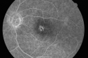
Congenital Achromatopsia 756 views26-year-old man was diagnosed with congenital achromatopsia by ERG as a child. He has family members who also have this disorder. OD 20/200, OS 20/200.
(0 votes)
|
|
|
|
|
|
|
26-year-old man who I saw in the office on June 25, 2009. He lived in Knoxville, Tennessee and was diagnosed there with congenital achromatopsia. He does have family members who also have this disorder. He says he sees better in dim lighting than in bright lighting. He believes as a child he might have had electroretinography testing. He has not had any testing done lately.
VISUAL ACUITY: OD 20/200, OS 20/200. IOP: OD 17, OS 18.
SLIT EXAMINATION: Normal with clear lenses.
EXTENDED OPHTHALMOSCOPY:
OD: Vertical C/D ratio is 0.1. There is no posterior vitreous separation. The macula looks normal.
OS: Vertical C/D ratio is 0.1. There is no posterior vitreous separation. The macula and periphery look normal.
OCT SCAN: The OCT scan shows a normal foveal contour with normal retinal architecture. Photos confirm clinical findings.
FLUORESCEIN ANGIOGRAPHY: Fluorescein angiography shows a very minimal patchy hyperfluorescence in the macula, which is not significantly different than normal.
IMPRESSION:
1. PROBABLE CONGENITAL ACHROMATOPSIA
DISCUSSION: I explained to the patient that I do not see any reason why any other diagnosis would fit his particular condition. Of course electroretinography would be diagnostic in a case such as this and I suggested he continue using dim lightning and sun glasses, which he is already doing.


