Pigmented Growing Macular Lesion - Melanoma
|
|
Initial Visit and Photos:
71-year-old man was seen in the office on 8/26/2011. He has been playing tennis and notices poor vision while he is playing. He states his vision has been bothering him for about a month. He does not notice one eye particularly worse than the other. He has cataracts but you were concerned about his macula and suggested he come here for an evaluation.
VISUAL ACUITY: Vision OD is 20/40, OS is 20/25. IOP: 18 in both eyes. His color vision is normal. There is 3+ nuclear sclerosis in both eyes.
EXTENDED OPHTHALMOSCOPY:
OD: Vertical C/D ratio is 0.3. There is an irregularly pigmented lesion in the macula in the right eye. It is a total of 6 mm across and about 6 mm top to bottom. In addition to the pigmented lesion under the macula, which has some orange-looking pigment spots in the center of it, there is another area of atrophy inferior to the optic nerve, a disc diameter away and a disc diameter in size. Then inferonasal to the optic nerve, there is a nevus a disc diameter in size with drusen over the surface.
OS: Vertical C/D ratio is 0.3. There is 2+ asteroid hyalosis and 2+ epiretinal membrane.
ULTRASOUND: The right eye shows the lesion to be about 1 mm thick.
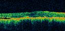
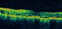
OCT SCAN: The OCT scan of the right eye shows what looks to be a shallow area of subretinal fluid inferior to the fovea. The left eye shows flattening of the macula from the macular pucker.
PHOTOGRAPHS: Photos confirm clinical findings.
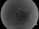
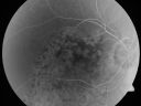
FLUORESCEIN ANGIOGRAPHY: The right eye shows hyperfluorescence corresponding to a window defect overlying the nevus. The choroidal vasculature can be easily seen throughout the area in a dependent fashion, suggestive of chronic subretinal fluid. There is also hyperfluorescence inferior to the optic nerve with hypofluorescence over the area of geographic atrophy. There is some stippled hyperfluorescence within the lesion in the late frame. The hyperfluorescence in general fades in the late frames. There is no evidence of any active leakage. The drusen over the surface of the nevus inferonasal to the optic nerve, which is overall dark, do stain in the late frames as well. The left eye is angiographically normal throughout the studies.
IMPRESSION:
1. MACULAR PUCKER – LEFT EYE
2. PROBABLE CHRONIC CHOROIDAL NEVUS WITH CHRONIC SEROUS RETINAL DETACHMENT AND PSEUDORETINITIS PIGMENTOSA CHANGES OVER THE SURFACE OF IT – RIGHT EYE
3. SECOND FLAT BENIGN CHOROIDAL NEVUS INFERONASAL TO THE OPTIC NERVE
4. AREA OF GEOGRAPHIC ATROPHY INFERIOR TO THE OPTIC NERVE, SUGGESTIVE OF PROBABLE CHRONIC SUBRETINAL FLUID
DISCUSSION: I explained to the patient the pigmented lesion in the right eye is atypical looking, but it does look like a benign choroidal nevus which has been there a long time causing pigment epithelial irritation and irregular pigmentation in the macula. There is subretinal fluid associated with it. For now, until we can be completely certain what it is, I suggest we leave the eye alone. I did ask him to return for check in a month. I need to watch it for a few visits to make sure it is not growing, but I doubt it will grow. I am about 99% confident that this is not a malignancy. If it does not grow and we need to deal with the subretinal fluid, it is possible we might be able to do some sort of treatment there, but if the subretinal fluid is not central, I do not know that the risk of that would be worth the potential benefit.
In the meantime, he was asking me about his cataracts. With 20/25 vision in the left eye and a macular pucker and 20/40 vision in the right eye and atrophy from the chronic subretinal fluid, I think the chance of cataract surgery of substantially improving his vision is relatively low and it may not be worth the potential risk. He will see you back regularly as well.
Subsequent Visit - January 3, 2012 (5 months later) - vision is a little worse and the lesion has grown and the patient is seeing more flashing lights. Possible Melanoma v. Unilateral Melanocytic Proliferation
|

Pigmented Choroidal Mass Right Eye - Melanoma vs. Nevus vs. Unilateral Melanocytic Proliferation - Initial Visit and 4 Month Follow-Up - Color Photovista 1043 vecesPigmented lesion is visible in the superior half of the macular on the follow-up visit.
(0 votos)
|
|

Pigmented Choroidal Mass Right Eye - Melanoma vs. Nevus vs. Unilateral Melanocytic Proliferation - 4 Month Follow-Up Imagesvista 750 veces71-year-old man his vision 4 month follow-up. Vision is 20/60 and patient sees flashing lights. Lesion has grown slightly in the superior half of the macula.
(0 votos)
|
|
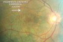
Pigmented Choroidal Mass Right Eye - Melanoma vs. Nevus vs. Unilateral Melanocytic Proliferation - 4 Month Follow-Up Imagesvista 716 veces71-year-old man his vision 4 month follow-up. Vision is 20/60 and patient sees flashing lights. Lesion has grown slightly in the superior half of the macula.
(0 votos)
|
|
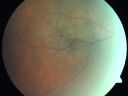
Pigmented Choroidal Mass Right Eye - Melanoma vs. Nevus vs. Unilateral Melanocytic Proliferation - 4 Month Follow-Up Imagesvista 803 veces71-year-old man his vision 4 month follow-up. Vision is 20/60 and patient sees flashing lights. Lesion has grown slightly in the superior half of the macula.
(0 votos)
|
|
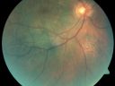
Pigmented Choroidal Mass Right Eye - Melanoma vs. Nevus vs. Unilateral Melanocytic Proliferation - 4 Month Follow-Up Imagesvista 726 veces71-year-old man his vision 4 month follow-up. Vision is 20/60 and patient sees flashing lights. Lesion has grown slightly in the superior half of the macula.
(0 votos)
|
|
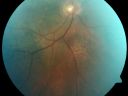
Pigmented Choroidal Mass Right Eye - Melanoma vs. Nevus vs. Unilateral Melanocytic Proliferation - 4 Month Follow-Up Imagesvista 668 veces71-year-old man his vision 4 month follow-up. Vision is 20/60 and patient sees flashing lights. Lesion has grown slightly in the superior half of the macula.
(0 votos)
|
|
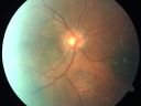
Pigmented Choroidal Mass Right Eye - Melanoma vs. Nevus vs. Unilateral Melanocytic Proliferation - 4 Month Follow-Up Imagesvista 713 veces71-year-old man his vision 4 month follow-up. Vision is 20/60 and patient sees flashing lights. Lesion has grown slightly in the superior half of the macula.
(0 votos)
|
|
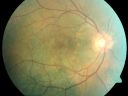
Pigmented Choroidal Mass Right Eye - Melanoma vs. Nevus vs. Unilateral Melanocytic Proliferation - 4 Month Follow-Up Imagesvista 662 veces71-year-old man his vision 4 month follow-up. Vision is 20/60 and patient sees flashing lights. Lesion has grown slightly in the superior half of the macula.
(0 votos)
|
|
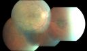
Pigmented Choroidal Mass Right Eye - Melanoma vs. Nevus vs. Unilateral Melanocytic Proliferation - 4 Month Follow-Up Imagesvista 614 veces71-year-old man his vision 4 month follow-up. Vision is 20/60 and patient sees flashing lights. Lesion has grown slightly in the superior half of the macula.
(0 votos)
|
|

Pigmented Choroidal Mass Right Eye - Melanoma vs. Nevus vs. Unilateral Melanocytic Proliferation - 4 Month Follow-Up Imagesvista 624 veces71-year-old man his vision 4 month follow-up. Vision is 20/60 and patient sees flashing lights. Lesion has grown slightly in the superior half of the macula.
(0 votos)
|
|
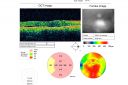
Pigmented Choroidal Mass Right Eye - Melanoma vs. Nevus vs. Unilateral Melanocytic Proliferation - 4 Month Follow-Up Imagesvista 660 veces71-year-old man his vision 4 month follow-up. Vision is 20/60 and patient sees flashing lights. Lesion has grown slightly in the superior half of the macula.
(0 votos)
|
|
Pigmented Choroidal Mass Right Eye - Melanoma vs. Nevus vs. Unilateral Melanocytic Proliferation -4 Month Follow-Up Imagesvista 552 veces71-year-old man his vision 4 month follow-up. Vision is 20/60 and patient sees flashing lights. Lesion has grown slightly in the superior half of the macula.
(0 votos)
|
|
Pigmented Choroidal Mass Right Eye - Melanoma vs. Nevus vs. Unilateral Melanocytic Proliferation - 4 Month Follow-Up Imagesvista 556 veces71-year-old man his vision 4 month follow-up. Vision is 20/60 and patient sees flashing lights. Lesion has grown slightly in the superior half of the macula.
(0 votos)
|
|
Pigmented Choroidal Mass Right Eye - Melanoma vs. Nevus vs. Unilateral Melanocytic Proliferation - 4 Month Follow-Up Imagesvista 589 veces71-year-old man his vision 4 month follow-up. Vision is 20/60 and patient sees flashing lights. Lesion has grown slightly in the superior half of the macula.
(0 votos)
|
|
Pigmented Choroidal Mass Right Eye - Melanoma vs. Nevus vs. Unilateral Melanocytic Proliferation - 4 Month Follow-Up Imagesvista 603 veces71-year-old man his vision 4 month follow-up. Vision is 20/60 and patient sees flashing lights. Lesion has grown slightly in the superior half of the macula.
(0 votos)
|
|
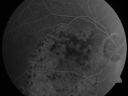
Pigmented Choroidal Mass Right Eye - Melanoma vs. Nevus vs. Unilateral Melanocytic Proliferation - 4 Month Follow-Up Imagesvista 556 veces71-year-old man his vision 4 month follow-up. Vision is 20/60 and patient sees flashing lights. Lesion has grown slightly in the superior half of the macula.
(0 votos)
|
|

Pigmented Choroidal Mass Right Eye - Melanoma vs. Nevus vs. Unilateral Melanocytic Proliferation - 4 Month Follow-Up Imagesvista 550 veces71-year-old man his vision 4 month follow-up. Vision is 20/60 and patient sees flashing lights. Lesion has grown slightly in the superior half of the macula.
(0 votos)
|
|
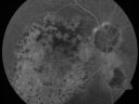
Pigmented Choroidal Mass Right Eye - Melanoma vs. Nevus vs. Unilateral Melanocytic Proliferation - 4 Month Follow-Up Imagesvista 526 veces71-year-old man his vision 4 month follow-up. Vision is 20/60 and patient sees flashing lights. Lesion has grown slightly in the superior half of the macula.
(0 votos)
|
|
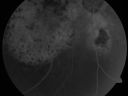
Pigmented Choroidal Mass Right Eye - Melanoma vs. Nevus vs. Unilateral Melanocytic Proliferation - 4 Month Follow-Up Imagesvista 528 veces71-year-old man his vision 4 month follow-up. Vision is 20/60 and patient sees flashing lights. Lesion has grown slightly in the superior half of the macula.
(0 votos)
|
|
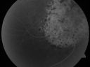
Pigmented Choroidal Mass Right Eye - Melanoma vs. Nevus vs. Unilateral Melanocytic Proliferation - 4 Month Follow-Up Images vista 558 veces71-year-old man his vision 4 month follow-up. Vision is 20/60 and patient sees flashing lights. Lesion has grown slightly in the superior half of the macula.
(0 votos)
|
|
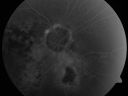
Pigmented Choroidal Mass Right Eye - Melanoma vs. Nevus vs. Unilateral Melanocytic Proliferation - 4 Month Follow-Up Imagesvista 500 veces71-year-old man his vision 4 month follow-up. Vision is 20/60 and patient sees flashing lights. Lesion has grown slightly in the superior half of the macula.
(0 votos)
|
|

Pigmented Choroidal Mass Right Eye - Melanoma vs. Nevus vs. Unilateral Melanocytic Proliferation - 4 Month Follow-Up Imagesvista 494 veces71-year-old man his vision 4 month follow-up. Vision is 20/60 and patient sees flashing lights. Lesion has grown slightly in the superior half of the macula.
(0 votos)
|
|
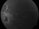
Pigmented Choroidal Mass Right Eye - Melanoma vs. Nevus vs. Unilateral Melanocytic Proliferation - 4 Month Follow-Up Imagesvista 505 veces71-year-old man his vision 4 month follow-up. Vision is 20/60 and patient sees flashing lights. Lesion has grown slightly in the superior half of the macula.
(0 votos)
|
|
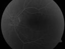
Pigmented Choroidal Mass Right Eye - Melanoma vs. Nevus vs. Unilateral Melanocytic Proliferation - 4 Month Follow-Up Imagesvista 492 veces71-year-old man his vision 4 month follow-up. Vision is 20/60 and patient sees flashing lights. Lesion has grown slightly in the superior half of the macula.
(0 votos)
|
|
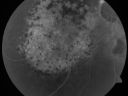
Pigmented Choroidal Mass Right Eye - Melanoma vs. Nevus vs. Unilateral Melanocytic Proliferation - 4 Month Follow-Up Imagesvista 572 veces71-year-old man his vision 4 month follow-up. Vision is 20/60 and patient sees flashing lights. Lesion has grown slightly in the superior half of the macula.
(0 votos)
|
|
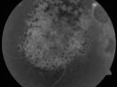
Pigmented Choroidal Mass Right Eye - Melanoma vs. Nevus vs. Unilateral Melanocytic Proliferation - 4 Month Follow-Up Imagesvista 566 veces71-year-old man his vision 4 month follow-up. Vision is 20/60 and patient sees flashing lights. Lesion has grown slightly in the superior half of the macula.
(0 votos)
|
|
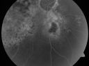
Pigmented Choroidal Mass Right Eye - Melanoma vs. Nevus vs. Unilateral Melanocytic Proliferation - 4 Month Follow-Up Imagesvista 518 veces71-year-old man his vision 4 month follow-up. Vision is 20/60 and patient sees flashing lights. Lesion has grown slightly in the superior half of the macula.
(0 votos)
|
|
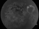
Pigmented Choroidal Mass Right Eye - Melanoma vs. Nevus vs. Unilateral Melanocytic Proliferation - 4 Month Follow-Up Images vista 498 veces71-year-old man his vision 4 month follow-up. Vision is 20/60 and patient sees flashing lights. Lesion has grown slightly in the superior half of the macula.
(0 votos)
|
|
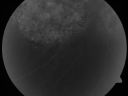
Pigmented Choroidal Mass Right Eye - Melanoma vs. Nevus vs. Unilateral Melanocytic Proliferation - 4 Month Follow-Up Imagesvista 507 veces71-year-old man his vision 4 month follow-up. Vision is 20/60 and patient sees flashing lights. Lesion has grown slightly in the superior half of the macula.
(0 votos)
|
|

Pigmented Choroidal Mass Right Eye - Melanoma vs. Nevus vs. Unilateral Melanocytic Proliferation - 4 Month Follow-Up Imagesvista 483 veces71-year-old man his vision 4 month follow-up. Vision is 20/60 and patient sees flashing lights. Lesion has grown slightly in the superior half of the macula.
(0 votos)
|
|
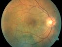
Pigmented Choroidal Mass Right Eye - Melanoma vs. Nevus vs. Unilateral Melanocytic Proliferation - Initial Visitvista 572 veces71-year-old man is vision has been bothering him for about a month. He does not notice one eye particularly worse than the other. OD is 20/40, OS is 20/25 - Initial Visit
(0 votos)
|
|
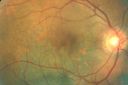
Pigmented Choroidal Mass Right Eye - Melanoma vs. Nevus vs. Unilateral Melanocytic Proliferation - Initial Visitvista 542 veces71-year-old man is vision has been bothering him for about a month. He does not notice one eye particularly worse than the other. OD is 20/40, OS is 20/25 - Initial Visit
(0 votos)
|
|
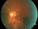
Pigmented Choroidal Mass Right Eye - Melanoma vs. Nevus vs. Unilateral Melanocytic Proliferation - Initial Visitvista 539 veces71-year-old man is vision has been bothering him for about a month. He does not notice one eye particularly worse than the other. OD is 20/40, OS is 20/25 - Initial Visit
(0 votos)
|
|
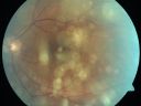
Pigmented Choroidal Mass Right Eye - Melanoma vs. Nevus vs. Unilateral Melanocytic Proliferation - Initial Visitvista 529 veces71-year-old man is vision has been bothering him for about a month. He does not notice one eye particularly worse than the other. OD is 20/40, OS is 20/25 - Initial Visit
(0 votos)
|
|
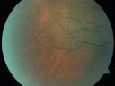
Pigmented Choroidal Mass Right Eye - Melanoma vs. Nevus vs. Unilateral Melanocytic Proliferation - Initial Visitvista 501 veces71-year-old man is vision has been bothering him for about a month. He does not notice one eye particularly worse than the other. OD is 20/40, OS is 20/25 - Initial Visit
(0 votos)
|
|
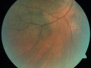
Pigmented Choroidal Mass Right Eye - Melanoma vs. Nevus vs. Unilateral Melanocytic Proliferation - Initial Visitvista 589 veces71-year-old man is vision has been bothering him for about a month. He does not notice one eye particularly worse than the other. OD is 20/40, OS is 20/25 - Initial Visit
(0 votos)
|
|
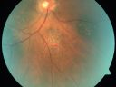
Pigmented Choroidal Mass Right Eye - Melanoma vs. Nevus vs. Unilateral Melanocytic Proliferation - Initial Visitvista 591 veces71-year-old man is vision has been bothering him for about a month. He does not notice one eye particularly worse than the other. OD is 20/40, OS is 20/25 - Initial Visit
(0 votos)
|
|
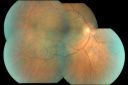
Pigmented Choroidal Mass Right Eye - Melanoma vs. Nevus vs. Unilateral Melanocytic Proliferation - Initial Visitvista 564 veces71-year-old man is vision has been bothering him for about a month. He does not notice one eye particularly worse than the other. OD is 20/40, OS is 20/25 - Initial Visit
(0 votos)
|
|

Pigmented Choroidal Mass Right Eye - Melanoma vs. Nevus vs. Unilateral Melanocytic Proliferation - Initial Visitvista 513 veces71-year-old man is vision has been bothering him for about a month. He does not notice one eye particularly worse than the other. OD is 20/40, OS is 20/25 - Initial Visit
(0 votos)
|
|

Pigmented Choroidal Mass Right Eye - Melanoma vs. Nevus vs. Unilateral Melanocytic Proliferation - Initial Visitvista 477 veces71-year-old man is vision has been bothering him for about a month. He does not notice one eye particularly worse than the other. OD is 20/40, OS is 20/25 - Initial Visit
(0 votos)
|
|
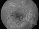
Pigmented Choroidal Mass Right Eye - Melanoma vs. Nevus vs. Unilateral Melanocytic Proliferation - Initial Visitvista 484 veces71-year-old man is vision has been bothering him for about a month. He does not notice one eye particularly worse than the other. OD is 20/40, OS is 20/25 - Initial Visit
(0 votos)
|
|
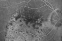
Pigmented Choroidal Mass Right Eye - Melanoma vs. Nevus vs. Unilateral Melanocytic Proliferation - Initial Visitvista 593 veces71-year-old man is vision has been bothering him for about a month. He does not notice one eye particularly worse than the other. OD is 20/40, OS is 20/25 - Initial Visit
(0 votos)
|
|
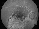
Pigmented Choroidal Mass Right Eye - Melanoma vs. Nevus vs. Unilateral Melanocytic Proliferation - Initial Visitvista 516 veces71-year-old man is vision has been bothering him for about a month. He does not notice one eye particularly worse than the other. OD is 20/40, OS is 20/25 - Initial Visit
(0 votos)
|
|
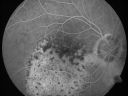
Pigmented Choroidal Mass Right Eye - Melanoma vs. Nevus vs. Unilateral Melanocytic Proliferation - Initial Visitvista 513 veces71-year-old man is vision has been bothering him for about a month. He does not notice one eye particularly worse than the other. OD is 20/40, OS is 20/25 - Initial Visit
(0 votos)
|
|
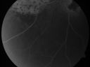
Pigmented Choroidal Mass Right Eye - Melanoma vs. Nevus vs. Unilateral Melanocytic Proliferation - Initial Visitvista 521 veces71-year-old man is vision has been bothering him for about a month. He does not notice one eye particularly worse than the other. OD is 20/40, OS is 20/25 - Initial Visit
(0 votos)
|
|
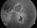
Pigmented Choroidal Mass Right Eye - Melanoma vs. Nevus vs. Unilateral Melanocytic Proliferation - Initial Visitvista 517 veces71-year-old man is vision has been bothering him for about a month. He does not notice one eye particularly worse than the other. OD is 20/40, OS is 20/25 - Initial Visit
(0 votos)
|
|
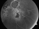
Pigmented Choroidal Mass Right Eye - Melanoma vs. Nevus vs. Unilateral Melanocytic Proliferation - Initial Visitvista 513 veces71-year-old man is vision has been bothering him for about a month. He does not notice one eye particularly worse than the other. OD is 20/40, OS is 20/25 - Initial Visit
(0 votos)
|
|

Pigmented Choroidal Mass Right Eye - Melanoma vs. Nevus vs. Unilateral Melanocytic Proliferation - Initial Visitvista 510 veces71-year-old man is vision has been bothering him for about a month. He does not notice one eye particularly worse than the other. OD is 20/40, OS is 20/25 - Initial Visit
(0 votos)
|
|
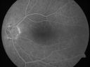
Pigmented Choroidal Mass Right Eye - Melanoma vs. Nevus vs. Unilateral Melanocytic Proliferation - Initial Visitvista 571 veces71-year-old man is vision has been bothering him for about a month. He does not notice one eye particularly worse than the other. OD is 20/40, OS is 20/25 - Initial Visit
(0 votos)
|
|
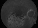
Pigmented Choroidal Mass Right Eye - Melanoma vs. Nevus vs. Unilateral Melanocytic Proliferation - Initial Visitvista 537 veces71-year-old man is vision has been bothering him for about a month. He does not notice one eye particularly worse than the other. OD is 20/40, OS is 20/25 - Initial Visit
(0 votos)
|
|
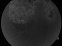
Pigmented Choroidal Mass Right Eye - Melanoma vs. Nevus vs. Unilateral Melanocytic Proliferation - Initial Visitvista 566 veces71-year-old man is vision has been bothering him for about a month. He does not notice one eye particularly worse than the other. OD is 20/40, OS is 20/25 - Initial Visit71-year-old man is vision has been bothering him for about a month. He does not notice one eye particularly worse than the other. OD is 20/40, OS is 20/25 - Initial Visit
(0 votos)
|
|
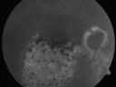
Pigmented Choroidal Mass Right Eye - Melanoma vs. Nevus vs. Unilateral Melanocytic Proliferation - Initial Visitvista 546 veces71-year-old man is vision has been bothering him for about a month. He does not notice one eye particularly worse than the other. OD is 20/40, OS is 20/25 - Initial Visit
(0 votos)
|
|
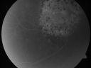
Pigmented Choroidal Mass Right Eye - Melanoma vs. Nevus vs. Unilateral Melanocytic Proliferation - Initial Visitvista 581 veces71-year-old man is vision has been bothering him for about a month. He does not notice one eye particularly worse than the other. OD is 20/40, OS is 20/25 - Initial Visit
(0 votos)
|
|
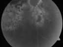
Pigmented Choroidal Mass Right Eye - Melanoma vs. Nevus vs. Unilateral Melanocytic Proliferation - Initial Visitvista 661 veces71-year-old man is vision has been bothering him for about a month. He does not notice one eye particularly worse than the other. OD is 20/40, OS is 20/25 - Initial Visit71-year-old man is vision has been bothering him for about a month. He does not notice one eye particularly worse than the other. OD is 20/40, OS is 20/25 - Initial Visit
(0 votos)
|
|
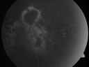
Pigmented Choroidal Mass Right Eye - Melanoma vs. Nevus vs. Unilateral Melanocytic Proliferation - Initial Visitvista 724 veces71-year-old man is vision has been bothering him for about a month. He does not notice one eye particularly worse than the other. OD is 20/40, OS is 20/25 - Initial Visit
(0 votos)
|
|
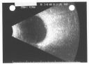
Pigmented Choroidal Mass Right Eye - Melanoma vs. Nevus vs. Unilateral Melanocytic Proliferation - Initial Visit B Scanvista 904 veces71-year-old man is vision has been bothering him for about a month. He does not notice one eye particularly worse than the other. OD is 20/40, OS is 20/25 - Initial Visit
(0 votos)
|
|
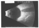
Pigmented Choroidal Mass Right Eye - Melanoma vs. Nevus vs. Unilateral Melanocytic Proliferation - Initial Visit B Scanvista 1437 veces71-year-old man is vision has been bothering him for about a month. He does not notice one eye particularly worse than the other. OD is 20/40, OS is 20/25 - Initial Visit
(0 votos)
|
|
|
|
|
| 57 archivos en 1 página(s) |
|
Initial Visit and Photos:
71-year-old man was seen in the office on 8/26/2011. He has been playing tennis and notices poor vision while he is playing. He states his vision has been bothering him for about a month. He does not notice one eye particularly worse than the other. He has cataracts but you were concerned about his macula and suggested he come here for an evaluation.
VISUAL ACUITY: Vision OD is 20/40, OS is 20/25. IOP: 18 in both eyes. His color vision is normal. There is 3+ nuclear sclerosis in both eyes.
EXTENDED OPHTHALMOSCOPY:
OD: Vertical C/D ratio is 0.3. There is an irregularly pigmented lesion in the macula in the right eye. It is a total of 6 mm across and about 6 mm top to bottom. In addition to the pigmented lesion under the macula, which has some orange-looking pigment spots in the center of it, there is another area of atrophy inferior to the optic nerve, a disc diameter away and a disc diameter in size. Then inferonasal to the optic nerve, there is a nevus a disc diameter in size with drusen over the surface.
OS: Vertical C/D ratio is 0.3. There is 2+ asteroid hyalosis and 2+ epiretinal membrane.
ULTRASOUND: The right eye shows the lesion to be about 1 mm thick.
OCT SCAN: The OCT scan of the right eye shows what looks to be a shallow area of subretinal fluid inferior to the fovea. The left eye shows flattening of the macula from the macular pucker.
PHOTOGRAPHS: Photos confirm clinical findings.
FLUORESCEIN ANGIOGRAPHY: The right eye shows hyperfluorescence corresponding to a window defect overlying the nevus. The choroidal vasculature can be easily seen throughout the area in a dependent fashion, suggestive of chronic subretinal fluid. There is also hyperfluorescence inferior to the optic nerve with hypofluorescence over the area of geographic atrophy. There is some stippled hyperfluorescence within the lesion in the late frame. The hyperfluorescence in general fades in the late frames. There is no evidence of any active leakage. The drusen over the surface of the nevus inferonasal to the optic nerve, which is overall dark, do stain in the late frames as well. The left eye is angiographically normal throughout the studies.
IMPRESSION:
1. MACULAR PUCKER – LEFT EYE
2. PROBABLE CHRONIC CHOROIDAL NEVUS WITH CHRONIC SEROUS RETINAL DETACHMENT AND PSEUDORETINITIS PIGMENTOSA CHANGES OVER THE SURFACE OF IT – RIGHT EYE
3. SECOND FLAT BENIGN CHOROIDAL NEVUS INFERONASAL TO THE OPTIC NERVE
4. AREA OF GEOGRAPHIC ATROPHY INFERIOR TO THE OPTIC NERVE, SUGGESTIVE OF PROBABLE CHRONIC SUBRETINAL FLUID
DISCUSSION: I explained to the patient the pigmented lesion in the right eye is atypical looking, but it does look like a benign choroidal nevus which has been there a long time causing pigment epithelial irritation and irregular pigmentation in the macula. There is subretinal fluid associated with it. For now, until we can be completely certain what it is, I suggest we leave the eye alone. I did ask him to return for check in a month. I need to watch it for a few visits to make sure it is not growing, but I doubt it will grow. I am about 99% confident that this is not a malignancy. If it does not grow and we need to deal with the subretinal fluid, it is possible we might be able to do some sort of treatment there, but if the subretinal fluid is not central, I do not know that the risk of that would be worth the potential benefit.
In the meantime, he was asking me about his cataracts. With 20/25 vision in the left eye and a macular pucker and 20/40 vision in the right eye and atrophy from the chronic subretinal fluid, I think the chance of cataract surgery of substantially improving his vision is relatively low and it may not be worth the potential risk. He will see you back regularly as well.
Subsequent Visit - January 3, 2012 (5 months later) - vision is a little worse and the lesion has grown and the patient is seeing more flashing lights. Possible Melanoma v. Unilateral Melanocytic Proliferation


