Acute Central Serous Retinopathy - Fundus Autofluorescence and ICG
|
|
42-year-old man was seen in the office on October 5, 2011. He had noticed starting in August after a course of antibiotic and steroids, that he developed new spots in his vision in the right eye. He may have had an episode like this sometime in the past. He did take steroids a few years ago and his vision did change at that time, but then returned.
VISUAL ACUITY: OD 20/32, OS 20/32. IOP: OD 10, OS 15.
SLIT EXAMINATION: There is 1+ nuclear sclerosis in both eyes.
EXTENDED OPHTHALMOSCOPY:
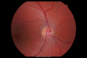
OD: Vertical C/D ratio is 0.1. There is a serous retinal detachment temporal to the fovea.
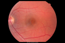
OS: Vertical C/D ratio is 0.1. There are macular pigment spots. There is no subretinal fluid.
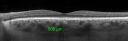
SPECTRALIS-SD-OCT SCAN: The OCT scan of the right eye confirms subretinal fluid. The left eye shows no subretinal fluid, but there are areas of pigment epithelial irregularities and there is one under the fovea. Photos confirm clinical findings. The enhanced step imaging of the right eye shows a choroidal thickness centrally of 537 microns and the left eye has 505 microns. These numbers are about double to normal.
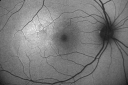
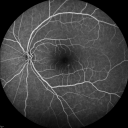
FUNDUS AUTO FLUORESCENCE: The fundus auto fluorescence shows typical hyper auto fluorescence inside the serous macular detachment from shed photoreceptor outer segments, the left eye on the fundus auto fluorescence is nearly normal. FLUORESCEIN ANGIOGRAPHY: Fluorescein angiography shows an area of hyperfluorescence superotemporal to the fovea, which minimally leaks in the late frames. The left eye shows some hyperfluorescence centrally, which stains.
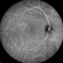
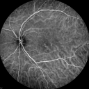
INDOCYANINE GREEN ANGIOGRAPHY: The indocyanine green angiogram in both eyes shows areas of capillary hyper permeability in the right eye superotemporally and the left eye centrally.
IMPRESSION:
1. CENTRAL SEROUS RETINOPATHY – BOTH EYES
2. SEROUS RETINAL DETACHMENT – RIGHT EYE
DISCUSSION: I explained to the patient he does have central serous retinopathy and that 95 percent of patients with this problem do recover vision without any incident. I warned him that sometimes the cases become chronic and they need treatment, but I think since this was probably steroid induced that he should recover over the next few months. I asked him to return in three months. I told him if his vision is fine then though, he doesn’t need to keep the appointment.
|

Central Serous Retinopathy Acute 1182 views42-year-old man was seen in the office on October 5, 2011. He had noticed starting in August after a course of antibiotic and steroids, that he developed new spots in his vision in the right eye. He may have had an episode like this sometime in the past. He did take steroids a few years ago and his vision did change at that time, but then returned.
VISUAL ACUITY: OD 20/32, OS 20/32
(0 votes)
|
|
Central Serous Retinopathy Acute 824 views42-year-old man was seen in the office on October 5, 2011. He had noticed starting in August after a course of antibiotic and steroids, that he developed new spots in his vision in the right eye. He may have had an episode like this sometime in the past. He did take steroids a few years ago and his vision did change at that time, but then returned.
VISUAL ACUITY: OD 20/32, OS 20/32
(0 votes)
|
|
Central Serous Retinopathy Acute 769 views42-year-old man was seen in the office on October 5, 2011. He had noticed starting in August after a course of antibiotic and steroids, that he developed new spots in his vision in the right eye. He may have had an episode like this sometime in the past. He did take steroids a few years ago and his vision did change at that time, but then returned.
VISUAL ACUITY: OD 20/32, OS 20/32
(0 votes)
|
|

Central Serous Retinopathy Acute 768 views42-year-old man was seen in the office on October 5, 2011. He had noticed starting in August after a course of antibiotic and steroids, that he developed new spots in his vision in the right eye. He may have had an episode like this sometime in the past. He did take steroids a few years ago and his vision did change at that time, but then returned.
VISUAL ACUITY: OD 20/32, OS 20/32
(0 votes)
|
|

Central Serous Retinopathy Acute 917 views42-year-old man was seen in the office on October 5, 2011. He had noticed starting in August after a course of antibiotic and steroids, that he developed new spots in his vision in the right eye. He may have had an episode like this sometime in the past. He did take steroids a few years ago and his vision did change at that time, but then returned.
VISUAL ACUITY: OD 20/32, OS 20/32
(0 votes)
|
|

Central Serous Retinopathy Acute - Enhanced Depth Imaging OCT - Thickened Choriod917 views42-year-old man was seen in the office on October 5, 2011. He had noticed starting in August after a course of antibiotic and steroids, that he developed new spots in his vision in the right eye. He may have had an episode like this sometime in the past. He did take steroids a few years ago and his vision did change at that time, but then returned.
VISUAL ACUITY: OD 20/32, OS 20/32
(0 votes)
|
|
Central Serous Retinopathy Acute 817 views42-year-old man was seen in the office on October 5, 2011. He had noticed starting in August after a course of antibiotic and steroids, that he developed new spots in his vision in the right eye. He may have had an episode like this sometime in the past. He did take steroids a few years ago and his vision did change at that time, but then returned.
VISUAL ACUITY: OD 20/32, OS 20/32
(0 votes)
|
|

Central Serous Retinopathy Acute - Enhanced Depth Imaging OCT - Thickened Choriod981 views42-year-old man was seen in the office on October 5, 2011. He had noticed starting in August after a course of antibiotic and steroids, that he developed new spots in his vision in the right eye. He may have had an episode like this sometime in the past. He did take steroids a few years ago and his vision did change at that time, but then returned.
VISUAL ACUITY: OD 20/32, OS 20/32
(0 votes)
|
|

Central Serous Retinopathy Acute - Fundus Autofluorescence1211 views42-year-old man was seen in the office on October 5, 2011. He had noticed starting in August after a course of antibiotic and steroids, that he developed new spots in his vision in the right eye. He may have had an episode like this sometime in the past. He did take steroids a few years ago and his vision did change at that time, but then returned.
VISUAL ACUITY: OD 20/32, OS 20/32
(0 votes)
|
|
Central Serous Retinopathy Acute - Fundus Autofluorescence1193 views42-year-old man was seen in the office on October 5, 2011. He had noticed starting in August after a course of antibiotic and steroids, that he developed new spots in his vision in the right eye. He may have had an episode like this sometime in the past. He did take steroids a few years ago and his vision did change at that time, but then returned.
VISUAL ACUITY: OD 20/32, OS 20/32
(1 votes)
|
|
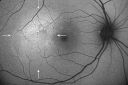
Central Serous Retinopathy Acute - Fundus Autofluorescence1464 views42-year-old man was seen in the office on October 5, 2011. He had noticed starting in August after a course of antibiotic and steroids, that he developed new spots in his vision in the right eye. He may have had an episode like this sometime in the past. He did take steroids a few years ago and his vision did change at that time, but then returned.
VISUAL ACUITY: OD 20/32, OS 20/32
(1 votes)
|
|

Central Serous Retinopathy Acute - Fundus Autofluorescence1300 views42-year-old man was seen in the office on October 5, 2011. He had noticed starting in August after a course of antibiotic and steroids, that he developed new spots in his vision in the right eye. He may have had an episode like this sometime in the past. He did take steroids a few years ago and his vision did change at that time, but then returned.
VISUAL ACUITY: OD 20/32, OS 20/32
(1 votes)
|
|
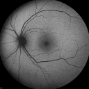
Central Serous Retinopathy Acute - Fundus Autofluorescence1374 views42-year-old man was seen in the office on October 5, 2011. He had noticed starting in August after a course of antibiotic and steroids, that he developed new spots in his vision in the right eye. He may have had an episode like this sometime in the past. He did take steroids a few years ago and his vision did change at that time, but then returned.
VISUAL ACUITY: OD 20/32, OS 20/32
(0 votes)
|
|
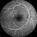
Central Serous Retinopathy Acute - Fluorescein Angiogram1316 views42-year-old man was seen in the office on October 5, 2011. He had noticed starting in August after a course of antibiotic and steroids, that he developed new spots in his vision in the right eye. He may have had an episode like this sometime in the past. He did take steroids a few years ago and his vision did change at that time, but then returned.
VISUAL ACUITY: OD 20/32, OS 20/32
(0 votes)
|
|

Central Serous Retinopathy Acute - Fluorescein Angiogram1013 views42-year-old man was seen in the office on October 5, 2011. He had noticed starting in August after a course of antibiotic and steroids, that he developed new spots in his vision in the right eye. He may have had an episode like this sometime in the past. He did take steroids a few years ago and his vision did change at that time, but then returned.
VISUAL ACUITY: OD 20/32, OS 20/32
(0 votes)
|
|
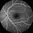
Central Serous Retinopathy Acute - Fluorescein Angiogram1213 views42-year-old man was seen in the office on October 5, 2011. He had noticed starting in August after a course of antibiotic and steroids, that he developed new spots in his vision in the right eye. He may have had an episode like this sometime in the past. He did take steroids a few years ago and his vision did change at that time, but then returned.
VISUAL ACUITY: OD 20/32, OS 20/32
(0 votes)
|
|
Central Serous Retinopathy Acute - Fluorescein Angiogram866 views42-year-old man was seen in the office on October 5, 2011. He had noticed starting in August after a course of antibiotic and steroids, that he developed new spots in his vision in the right eye. He may have had an episode like this sometime in the past. He did take steroids a few years ago and his vision did change at that time, but then returned.
VISUAL ACUITY: OD 20/32, OS 20/32
(0 votes)
|
|

Central Serous Retinopathy Acute - Indocyanine Green Angiogram - Leakage Choriocapillaris 1148 views42-year-old man was seen in the office on October 5, 2011. He had noticed starting in August after a course of antibiotic and steroids, that he developed new spots in his vision in the right eye. He may have had an episode like this sometime in the past. He did take steroids a few years ago and his vision did change at that time, but then returned.
VISUAL ACUITY: OD 20/32, OS 20/32
(0 votes)
|
|
Central Serous Retinopathy Acute - Indocyanine Green Angiogram - Leakage Choriocapillaris 1006 views42-year-old man was seen in the office on October 5, 2011. He had noticed starting in August after a course of antibiotic and steroids, that he developed new spots in his vision in the right eye. He may have had an episode like this sometime in the past. He did take steroids a few years ago and his vision did change at that time, but then returned.
VISUAL ACUITY: OD 20/32, OS 20/32
(0 votes)
|
|

Central Serous Retinopathy Acute - Indocyanine Green Angiogram - Leakage Choriocapillaris 1609 views42-year-old man was seen in the office on October 5, 2011. He had noticed starting in August after a course of antibiotic and steroids, that he developed new spots in his vision in the right eye. He may have had an episode like this sometime in the past. He did take steroids a few years ago and his vision did change at that time, but then returned.
VISUAL ACUITY: OD 20/32, OS 20/32
(1 votes)
|
|

Central Serous Retinopathy Acute - Indocyanine Green Angiogram - Leakage Choriocapillaris 1065 views42-year-old man was seen in the office on October 5, 2011. He had noticed starting in August after a course of antibiotic and steroids, that he developed new spots in his vision in the right eye. He may have had an episode like this sometime in the past. He did take steroids a few years ago and his vision did change at that time, but then returned.
VISUAL ACUITY: OD 20/32, OS 20/32
(0 votes)
|
|

Central Serous Retinopathy Acute - Indocyanine Green Angiogram - Leakage Choriocapillaris 1067 views42-year-old man was seen in the office on October 5, 2011. He had noticed starting in August after a course of antibiotic and steroids, that he developed new spots in his vision in the right eye. He may have had an episode like this sometime in the past. He did take steroids a few years ago and his vision did change at that time, but then returned.
VISUAL ACUITY: OD 20/32, OS 20/32
(0 votes)
|
|
Central Serous Retinopathy Acute - Indocyanine Green Angiogram - Leakage Choriocapillaris 1193 views42-year-old man was seen in the office on October 5, 2011. He had noticed starting in August after a course of antibiotic and steroids, that he developed new spots in his vision in the right eye. He may have had an episode like this sometime in the past. He did take steroids a few years ago and his vision did change at that time, but then returned.
VISUAL ACUITY: OD 20/32, OS 20/32
(0 votes)
|
|

Central Serous Retinopathy Acute - Indocyanine Green Angiogram - Leakage Choriocapillaris 1066 views42-year-old man was seen in the office on October 5, 2011. He had noticed starting in August after a course of antibiotic and steroids, that he developed new spots in his vision in the right eye. He may have had an episode like this sometime in the past. He did take steroids a few years ago and his vision did change at that time, but then returned.
VISUAL ACUITY: OD 20/32, OS 20/32
(0 votes)
|
|

Central Serous Retinopathy Acute - Indocyanine Green Angiogram - Leakage Choriocapillaris 937 views42-year-old man was seen in the office on October 5, 2011. He had noticed starting in August after a course of antibiotic and steroids, that he developed new spots in his vision in the right eye. He may have had an episode like this sometime in the past. He did take steroids a few years ago and his vision did change at that time, but then returned.
VISUAL ACUITY: OD 20/32, OS 20/32
(0 votes)
|
|

Central Serous Retinopathy Acute - Indocyanine Green Angiogram - Leakage Choriocapillaris 1152 views42-year-old man was seen in the office on October 5, 2011. He had noticed starting in August after a course of antibiotic and steroids, that he developed new spots in his vision in the right eye. He may have had an episode like this sometime in the past. He did take steroids a few years ago and his vision did change at that time, but then returned.
VISUAL ACUITY: OD 20/32, OS 20/32
(0 votes)
|
|
|
|
|
|
42-year-old man was seen in the office on October 5, 2011. He had noticed starting in August after a course of antibiotic and steroids, that he developed new spots in his vision in the right eye. He may have had an episode like this sometime in the past. He did take steroids a few years ago and his vision did change at that time, but then returned.
VISUAL ACUITY: OD 20/32, OS 20/32. IOP: OD 10, OS 15.
SLIT EXAMINATION: There is 1+ nuclear sclerosis in both eyes.
EXTENDED OPHTHALMOSCOPY:
OD: Vertical C/D ratio is 0.1. There is a serous retinal detachment temporal to the fovea.
OS: Vertical C/D ratio is 0.1. There are macular pigment spots. There is no subretinal fluid.
SPECTRALIS-SD-OCT SCAN: The OCT scan of the right eye confirms subretinal fluid. The left eye shows no subretinal fluid, but there are areas of pigment epithelial irregularities and there is one under the fovea. Photos confirm clinical findings. The enhanced step imaging of the right eye shows a choroidal thickness centrally of 537 microns and the left eye has 505 microns. These numbers are about double to normal.
FUNDUS AUTO FLUORESCENCE: The fundus auto fluorescence shows typical hyper auto fluorescence inside the serous macular detachment from shed photoreceptor outer segments, the left eye on the fundus auto fluorescence is nearly normal. FLUORESCEIN ANGIOGRAPHY: Fluorescein angiography shows an area of hyperfluorescence superotemporal to the fovea, which minimally leaks in the late frames. The left eye shows some hyperfluorescence centrally, which stains.
INDOCYANINE GREEN ANGIOGRAPHY: The indocyanine green angiogram in both eyes shows areas of capillary hyper permeability in the right eye superotemporally and the left eye centrally.
IMPRESSION:
1. CENTRAL SEROUS RETINOPATHY – BOTH EYES
2. SEROUS RETINAL DETACHMENT – RIGHT EYE
DISCUSSION: I explained to the patient he does have central serous retinopathy and that 95 percent of patients with this problem do recover vision without any incident. I warned him that sometimes the cases become chronic and they need treatment, but I think since this was probably steroid induced that he should recover over the next few months. I asked him to return in three months. I told him if his vision is fine then though, he doesn’t need to keep the appointment.


