83-year-old woman was seen in the office on 8/6/2010. She is a high myope with myopic macular degeneration. She had a retinal detachment repair in the right eye in 1968. I saw her in June of 2003. She had posterior colobomas and areas of pigment epithelial atrophy. She has noticed over the last six weeks substantial declining vision in the right eye. Prior to that, the vision in the two eyes was about the same. She uses Xalatan in the left eye and Alphagan in both eyes.
VISUAL ACUITY: Vision OD is 20/160, OS is 20/40. IOP: OD 17, OS 21.
SLIT LAMP EXAM: The posterior chamber intraocular lens is in good position in both eyes with open capsules.
EXTENDED OPHTHALMOSCOPY:
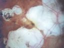
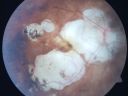
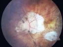
OD: Vertical C/D ratio is 0.2. There is a posterior vitreous separation. There is geographic atrophy inferior to the fovea and nasal to the fovea, squeezing the fovea, leaving about a disc diameter central pigment epithelial tissue which does look thickened. There is no frank blood. There is some chorioretinal scarring inferiorly where the retinal detachment was repaired. The retina is attached.
OS: Vertical C/D ratio is 0.8. The nerve is pale. There is atrophy temporal to the optic nerve, and inferior to the fovea there is much more pigment epithelium spared centrally in the left eye. The retinas are both attached.
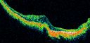
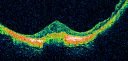
OCT SCAN: The OCT scan of the right eye shows macular edema. The left eye shows the macula to be dry.
PHOTOGRAPHS: Photos confirm clinical findings.
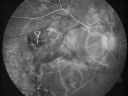
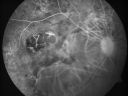
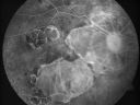
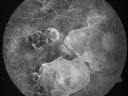
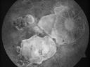
FLUORESCEIN ANGIOGRAPHY: The FA of the right eye shows hyperfluorescence but it is very subtle central hyperfluorescence in the late frames, representing a probable occult subfoveal choroidal neovascular membrane. The left eye has staining of the areas of atrophy but no increasing hyperfluorescence.
IMPRESSION:
1. MYOPIC MACULAR DEGENERATION – BOTH EYES
2. WET AGE-RELATED MACULAR DEGENERATION – RIGHT EYE
3. GLAUCOMA – WORSE IN THE LEFT EYE THAN THE RIGHT EYE
4. RECENT VISION LOSS – RIGHT EYE
5. PREVIOUS RETINAL DETACHMENT REPAIR – RIGHT EYE
6. POSTERIOR STAPHYLOMA – BOTH EYES
DISCUSSION: I explained to the patient that the right eye does appear to have macular edema from a probable occult subfoveal choroidal neovascular membrane. With intravitreal Lucentis, there is about a 90% chance of improving her vision some and a 50% chance of improving her vision a lot.
I injected the right eye with Lucentis (0.5 mg/0.05 ml) today without any difficulty. I warned her this is a monthly ongoing treatment. In her case, I would try at least three months of therapy to see if her vision might improve, and if she does improve, I would probably continue treatment. If she does not, we could talk about other possible therapies
1 year later -
Vision 20/70 in right eye and receiving every 3 month Lucentis Therapy
|
Myopic Macular Degeneration - Staphyloma - Recent Vision Loss Right Eye - Possible CNVMvu 666 fois83-year-old woman is a high myope with myopic macular degeneration. She had a retinal detachment repair in the right eye in 1968. I saw her in June of 2003. She had posterior colobomas and areas of pigment epithelial atrophy. She has noticed over the last six weeks substantial declining vision in the right eye. Prior to that, the vision in the two eyes was about the same. She uses Xalatan in the left eye and Alphagan in both eyes.
VISUAL ACUITY: Vision OD is 20/160, OS is 20/40
(0 votes)
|
|
Myopic Macular Degeneration - Staphyloma - Recent Vision Loss Right Eye - Possible CNVMvu 644 fois83-year-old woman is a high myope with myopic macular degeneration. She had a retinal detachment repair in the right eye in 1968. I saw her in June of 2003. She had posterior colobomas and areas of pigment epithelial atrophy. She has noticed over the last six weeks substantial declining vision in the right eye. Prior to that, the vision in the two eyes was about the same. She uses Xalatan in the left eye and Alphagan in both eyes.
VISUAL ACUITY: Vision OD is 20/160, OS is 20/40
(0 votes)
|
|

Myopic Macular Degeneration - Staphyloma - Recent Vision Loss Right Eye - Possible CNVMvu 726 fois83-year-old woman is a high myope with myopic macular degeneration. She had a retinal detachment repair in the right eye in 1968. I saw her in June of 2003. She had posterior colobomas and areas of pigment epithelial atrophy. She has noticed over the last six weeks substantial declining vision in the right eye. Prior to that, the vision in the two eyes was about the same. She uses Xalatan in the left eye and Alphagan in both eyes.
VISUAL ACUITY: Vision OD is 20/160, OS is 20/40
(1 votes)
|
|
Myopic Macular Degeneration - Staphyloma - Recent Vision Loss Right Eye - Possible CNVMvu 511 fois83-year-old woman is a high myope with myopic macular degeneration. She had a retinal detachment repair in the right eye in 1968. I saw her in June of 2003. She had posterior colobomas and areas of pigment epithelial atrophy. She has noticed over the last six weeks substantial declining vision in the right eye. Prior to that, the vision in the two eyes was about the same. She uses Xalatan in the left eye and Alphagan in both eyes.
VISUAL ACUITY: Vision OD is 20/160, OS is 20/40
(0 votes)
|
|
Myopic Macular Degeneration - Staphyloma - Recent Vision Loss Right Eye - Possible CNVMvu 490 fois83-year-old woman is a high myope with myopic macular degeneration. She had a retinal detachment repair in the right eye in 1968. I saw her in June of 2003. She had posterior colobomas and areas of pigment epithelial atrophy. She has noticed over the last six weeks substantial declining vision in the right eye. Prior to that, the vision in the two eyes was about the same. She uses Xalatan in the left eye and Alphagan in both eyes.
VISUAL ACUITY: Vision OD is 20/160, OS is 20/40
(0 votes)
|
|
Myopic Macular Degeneration - Staphyloma - Recent Vision Loss Right Eye - Possible CNVMvu 514 fois83-year-old woman is a high myope with myopic macular degeneration. She had a retinal detachment repair in the right eye in 1968. I saw her in June of 2003. She had posterior colobomas and areas of pigment epithelial atrophy. She has noticed over the last six weeks substantial declining vision in the right eye. Prior to that, the vision in the two eyes was about the same. She uses Xalatan in the left eye and Alphagan in both eyes.
VISUAL ACUITY: Vision OD is 20/160, OS is 20/40
(0 votes)
|
|
Myopic Macular Degeneration - Staphyloma - Recent Vision Loss Right Eye - Possible CNVMvu 505 fois83-year-old woman is a high myope with myopic macular degeneration. She had a retinal detachment repair in the right eye in 1968. I saw her in June of 2003. She had posterior colobomas and areas of pigment epithelial atrophy. She has noticed over the last six weeks substantial declining vision in the right eye. Prior to that, the vision in the two eyes was about the same. She uses Xalatan in the left eye and Alphagan in both eyes.
VISUAL ACUITY: Vision OD is 20/160, OS is 20/40
(0 votes)
|
|

Myopic Macular Degeneration - Staphyloma - Recent Vision Loss Right Eye - Possible CNVMvu 473 fois83-year-old woman is a high myope with myopic macular degeneration. She had a retinal detachment repair in the right eye in 1968. I saw her in June of 2003. She had posterior colobomas and areas of pigment epithelial atrophy. She has noticed over the last six weeks substantial declining vision in the right eye. Prior to that, the vision in the two eyes was about the same. She uses Xalatan in the left eye and Alphagan in both eyes.
VISUAL ACUITY: Vision OD is 20/160, OS is 20/40
(0 votes)
|
|

Myopic Macular Degeneration - Staphyloma - Recent Vision Loss Right Eye - Possible CNVMvu 500 fois83-year-old woman is a high myope with myopic macular degeneration. She had a retinal detachment repair in the right eye in 1968. I saw her in June of 2003. She had posterior colobomas and areas of pigment epithelial atrophy. She has noticed over the last six weeks substantial declining vision in the right eye. Prior to that, the vision in the two eyes was about the same. She uses Xalatan in the left eye and Alphagan in both eyes.
VISUAL ACUITY: Vision OD is 20/160, OS is 20/40
(0 votes)
|
|
Myopic Macular Degeneration - Staphyloma - Recent Vision Loss Right Eye - Possible CNVMvu 541 fois83-year-old woman is a high myope with myopic macular degeneration. She had a retinal detachment repair in the right eye in 1968. I saw her in June of 2003. She had posterior colobomas and areas of pigment epithelial atrophy. She has noticed over the last six weeks substantial declining vision in the right eye. Prior to that, the vision in the two eyes was about the same. She uses Xalatan in the left eye and Alphagan in both eyes.
VISUAL ACUITY: Vision OD is 20/160, OS is 20/40
(0 votes)
|
|
Myopic Macular Degeneration - Staphyloma - Recent Vision Loss Right Eye - Possible CNVMvu 491 fois83-year-old woman is a high myope with myopic macular degeneration. She had a retinal detachment repair in the right eye in 1968. I saw her in June of 2003. She had posterior colobomas and areas of pigment epithelial atrophy. She has noticed over the last six weeks substantial declining vision in the right eye. Prior to that, the vision in the two eyes was about the same. She uses Xalatan in the left eye and Alphagan in both eyes.
VISUAL ACUITY: Vision OD is 20/160, OS is 20/40
(0 votes)
|
|
Myopic Macular Degeneration - Staphyloma - Recent Vision Loss Right Eye - Possible CNVMvu 471 fois83-year-old woman is a high myope with myopic macular degeneration. She had a retinal detachment repair in the right eye in 1968. I saw her in June of 2003. She had posterior colobomas and areas of pigment epithelial atrophy. She has noticed over the last six weeks substantial declining vision in the right eye. Prior to that, the vision in the two eyes was about the same. She uses Xalatan in the left eye and Alphagan in both eyes.
VISUAL ACUITY: Vision OD is 20/160, OS is 20/40
(0 votes)
|
|

Myopic Macular Degeneration - Staphyloma - Recent Vision Loss Right Eye - Possible CNVMvu 462 fois83-year-old woman is a high myope with myopic macular degeneration. She had a retinal detachment repair in the right eye in 1968. I saw her in June of 2003. She had posterior colobomas and areas of pigment epithelial atrophy. She has noticed over the last six weeks substantial declining vision in the right eye. Prior to that, the vision in the two eyes was about the same. She uses Xalatan in the left eye and Alphagan in both eyes.
VISUAL ACUITY: Vision OD is 20/160, OS is 20/40
(0 votes)
|
|

Myopic Macular Degeneration - Staphyloma - Recent Vision Loss Right Eye - Possible CNVMvu 582 fois83-year-old woman is a high myope with myopic macular degeneration. She had a retinal detachment repair in the right eye in 1968. I saw her in June of 2003. She had posterior colobomas and areas of pigment epithelial atrophy. She has noticed over the last six weeks substantial declining vision in the right eye. Prior to that, the vision in the two eyes was about the same. She uses Xalatan in the left eye and Alphagan in both eyes.
VISUAL ACUITY: Vision OD is 20/160, OS is 20/40
(0 votes)
|
|
Myopic Macular Degeneration - Staphyloma - Recent Vision Loss Right Eye - Possible CNVMvu 571 fois83-year-old woman is a high myope with myopic macular degeneration. She had a retinal detachment repair in the right eye in 1968. I saw her in June of 2003. She had posterior colobomas and areas of pigment epithelial atrophy. She has noticed over the last six weeks substantial declining vision in the right eye. Prior to that, the vision in the two eyes was about the same. She uses Xalatan in the left eye and Alphagan in both eyes.
VISUAL ACUITY: Vision OD is 20/160, OS is 20/40
(0 votes)
|
|

Myopic Macular Degeneration - Staphyloma - Recent Vision Loss Right Eye - Possible CNVMvu 630 fois83-year-old woman is a high myope with myopic macular degeneration. She had a retinal detachment repair in the right eye in 1968. I saw her in June of 2003. She had posterior colobomas and areas of pigment epithelial atrophy. She has noticed over the last six weeks substantial declining vision in the right eye. Prior to that, the vision in the two eyes was about the same. She uses Xalatan in the left eye and Alphagan in both eyes.
VISUAL ACUITY: Vision OD is 20/160, OS is 20/40
(1 votes)
|
|

Myopic Macular Degeneration - Staphyloma - Recent Vision Loss Right Eye - Possible CNVMvu 604 fois83-year-old woman is a high myope with myopic macular degeneration. She had a retinal detachment repair in the right eye in 1968. I saw her in June of 2003. She had posterior colobomas and areas of pigment epithelial atrophy. She has noticed over the last six weeks substantial declining vision in the right eye. Prior to that, the vision in the two eyes was about the same. She uses Xalatan in the left eye and Alphagan in both eyes.
VISUAL ACUITY: Vision OD is 20/160, OS is 20/40
(0 votes)
|
|

Myopic Macular Degeneration - Staphyloma - Recent Vision Loss Right Eye - Possible CNVMvu 569 fois83-year-old woman is a high myope with myopic macular degeneration. She had a retinal detachment repair in the right eye in 1968. I saw her in June of 2003. She had posterior colobomas and areas of pigment epithelial atrophy. She has noticed over the last six weeks substantial declining vision in the right eye. Prior to that, the vision in the two eyes was about the same. She uses Xalatan in the left eye and Alphagan in both eyes.
VISUAL ACUITY: Vision OD is 20/160, OS is 20/40
(0 votes)
|
|

Myopic Macular Degeneration - Staphyloma - Recent Vision Loss Right Eye - Possible CNVMvu 578 fois83-year-old woman is a high myope with myopic macular degeneration. She had a retinal detachment repair in the right eye in 1968. I saw her in June of 2003. She had posterior colobomas and areas of pigment epithelial atrophy. She has noticed over the last six weeks substantial declining vision in the right eye. Prior to that, the vision in the two eyes was about the same. She uses Xalatan in the left eye and Alphagan in both eyes.
VISUAL ACUITY: Vision OD is 20/160, OS is 20/40
(0 votes)
|
|
|
|
|
83-year-old woman was seen in the office on 8/6/2010. She is a high myope with myopic macular degeneration. She had a retinal detachment repair in the right eye in 1968. I saw her in June of 2003. She had posterior colobomas and areas of pigment epithelial atrophy. She has noticed over the last six weeks substantial declining vision in the right eye. Prior to that, the vision in the two eyes was about the same. She uses Xalatan in the left eye and Alphagan in both eyes.
VISUAL ACUITY: Vision OD is 20/160, OS is 20/40. IOP: OD 17, OS 21.
SLIT LAMP EXAM: The posterior chamber intraocular lens is in good position in both eyes with open capsules.
EXTENDED OPHTHALMOSCOPY:
OD: Vertical C/D ratio is 0.2. There is a posterior vitreous separation. There is geographic atrophy inferior to the fovea and nasal to the fovea, squeezing the fovea, leaving about a disc diameter central pigment epithelial tissue which does look thickened. There is no frank blood. There is some chorioretinal scarring inferiorly where the retinal detachment was repaired. The retina is attached.
OS: Vertical C/D ratio is 0.8. The nerve is pale. There is atrophy temporal to the optic nerve, and inferior to the fovea there is much more pigment epithelium spared centrally in the left eye. The retinas are both attached.
OCT SCAN: The OCT scan of the right eye shows macular edema. The left eye shows the macula to be dry.
PHOTOGRAPHS: Photos confirm clinical findings.
FLUORESCEIN ANGIOGRAPHY: The FA of the right eye shows hyperfluorescence but it is very subtle central hyperfluorescence in the late frames, representing a probable occult subfoveal choroidal neovascular membrane. The left eye has staining of the areas of atrophy but no increasing hyperfluorescence.
IMPRESSION:
1. MYOPIC MACULAR DEGENERATION – BOTH EYES
2. WET AGE-RELATED MACULAR DEGENERATION – RIGHT EYE
3. GLAUCOMA – WORSE IN THE LEFT EYE THAN THE RIGHT EYE
4. RECENT VISION LOSS – RIGHT EYE
5. PREVIOUS RETINAL DETACHMENT REPAIR – RIGHT EYE
6. POSTERIOR STAPHYLOMA – BOTH EYES
DISCUSSION: I explained to the patient that the right eye does appear to have macular edema from a probable occult subfoveal choroidal neovascular membrane. With intravitreal Lucentis, there is about a 90% chance of improving her vision some and a 50% chance of improving her vision a lot.
I injected the right eye with Lucentis (0.5 mg/0.05 ml) today without any difficulty. I warned her this is a monthly ongoing treatment. In her case, I would try at least three months of therapy to see if her vision might improve, and if she does improve, I would probably continue treatment. If she does not, we could talk about other possible therapies
1 year later -
Vision 20/70 in right eye and receiving every 3 month Lucentis Therapy


