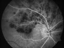
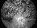
84-year-old man has a hemicentral retinal vein occlusion in the right eye. His vision has declined since then. VISUAL ACUITY: Vision OD is 20/60. IOP: OD 20. The posterior chamber intraocular lens is in good position. EXTENDED OPHTHALMOSCOPY: OD: Vertical C/D ratio is 0.5. There is a superotemporal hemicentral retinal vein occlusion with hemorrhage and edema. OCT SCAN: The OCT scan shows increased macular edema today. The average central foveal thickness today in the right eye is 386 microns compared to 213 micros. PHOTOGRAPHS: Photos confirm clinical findings. FLUORESCEIN ANGIOGRAPHY: The FA shows poor perfusion of the superior hemicentral retinal vein with some leakage in the macula in the late frames. There is also hypofluorescence corresponding to the hemorrhage. IMPRESSION: 1. HEMICENTRAL RETINAL VEIN OCCLUSION WITH MACULAR EDEMA – RIGHT EYE DISCUSSION: I explained to the patient with intravitreal injections there is about a 60% chance of his vision improving over the next six months; however, without them there is almost a 30% chance of him improving. He is not completely enthusiastic about the treatment. I think it is reasonable in his case, because his macula looked so good a few months ago, to see if possibly it might improve on its own. I asked him to return for check in one month. If the macula is no better, I will probably start Lucentis injections. If the macula looks better, then we can watch him conservatively. He will return urgently if there is a problem. In the meantime, I suggested he may want to see their medical doctor to see if there are any health problems that could be tuned up. He does have blood pressure problems, but it is under good control now. |
|||||||
|
|
|
|
||||
|
|
|
|
||||
|
|
||||||
|
|||||||
84-year-old man has a hemicentral retinal vein occlusion in the right eye. His vision has declined since then. VISUAL ACUITY: Vision OD is 20/60. IOP: OD 20. The posterior chamber intraocular lens is in good position. EXTENDED OPHTHALMOSCOPY: OD: Vertical C/D ratio is 0.5. There is a superotemporal hemicentral retinal vein occlusion with hemorrhage and edema. OCT SCAN: The OCT scan shows increased macular edema today. The average central foveal thickness today in the right eye is 386 microns compared to 213 micros. PHOTOGRAPHS: Photos confirm clinical findings. FLUORESCEIN ANGIOGRAPHY: The FA shows poor perfusion of the superior hemicentral retinal vein with some leakage in the macula in the late frames. There is also hypofluorescence corresponding to the hemorrhage. IMPRESSION: 1. HEMICENTRAL RETINAL VEIN OCCLUSION WITH MACULAR EDEMA – RIGHT EYE DISCUSSION: I explained to the patient with intravitreal injections there is about a 60% chance of his vision improving over the next six months; however, without them there is almost a 30% chance of him improving. He is not completely enthusiastic about the treatment. I think it is reasonable in his case, because his macula looked so good a few months ago, to see if possibly it might improve on its own. I asked him to return for check in one month. If the macula is no better, I will probably start Lucentis injections. If the macula looks better, then we can watch him conservatively. He will return urgently if there is a problem. In the meantime, I suggested he may want to see their medical doctor to see if there are any health problems that could be tuned up. He does have blood pressure problems, but it is under good control now.