Optic Nerve Pit - Retinal Detachment - 11 Year Old Child
|
|
This pleasant 8-year-old child was seen in the office on May 12, 2011.
It was picked up on two school exams that his vision was poor in the left eye and his last normal exam was in preschool. You saw him for an evaluation, noticed a problem with the vision in the left eye and with the macular
in the left eye, suggest he come here for an evaluation.
VISUAL ACUITY: OD 20/30, OS 5/200. IOP: 14 OU.
SLIT EXAMINATION: Normal with clear lenses.
EXTENDED OPHTHALMOSCOPY:
OD: Vertical C/D ratio is 0.1. The macula is flat and healthy.
OS: Vertical C/D ratio is 0.2. There is a whitening on the temporal edge of the optic nerve consistent with an optic disc pit and there is a serous macular detachment.
OCT SCAN: The OCT scan confirms a serous macular detachment with what looks to be inner retinal schisis, which is typical around the optic nerve pits. Photos confirm clinical findings and I did not do a fluorescein angiogram.
IMPRESSION:
1. OPTIC DISC PIT/COLOBOMA WITH SEROUS MACULAR DETACHMENT
DISCUSSION: I explained to the patient’s mother that these optic disc pits will sometimes spontaneously close and the serous macular detachment will resolve on their own. I suggest for at least a few months that we watch this. Based on the pigment epithelium in the back of the eye it does look like the retinal detachment is relatively fresh. If in two months it is no better, there are two treatments that are currently accepted, are laser or vitrectomy, both of which are reasonably effective at closing the pit. I think given the risk benefits of the two, especially given his age, it is reasonable to start with a little bit of laser around the optic nerve to try to seal up the area. He will return urgently if there is a problem and see you back regularly.
|

Optic Nerve Pit Retinal Detachment - Macular Detachment - 8 Year Old - 5/200 Vision OSvista 944 veces8-year-old child on two school exams that his vision was poor in the left eye and his last normal exam was in preschool.
VISUAL ACUITY: OD 20/30, OS 5/200. OS: Vertical C/D ratio is 0.2. There is a whitening on the temporal edge of the optic nerve consistent with an optic disc pit and there is a serous macular detachment.

(0 votos)
|
|

Optic Nerve Pit Retinal Detachment - Macular Detachment - 8 Year Old - 5/200 Vision OSvista 752 veces8-year-old child on two school exams that his vision was poor in the left eye and his last normal exam was in preschool.
VISUAL ACUITY: OD 20/30, OS 5/200. OS: Vertical C/D ratio is 0.2. There is a whitening on the temporal edge of the optic nerve consistent with an optic disc pit and there is a serous macular detachment.
(0 votos)
|
|

Optic Nerve Pit Retinal Detachment - Macular Detachment - 8 Year Old - 5/200 Vision OSvista 1094 veces8-year-old child on two school exams that his vision was poor in the left eye and his last normal exam was in preschool.
VISUAL ACUITY: OD 20/30, OS 5/200. OS: Vertical C/D ratio is 0.2. There is a whitening on the temporal edge of the optic nerve consistent with an optic disc pit and there is a serous macular detachment.
(0 votos)
|
|

Optic Nerve Pit Retinal Detachment - Macular Detachment - 8 Year Old - 5/200 Vision OSvista 842 veces8-year-old child on two school exams that his vision was poor in the left eye and his last normal exam was in preschool.
VISUAL ACUITY: OD 20/30, OS 5/200. OS: Vertical C/D ratio is 0.2. There is a whitening on the temporal edge of the optic nerve consistent with an optic disc pit and there is a serous macular detachment.

(1 votos)
|
|

Optic Nerve Pit Retinal Detachment - Macular Detachment - 8 Year Old - 5/200 Vision OSvista 785 veces8-year-old child on two school exams that his vision was poor in the left eye and his last normal exam was in preschool.
VISUAL ACUITY: OD 20/30, OS 5/200. OS: Vertical C/D ratio is 0.2. There is a whitening on the temporal edge of the optic nerve consistent with an optic disc pit and there is a serous macular detachment.
(0 votos)
|
|
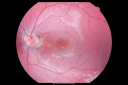
Optic Nerve Pit Retinal Detachment - Macular Detachment - 8 Year Old - 5/200 Vision OSvista 766 veces8-year-old child on two school exams that his vision was poor in the left eye and his last normal exam was in preschool.
VISUAL ACUITY: OD 20/30, OS 5/200. OS: Vertical C/D ratio is 0.2. There is a whitening on the temporal edge of the optic nerve consistent with an optic disc pit and there is a serous macular detachment.
(0 votos)
|
|

Optic Nerve Pit Retinal Detachment - Macular Detachment - 8 Year Old - 5/200 Vision OSvista 668 veces8-year-old child on two school exams that his vision was poor in the left eye and his last normal exam was in preschool.
VISUAL ACUITY: OD 20/30, OS 5/200. OS: Vertical C/D ratio is 0.2. There is a whitening on the temporal edge of the optic nerve consistent with an optic disc pit and there is a serous macular detachment.
(0 votos)
|
|

Optic Nerve Pit Retinal Detachment - Macular Detachment - 8 Year Old - 5/200 Vision OSvista 794 veces8-year-old child on two school exams that his vision was poor in the left eye and his last normal exam was in preschool.
VISUAL ACUITY: OD 20/30, OS 5/200. OS: Vertical C/D ratio is 0.2. There is a whitening on the temporal edge of the optic nerve consistent with an optic disc pit and there is a serous macular detachment.
(0 votos)
|
|

Optic Nerve Pit Retinal Detachment - Macular Detachment - 8 Year Old - 5/200 Vision OSvista 748 veces8-year-old child on two school exams that his vision was poor in the left eye and his last normal exam was in preschool.
VISUAL ACUITY: OD 20/30, OS 5/200. OS: Vertical C/D ratio is 0.2. There is a whitening on the temporal edge of the optic nerve consistent with an optic disc pit and there is a serous macular detachment.
(0 votos)
|
|

Optic Nerve Pit Retinal Detachment - Macular Detachment - 8 Year Old - 5/200 Vision OSvista 712 veces8-year-old child on two school exams that his vision was poor in the left eye and his last normal exam was in preschool.
VISUAL ACUITY: OD 20/30, OS 5/200. OS: Vertical C/D ratio is 0.2. There is a whitening on the temporal edge of the optic nerve consistent with an optic disc pit and there is a serous macular detachment.
(0 votos)
|
|

Optic Nerve Pit Retinal Detachment - Macular Detachment - 8 Year Old - 5/200 Vision OSvista 754 veces8-year-old child on two school exams that his vision was poor in the left eye and his last normal exam was in preschool.
VISUAL ACUITY: OD 20/30, OS 5/200. OS: Vertical C/D ratio is 0.2. There is a whitening on the temporal edge of the optic nerve consistent with an optic disc pit and there is a serous macular detachment.
(0 votos)
|
|
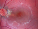
Optic Nerve Pit Retinal Detachment - Macular Detachment - 8 Year Old - Fresh Laservista 883 veces8-year-old child on two school exams that his vision was poor in the left eye and his last normal exam was in preschool.
VISUAL ACUITY: OD 20/30, OS 5/200. OS: Vertical C/D ratio is 0.2. There is a whitening on the temporal edge of the optic nerve consistent with an optic disc pit and there is a serous macular detachment.
No better after 3 months observation.
(0 votos)
|
|

Optic Nerve Pit Retinal Detachment - Macular Detachment - 8 Year Old - Fresh Laservista 794 veces8-year-old child on two school exams that his vision was poor in the left eye and his last normal exam was in preschool.
VISUAL ACUITY: OD 20/30, OS 5/200. OS: Vertical C/D ratio is 0.2. There is a whitening on the temporal edge of the optic nerve consistent with an optic disc pit and there is a serous macular detachment.
No better after 3 months observation.
(0 votos)
|
|

Optic Nerve Pit Retinal Detachment - Macular Detachment - 8 Year Old - Fresh Laservista 875 veces8-year-old child on two school exams that his vision was poor in the left eye and his last normal exam was in preschool.
VISUAL ACUITY: OD 20/30, OS 5/200. OS: Vertical C/D ratio is 0.2. There is a whitening on the temporal edge of the optic nerve consistent with an optic disc pit and there is a serous macular detachment.
No better after 3 months observation.
(0 votos)
|
|

Optic Nerve Pit Retinal Detachment - Macular Detachment - 8 Year Old - Fresh Laservista 769 veces8-year-old child on two school exams that his vision was poor in the left eye and his last normal exam was in preschool.
VISUAL ACUITY: OD 20/30, OS 5/200. OS: Vertical C/D ratio is 0.2. There is a whitening on the temporal edge of the optic nerve consistent with an optic disc pit and there is a serous macular detachment.
No better after 3 months observation.
(0 votos)
|
|
|
| 15 archivos en 1 página(s) |
|
This pleasant 8-year-old child was seen in the office on May 12, 2011.
It was picked up on two school exams that his vision was poor in the left eye and his last normal exam was in preschool. You saw him for an evaluation, noticed a problem with the vision in the left eye and with the macular
in the left eye, suggest he come here for an evaluation.
VISUAL ACUITY: OD 20/30, OS 5/200. IOP: 14 OU.
SLIT EXAMINATION: Normal with clear lenses.
EXTENDED OPHTHALMOSCOPY:
OD: Vertical C/D ratio is 0.1. The macula is flat and healthy.
OS: Vertical C/D ratio is 0.2. There is a whitening on the temporal edge of the optic nerve consistent with an optic disc pit and there is a serous macular detachment.
OCT SCAN: The OCT scan confirms a serous macular detachment with what looks to be inner retinal schisis, which is typical around the optic nerve pits. Photos confirm clinical findings and I did not do a fluorescein angiogram.
IMPRESSION:
1. OPTIC DISC PIT/COLOBOMA WITH SEROUS MACULAR DETACHMENT
DISCUSSION: I explained to the patient’s mother that these optic disc pits will sometimes spontaneously close and the serous macular detachment will resolve on their own. I suggest for at least a few months that we watch this. Based on the pigment epithelium in the back of the eye it does look like the retinal detachment is relatively fresh. If in two months it is no better, there are two treatments that are currently accepted, are laser or vitrectomy, both of which are reasonably effective at closing the pit. I think given the risk benefits of the two, especially given his age, it is reasonable to start with a little bit of laser around the optic nerve to try to seal up the area. He will return urgently if there is a problem and see you back regularly.


