NVD - Neovascularization of the Disc - Macular Edema
|
|
57-year-old man has diabetic retinopathy in both eyes.
Diabetic for 14 years with HgB A1C often over 10.
VISUAL ACUITY: OD 20/30, OS 20/40. IOP: OD 11, OS 12. The right eye has a posterior chamber intraocular lens in good position. The left eye has 1+ nuclear sclerosis.
EXTENDED OPHTHALMOSCOPY:
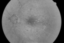
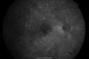
OD: Vertical C/D ratio is 0.1. There are multiple microaneurysms and light laser and patchy hemorrhages. There is also exudate inferotemporal to the fovea.
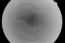
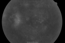
OS: Vertical C/D ratio is 0.0. There is neovascularization on the nerve extending about a disc and a half diameter off the nerve. There is also patchy microaneurysms and light laser.
OCT SCAN: The OCT scan of the right eye shows a normal central foveal thickness. The left eye however, does show macular edema centrally. Photos confirm clinical findings.
FLUORESCEIN ANGIOGRAPHY: Fluorescein angiography shows leaking microaneurysms in each eye, as well as leakage from the neovascularization in the left eye.
IMPRESSION:
1. PROLIFERATIVE DIABETIC RETINOPATHY – LEFT EYE
2. CLINICALLY SIGNIFICANT MACULAR EDEMA – LEFT EYE
3. BACKGROUND DIABETIC RETINOPATHY – RIGHT EYE
DISCUSSION: I explained to the patient that unfortunately the macula in the left eye has edema affecting the center of the fovea. I treated that eye with laser to try and control the edema and prevent further vision loss and possibly improve the vision. In addition, because of the proliferative diabetic retinopathy, the left eye needs to
start pan retinal laser. I would like to allow the focal laser to take affect though and therefore I am going to have him return in about a month for evaluation and probable further laser
|

BDR OD with Edemavu 666 fois57-year-old man has diabetic retinopathy in both eyes.
Diabetic for 14 years with HgB A1C often over 10.
VISUAL ACUITY: OD 20/30, OS 20/40. PDR OS BDR OD
(0 votes)
|
|
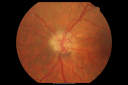
PDR OS with NVEvu 1295 fois57-year-old man has diabetic retinopathy in both eyes.
Diabetic for 14 years with HgB A1C often over 10.
VISUAL ACUITY: OD 20/30, OS 20/40. PDR OS BDR OD
(0 votes)
|
|

BDR OD with Edemavu 658 fois57-year-old man has diabetic retinopathy in both eyes.
Diabetic for 14 years with HgB A1C often over 10.
VISUAL ACUITY: OD 20/30, OS 20/40. PDR OS BDR OD
(0 votes)
|
|

PDR OS with NVEvu 793 fois57-year-old man has diabetic retinopathy in both eyes.
Diabetic for 14 years with HgB A1C often over 10.
VISUAL ACUITY: OD 20/30, OS 20/40. PDR OS BDR OD
(0 votes)
|
|

PDR OS with NVEvu 1149 fois57-year-old man has diabetic retinopathy in both eyes.
Diabetic for 14 years with HgB A1C often over 10.
VISUAL ACUITY: OD 20/30, OS 20/40. PDR OS BDR OD
(0 votes)
|
|

BDR OD with Edemavu 562 fois57-year-old man has diabetic retinopathy in both eyes.
Diabetic for 14 years with HgB A1C often over 10.
VISUAL ACUITY: OD 20/30, OS 20/40. PDR OS BDR OD
(0 votes)
|
|
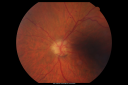
PDR OS with NVEvu 514 fois57-year-old man has diabetic retinopathy in both eyes.
Diabetic for 14 years with HgB A1C often over 10.
VISUAL ACUITY: OD 20/30, OS 20/40. PDR OS BDR OD
(0 votes)
|
|
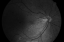
BDR OD with Edemavu 407 fois57-year-old man has diabetic retinopathy in both eyes.
Diabetic for 14 years with HgB A1C often over 10.
VISUAL ACUITY: OD 20/30, OS 20/40. PDR OS BDR OD
(0 votes)
|
|

PDR OS with NVEvu 449 fois57-year-old man has diabetic retinopathy in both eyes.
Diabetic for 14 years with HgB A1C often over 10.
VISUAL ACUITY: OD 20/30, OS 20/40. PDR OS BDR OD
(0 votes)
|
|
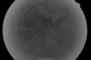
PDR OS with NVEvu 396 fois57-year-old man has diabetic retinopathy in both eyes.
Diabetic for 14 years with HgB A1C often over 10.
VISUAL ACUITY: OD 20/30, OS 20/40. PDR OS BDR OD
(0 votes)
|
|
PDR OS with NVEvu 388 fois57-year-old man has diabetic retinopathy in both eyes.
Diabetic for 14 years with HgB A1C often over 10.
VISUAL ACUITY: OD 20/30, OS 20/40. PDR OS BDR OD
(0 votes)
|
|

PDR OS with NVEvu 406 fois57-year-old man has diabetic retinopathy in both eyes.
Diabetic for 14 years with HgB A1C often over 10.
VISUAL ACUITY: OD 20/30, OS 20/40. PDR OS BDR OD
(0 votes)
|
|
PDR OS with NVEvu 356 fois57-year-old man has diabetic retinopathy in both eyes.
Diabetic for 14 years with HgB A1C often over 10.
VISUAL ACUITY: OD 20/30, OS 20/40. PDR OS BDR OD
(0 votes)
|
|

BDR OD with Edemavu 405 fois57-year-old man has diabetic retinopathy in both eyes.
Diabetic for 14 years with HgB A1C often over 10.
VISUAL ACUITY: OD 20/30, OS 20/40. PDR OS BDR OD
(0 votes)
|
|

PDR OS with NVEvu 388 fois57-year-old man has diabetic retinopathy in both eyes.
Diabetic for 14 years with HgB A1C often over 10.
VISUAL ACUITY: OD 20/30, OS 20/40. PDR OS BDR OD
(0 votes)
|
|
BDR OD with Edemavu 386 fois57-year-old man has diabetic retinopathy in both eyes.
Diabetic for 14 years with HgB A1C often over 10.
VISUAL ACUITY: OD 20/30, OS 20/40. PDR OS BDR OD
(0 votes)
|
|
PDR OS with NVEvu 387 fois57-year-old man has diabetic retinopathy in both eyes.
Diabetic for 14 years with HgB A1C often over 10.
VISUAL ACUITY: OD 20/30, OS 20/40. PDR OS BDR OD
(0 votes)
|
|
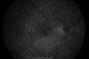
BDR OD with Edemavu 380 fois57-year-old man has diabetic retinopathy in both eyes.
Diabetic for 14 years with HgB A1C often over 10.
VISUAL ACUITY: OD 20/30, OS 20/40. PDR OS BDR OD
(0 votes)
|
|
|
|
|
|
57-year-old man has diabetic retinopathy in both eyes.
Diabetic for 14 years with HgB A1C often over 10.
VISUAL ACUITY: OD 20/30, OS 20/40. IOP: OD 11, OS 12. The right eye has a posterior chamber intraocular lens in good position. The left eye has 1+ nuclear sclerosis.
EXTENDED OPHTHALMOSCOPY:
OD: Vertical C/D ratio is 0.1. There are multiple microaneurysms and light laser and patchy hemorrhages. There is also exudate inferotemporal to the fovea.
OS: Vertical C/D ratio is 0.0. There is neovascularization on the nerve extending about a disc and a half diameter off the nerve. There is also patchy microaneurysms and light laser.
OCT SCAN: The OCT scan of the right eye shows a normal central foveal thickness. The left eye however, does show macular edema centrally. Photos confirm clinical findings.
FLUORESCEIN ANGIOGRAPHY: Fluorescein angiography shows leaking microaneurysms in each eye, as well as leakage from the neovascularization in the left eye.
IMPRESSION:
1. PROLIFERATIVE DIABETIC RETINOPATHY – LEFT EYE
2. CLINICALLY SIGNIFICANT MACULAR EDEMA – LEFT EYE
3. BACKGROUND DIABETIC RETINOPATHY – RIGHT EYE
DISCUSSION: I explained to the patient that unfortunately the macula in the left eye has edema affecting the center of the fovea. I treated that eye with laser to try and control the edema and prevent further vision loss and possibly improve the vision. In addition, because of the proliferative diabetic retinopathy, the left eye needs to
start pan retinal laser. I would like to allow the focal laser to take affect though and therefore I am going to have him return in about a month for evaluation and probable further laser


